. 2008 Apr;135(7):1223-34.
doi: 10.1242/dev.015933. Epub 2008 Feb 20.

Mesenchyme-dependent BMP signaling directs the timing of mandibular osteogenesis

Affiliations

Mesenchyme-dependent BMP signaling directs the timing of mandibular osteogenesis

Amy E Merrill et al. Development. 2008 Apr.

Abstract

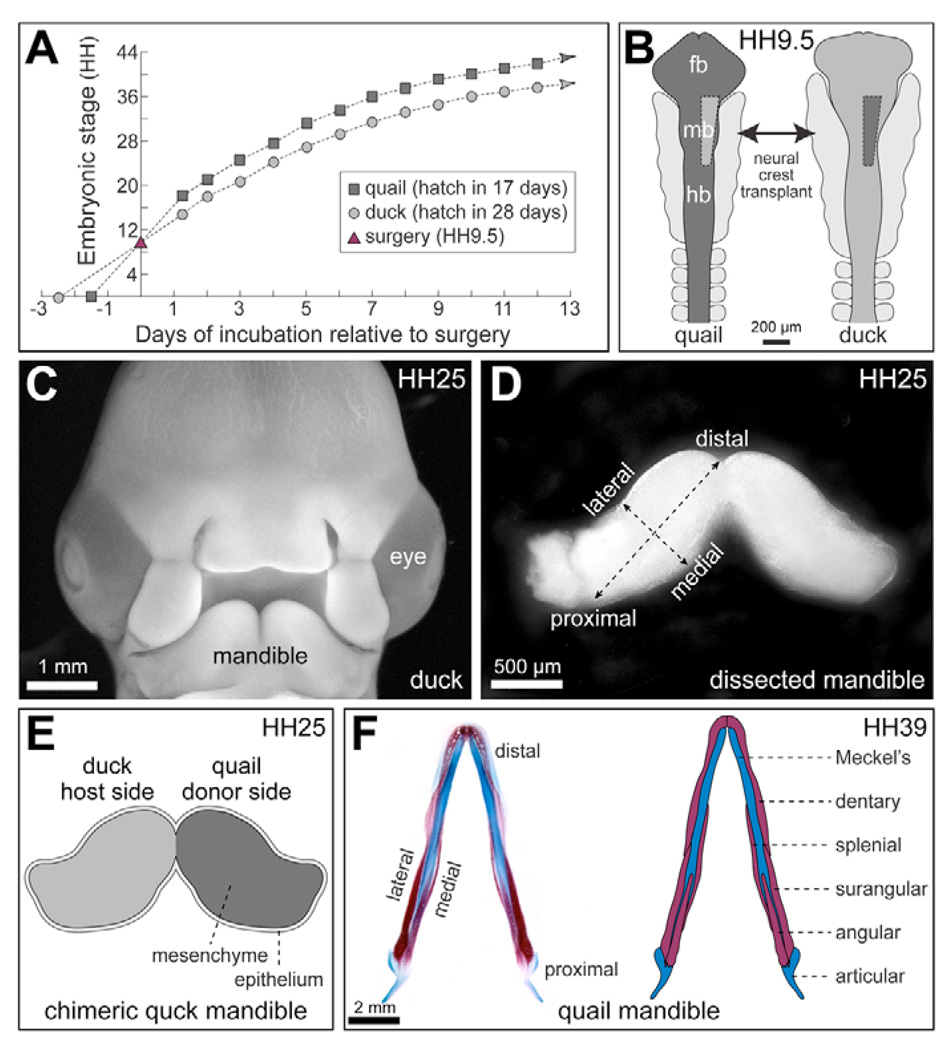
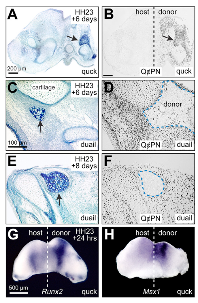
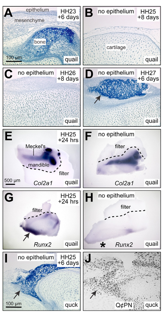
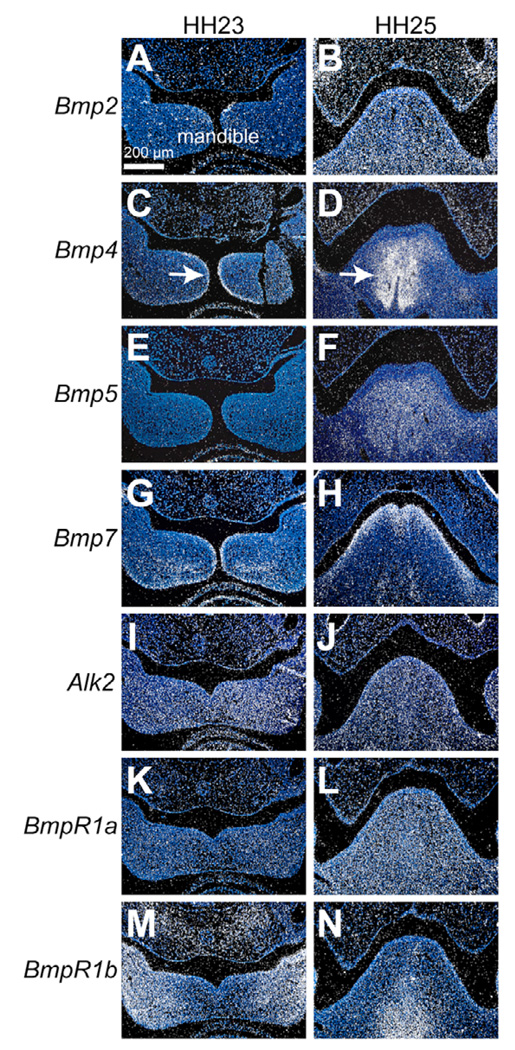
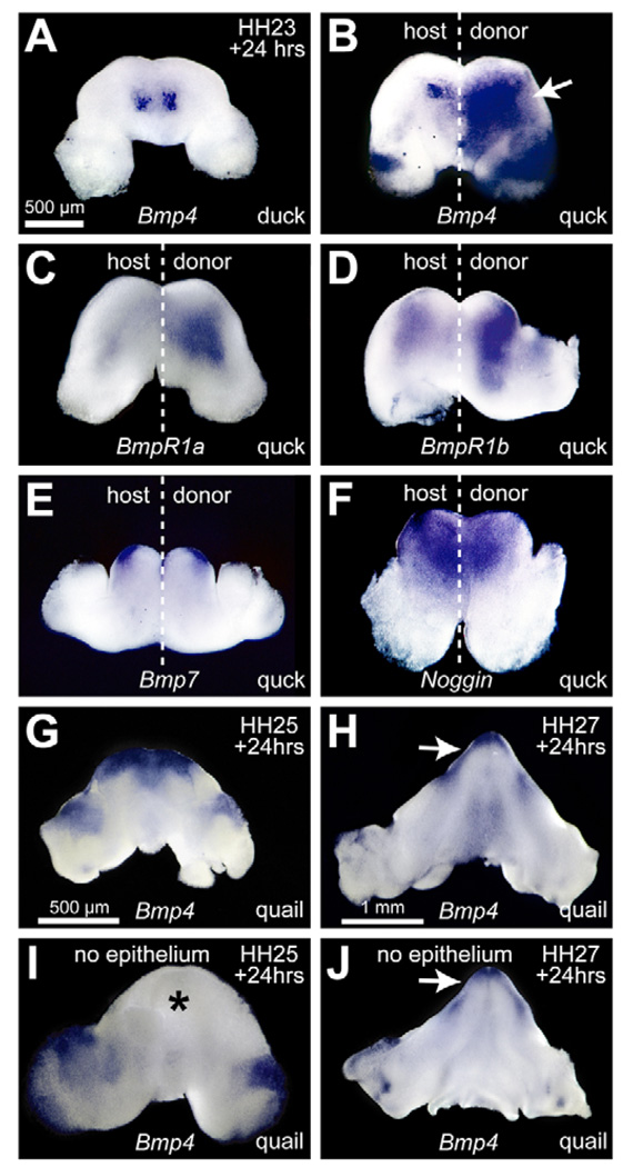
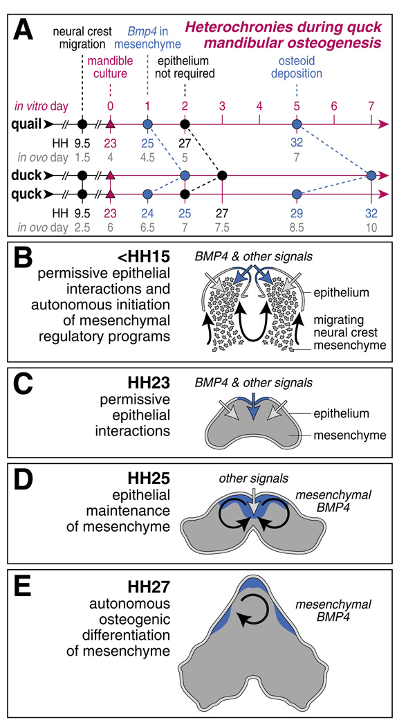
To identify molecular and cellular mechanisms that determine when bone forms, and to elucidate the role played by osteogenic mesenchyme, we employed an avian chimeric system that draws upon the divergent embryonic maturation rates of quail and duck. Pre-migratory neural crest mesenchyme destined to form bone in the mandible was transplanted from quail to duck. In resulting chimeras, quail donor mesenchyme established significantly faster molecular and histological programs for osteogenesis within the relatively slower-progressing duck host environment. To understand this phenotype, we assayed for changes in the timing of epithelial-mesenchymal interactions required for bone formation and found that such interactions were accelerated in chimeras. In situ hybridization analyses uncovered donor-dependent changes in the spatiotemporal expression of genes, including the osteo-inductive growth factor Bmp4. Mesenchymal expression of Bmp4 correlated with an ability of quail donor cells to form bone precociously without duck host epithelium, and also relied upon epithelial interactions until mesenchyme could form bone independently. Treating control mandibles with exogenous BMP4 recapitulated the capacity of chimeras to express molecular mediators of osteogenesis prematurely and led to the early differentiation of bone. Inhibiting BMP signaling delayed bone formation in a stage-dependent manner that was accelerated in chimeras. Thus, mandibular mesenchyme dictates when bone forms by temporally regulating its interactions with epithelium and its own expression of Bmp4. Our findings offer a developmental mechanism to explain how neural crest-derived mesenchyme and BMP signaling underlie the evolution of species-specific skeletal morphology.


Figures

Fig. 1
Fig. 1. Creation and culture of avian chimeric mandibles
(A) To manipulate temporal information being conveyed between mesenchyme and epithelium, we exploited the divergent maturation rates of quail and duck embryos. Embryos were incubated until stage-matched at Hamburger-Hamilton (HH) 9.5. (B) Unilateral grafts of neural crest from the hindbrain (hb) and midbrain (mb) were transplanted between quail and duck. (C) Quail, duck and chimeras were harvested at various stages. (D) Mandibular primordia were surgically excised and cultured. (E) Quck mandibles contained quail donor mesenchyme (dark gray) alongside duck host mesenchyme (gray) and epithelium (light gray). (F) Skeletal structures derived from the mandibular primordia, visualized by Alizarin Red (bone) and Alcian Blue (cartilage) staining in actual (left) and schematic (right) dorsal view.
Fig. 2
Fig. 2. Mesenchyme determines the timing of intramembranous ossification
(A) In quck mandibles, quail mesenchyme maintains its faster timetable for bone formation within the slower environment of duck hosts, based on histological detection of matrix (arrow) using Osteoid stain (blue). (B) Staining for bone is coincident only with Q¢PN positive cells (i.e. quail-derived black cells, arrow) on the donor side. (C) Reciprocal transplants that generate duail result in abundant quail host-derived bone in chimeric mandibles (arrow). (D) On the duck donor-derived side, bone has yet to form at 6 days of culture (dashed outline). Note the sporadic Q¢PN-positive angioblasts and endothelial cells, which are derived from quail host mesoderm, among duck donor mesenchyme (Q¢PN-negative). (E,F) After 8 days of culture, which is equivalent to the time required for bone to form in control duck, duck-derived mesenchyme (i.e. Q¢PN-negative) stains positively (dashed outline). (G,H) Whole-mount in situ hybridization reveals that Runx2 and Msx1 are upregulated on the donor-derived side.
Fig. 3
Fig. 3. Mesenchyme sets the timing of tissue interactions required for osteogenesis
(A) HH23 quail mandibles cultured for 6 days are positive for Osteoid (blue). (B,C) By contrast, quail mesenchyme harvested without epithelium at HH25 and HH26, and cultured for up to 8 days forms cartilage but not bone. (D) By HH27, mesenchyme cultured without epithelium makes bone (arrow). (E,F) Mandibles harvested from quail at HH25 and cultured with and without epithelium for 24 hours express Col2a1 in Meckel’s cartilage as shown in lateral view. (G) HH25 mesenchyme cultured with epithelium for 24 hours expresses Runx2 (arrow). (H) When mandibles are cultured without epithelium, Runx2 expression is significantly downregulated (asterisk). (I,J) Quck mandibles harvested at HH25 and cultured for 6 days without epithelium form bone (arrow) due to faster developing quail donor mesenchyme (arrow).
Fig. 4
Fig. 4. Spatiotemporal expression patterns of BMP pathway members in quail
(A,B) Bmp2 (white), detected at low levels in epithelium at HH23, is expressed across the mandible at HH25. (C) Bmp4 transcripts are restricted to epithelium at HH23 (arrow). (D) By HH25, Bmp4 transcripts are expressed only in mesenchyme (arrow). (E,F) Bmp5 is not expressed at HH23 but is detected at HH25 in mesenchyme. (G,H) Bmp7 expression is observed in both epithelium and mesenchyme at HH23 and HH25. (I,J) Alk2 is uniformly expressed throughout the mandible at HH23 and HH25. (K,L) Bmpr1a, although not detected at HH23, is expressed across the mandible by HH25. (M,N) Bmpr1b transcripts are concentrated in lateral mesenchyme and epithelium at HH23 and become localized to medial mesenchyme by HH25.
Fig. 5
Fig. 5. Donor-induced expression of BMP pathway members and epithelial maintenance of Bmp4
(A) Bmp4 expression is restricted to epithelial domains of HH23 duck control mandibles cultured for 24 hours as shown by whole-mount in situ hybridization. (B) In quck mandibles, Bmp4 is prematurely upregulated in a large domain across donor-derived mesenchyme (arrow). (C,D) BMP4 receptors, Bmpr1a and Bmpr1b, are upregulated in donor-derived mesenchyme. (E) Such expression changes are specific to Bmp4 as no changes are observed during these stages for other BMPs, such as Bmp7. (F) Similarly, no donor-mediated changes in Noggin expression are apparent. (G) In HH25 mandibles, Bmp4 expression is found broadly across distal mesenchyme. (H) By HH27, expression of Bmp4 in distal mesenchyme is further restricted along the lower jaw (arrow). (I) When epithelium is removed from HH25 mandibles, Bmp4 expression is lost in distal mesenchyme (asterisk). (J) By HH27, distal mesenchyme maintains expression of Bmp4 even without overlying epithelium (arrow).
Fig. 6
Fig. 6. Exogenous BMP4 induces premature differentiation of bone, whereas Noggin treatment delays differentiation of bone
(A) HH23 Quail mandibles treated with BMP4 were cultured for 24 hours and processed for whole-mount in situ hybridization. Control contralateral sides were treated with BSA. (B) Msx1 is expressed medially. Upon treatment with BMP4, Msx1 expression expands laterally. (C) Twist1 is expressed medially but BMP4 treatment causes a downregulation. (D) To evaluate the extent of BMP4-induced Runx2, mandibles were treated at HH21, which is a stage that lacks endogenous Runx2 expression. Runx2 is slightly upregulated following BMP4 treatment. (E) Quail explants were harvested at HH23, treated with BMP4, and cultured for 5 days. (F) Section through a quail mandible treated unilaterally with BMP4 at HH23, cultured for 6 days, and stained with Osteoid (dark blue bone within dashed box). (G) Pixels comprising bone (red) were quantified to determine matrix volume (MV). (H) Pixels within a given condensation (red line) were quantified to determine condensation volume (CV). (I) Following 24 hours of culture, Noggin treatments inhibit Msx1 expression in explants. (J) In control quail mandibles, Noggin delays bone formation prior to 48 hours of culture, but afterwards bone can form (arrow). (K,L) In quck chimeras, this period of responsiveness occurs before 24 hours and bone (arrow) forms thereafter due to the presence of the faster-developing donor quail cells (Q¢PN-positive). Tx, treatment.
Fig. 7
Fig. 7. Summary of experimental results and a model for the role of mesenchyme in regulating mandibular osteogenesis via BMP signaling
(A) The HH staging system operates independently of time, and instead is based on external morphological characters. Thus, quail and duck can be aligned at equivalent points during development by incubating them for prescribed lengths of time. Internal events such as Bmp4 expression and osteoid matrix deposition occur at equivalent stages in quail and duck but are accelerated on the donor side of quck relative to the duck host. (B) Neural crest-derived mesenchyme (dark gray) migrates from the midbrain and hindbrain around HH9.5 and settles in the mandibular primordia by HH15 (Tosney, 1982; Noden, 1991). During this early period, they establish the timing of interactions required for bone formation by signaling to adjacent epithelium (black arrows). Simultaneously, the epithelium is expressing growth factors such as Bmp4 (blue arrows) and other genes (light gray arrows) that support autonomous progression of mesenchymal regulatory programs. (C) At HH23, epithelium continues to express Bmp4 (blue domain) and other factors that enable mesenchymal cells to continue outgrowth. (D) By HH25, Bmp4 expression transitions from epithelium to mesenchyme. Mesenchymal expression of Bmp4 is auto-regulatory (black arrows), but requires permissive signals from epithelium. (E) By HH27, epithelium is no longer required to maintain Bmp4 expression and mesenchyme autonomously differentiates into bone.

References

    1. Aberg T, Wozney J, Thesleff I. Expression patterns of bone morphogenetic proteins (Bmps) in the developing mouse tooth suggest roles in morphogenesis and cell differentiation. Dev. Dyn. 1997;210:383–396. - PubMed
    1. Abzhanov A, Protas M, Grant BR, Grant PR, Tabin CJ. Bmp4 and morphological variation of beaks in Darwin's finches. Science. 2004;305:1462–1465. - PubMed
    1. Abzhanov A, Rodda SJ, McMahon AP, Tabin CJ. Regulation of skeletogenic differentiation in cranial dermal bone. Development. 2007;134:3133–3144. - PubMed
    1. Albertson RC, Kocher TD. Genetic and developmental basis of cichlid trophic diversity. Heredity. 2006;97:211–221. - PubMed
    1. Albertson RC, Streelman JT, Kocher TD, Yelick PC. Integration and evolution of the cichlid mandible: the molecular basis of alternate feeding strategies. Proc. Natl. Acad. Sci. USA. 2005;102:16287–16292. - PMC - PubMed

Publication types

MeSH terms

Substances