. 2008 May;82(9):4265-74.
doi: 10.1128/JVI.02602-07. Epub 2008 Feb 20.

Alveolar macrophages are indispensable for controlling influenza viruses in lungs of pigs

Affiliations

Alveolar macrophages are indispensable for controlling influenza viruses in lungs of pigs

Heui Man Kim et al. J Virol. 2008 May.

Abstract

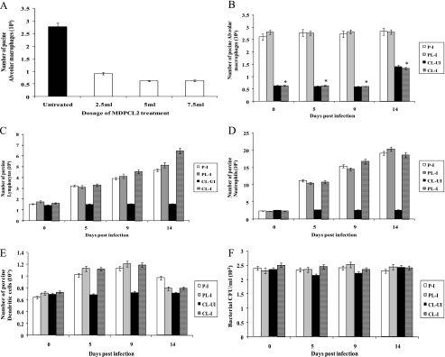
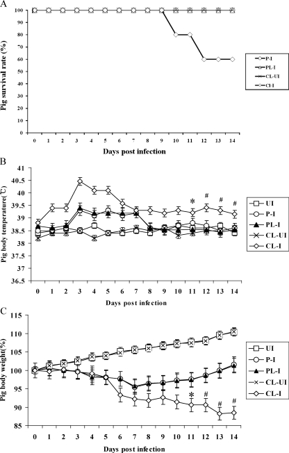
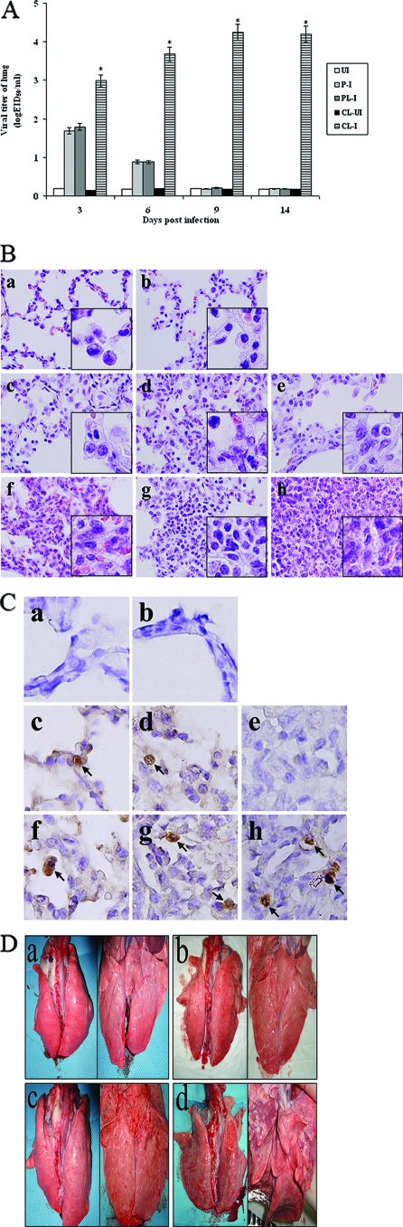
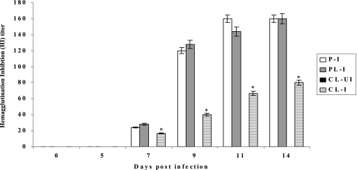
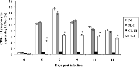
Alveolar macrophages constitutively reside in the respiratory tracts of pigs and humans. An in vivo role of alveolar macrophages in defending against influenza viruses in mice infected with a reassorted influenza virus, 1918 HA/NA:Tx/91, was reported, but there has been no report on an in vivo role of alveolar macrophages in a natural host such as a pig using currently circulating human influenza virus. Here we show that in vivo depletion of alveolar macrophages in pigs by dichloromethylene diphosphonate (MDPCL2) treatment results in 40% mortality when pigs are infected with currently circulating human H1N1 influenza viruses, while none of the infected control pigs died. All infected pigs depleted of alveolar macrophages suffered from more severe respiratory signs than infected control pigs. Induction of tumor necrosis factor alpha in the infected pigs depleted of alveolar macrophages was significantly lower than that in the lungs of infected control pigs, and the induction of interleukin-10, an immunosuppressive cytokine, significantly increased in the lungs of infected pigs depleted of alveolar macrophages compared to infected control pigs. When we measured antibody titers and CD8(+) T lymphocytes expressing gamma interferon (IFN-gamma), lower antibody titers and a lower percentage of CD8(+) T lymphocytes expressing IFN-gamma were detectable in MDPCL2-treated infected pigs than in phosphate-buffered saline- and liposome-treated and infected pigs. Taken together, our findings suggest that alveolar macrophages are essential for controlling H1N1 influenza viruses in pigs.


Figures

FIG. 1.
FIG. 1.
Efficiency of depletion of alveolar macrophages in the lungs of pigs by MDPCL2 treatment. (A) Determination of MDPCL2 dosage. Six-week-old pigs were i.m. anesthetized with Zoletil (2 mg/kg) and were daily i.n. administered 2.5 ml, 5 ml, or 7.5 ml of MDPCL2. Three pigs per time point were euthanized, and BAL was performed with 50 ml of PBS (pH 7.4). The lavaged samples were cytospun and stained with mouse anti-porcine macrophage antibody and FITC-labeled goat anti-mouse secondary antibody. Alveolar macrophages stained with antibody specific for porcine alveolar macrophages were counted under a fluorescence microscope. Data are the means for three pigs ± standard errors per time point. (B) Number of alveolar macrophages in MDPCL2-treated infected pigs. Six-week-old pigs were i.m. anesthetized with Zoletil (2 mg/kg) and were daily i.n. administered 5 ml of MDPCL2 or PBS-liposomes for 3 days before being infected with 2 ml of 104 EID50 of human H1N1 (A/New Caledonia/20/99) influenza virus. Three pigs per time point were euthanized, and BAL was performed with 50 ml of PBS (pH 7.4). The lavaged samples were cytospun and stained with mouse anti-porcine macrophage antibody and FITC-labeled goat anti-mouse secondary antibody. Alveolar macrophages stained with antibody specific for porcine alveolar macrophages were counted under a fluorescence microscope. Data are the means for three pigs ± standard errors per time point. We statistically compared group 1-4 with group 1-2. *, P < 0.05. P-I (group 1-1), PBS-treated infected pigs; PL-I (group 1-2), PBS-liposome-treated infected pigs; CL-UI (group 1-3), MDPCL2-treated uninfected pigs; CL-I (group 1-4), MDPCL2-treated infected pigs. (C) Number of porcine lymphocytes in MDPCL2-treated infected pigs. Lymphocytes were counted by Wright staining using the same samples as for panel B. Data are the means for three pigs ± standard errors per time point. (D) Number of neutrophils in MDPCL2-treated infected pigs. Neutrophils were counted by Wright staining using the same samples as for panel B. Data are the means for three pigs ± standard errors per time point. (E) Number of dendritic cells in MDPCL2-treated infected pigs. Dendritic cells were counted by Wright staining using the same samples as for panel B. Data are the means for three pigs ± standard errors per time point. (F) Number of bacteria in MDPCL2-treated infected pigs. Bacteria were counted using a MacConkey agar plate, a tryptic soy agar plate, or blood agar and the same samples as for panel B. Data are the means for three pigs ± standard errors per time point.
FIG. 2.
FIG. 2.
Clinical signs in pigs depleted of alveolar macrophages by MDPCL2 treatment. MDPCL2-treated pigs or PBS-liposome-treated pigs were i.n. infected with 2 ml of 104 EID50 of human H1N1 (A/New Caledonia/20/99) influenza virus. Clinical signs such as survival rates, change of body temperatures, change of pig body weights, and change of lung images were observed daily for 14 days after infection. This experiment was independent from the one described for Fig. 1. (A) Survival rates of pigs. Two MDPCL2-treated and infected pigs died at 10 days p.i., and an additional two MDPCL2-treated and infected pigs died at 12 days p.i. P-I (group 3-1), PBS-treated infected pigs; PL-I (group 3-2), PBS-liposome-treated infected pigs; CL-UI (group 3-3), MDPCL2-treated uninfected pigs; CL-I (group 3-4), MDPCL2-treated infected pigs. (B) Change of body temperature in pigs. Pig body temperatures were measured rectally. Data are the means for 10 pigs ± standard errors per time point, except at 10 and 12 days p.i., when two pigs at each time point died. *, mean for eight pigs ± standard errors; #, mean for six pigs ± standard errors. UI (group 3-5), untreated and uninfected pigs. (C) Change of body weight in pigs. Data are the means for 10 pigs ± standard errors per time point, except at 10 and 12 days p.i., when two pigs at each time point died. *, mean for eight pigs ± standard errors; #, mean for six pigs ± standard errors.
FIG. 3.
FIG. 3.
Viral titers, cytokine responses, and histopathological signs in the lungs of pigs. Pigs (20 pigs per group) were i.n. infected with 2 ml of 104 EID50 of human H1N1 (A/New Caledonia/20/99) influenza virus. Three pigs per time point were euthanized before the left cranial lobes of lungs were collected for determining viral titers, cytokine responses, and histopathology. This experiment was independent from those described for Fig. 1 and Fig. 2. (A) Viral titers in the lungs of pigs. Viral titers were determined by log10 EID50/ml. Data are the means for three pigs ± standard errors per time point. We statistically compared group 4-4 with group 4-2. *, P < 0.05. P-I (group 4-1), PBS-treated infected pigs; PL-I (group 4-2), PBS-liposome-treated infected pigs; CL-UI (group 4-3), MDPCL2-treated uninfected pigs; CL-I (group 4-4), MDPCL2-treated infected pigs; UI (group 4-5), untreated and uninfected pigs. (B) Staining of lungs of pigs with H&E (magnification, ×400). The left cranial lobes of lungs were cut by 5 micrometers and were stained with H&E. a, lung of an untreated and uninfected pig (group 4-5); b, lung of an MDPCL2-treated and uninfected pig (group 4-3); c to e, lungs of PBS-liposome-treated and infected pigs (group 4-2) at 3, 6, and 9 days p.i., respectively; f to h, lungs of MDPCL2-treated and infected pigs (group 4-4) at 3, 6, and 9 days p.i., respectively. The insets show magnifications of ×1,000. (C) Immunohistochemical staining of lungs of pigs using influenza A virus antinucleoprotein antibody (magnification, ×1,000). The left cranial lobes of lungs were cut by five micrometers and were stained with mouse influenza A virus antinucleoprotein, peroxidase-labeled goat anti-mouse immunoglobulin, and with stable diaminobenzidine peroxidase substrate. The stained tissue sections were counterstained with hematoxylin before they were evaluated under the microscope. a, lung of an untreated and uninfected pig (group 4-5); b, lung of an MDPCL2-treated and uninfected pig (group 4-3); c to e, lungs of PBS-liposome-treated and infected pigs (group 4-2) at 3, 6, and 9 days p.i.; f to h, lungs of MDPCL2-treated and infected pigs (group 4-4) at 3, 6, and 9 days p.i. Arrows, cells positive for influenza A virus nucleoprotein protein. (D) Photographs of lungs of pigs. Lungs of pigs were photographed at 9 days p.i. after surviving pigs were euthanized. a, lung of an untreated and uninfected pig (group 4-5); b, lung of an MDPCL2-treated and uninfected pig (group 4-3); c, lung of a PBS-liposome-treated and infected pig (group 4-2); d, lung of an MDPCL2-treated and infected pig (group 4-4). Left, dorsal position; right, ventral position.
FIG. 4.
FIG. 4.
Quantification of inflammatory cytokines in lungs. One-gram portions of left cranial lobes of lungs from the same pigs per time point described in Fig. 3 were suspended in 1 ml of PBS (pH 7.4) before being disrupted by homogenizing and then freezing and thawing three times. The supernatants from the disrupted tissues were collected to analyze the porcine inflammatory cytokines TNF-α (A), IFN-γ (B), IL-10 (C), and IL-4 (D) using porcine cytokine-specific ELISA kits. The quantity of cytokines was calculated and plotted based on a standard curve for each cytokine. Data are the mean for three pigs ± standard errors per time point. We statistically compared group 4-4 with group 4-2. **, P < 0.001; *, P < 0.05. P-I (group 4-1), PBS-treated infected pigs; PL-I (group 4-2), PBS-liposome-treated infected pigs; CL-UI (group 4-3), MDPCL2-treated uninfected pigs; CL-I (group 4-4), MDPCL2-treated infected pigs; UI (group 4-5), untreated and uninfected pigs.
FIG. 5.
FIG. 5.
Determination of antibody titers in pigs. Sera were collected from MDPCL2-treated or untreated pigs infected with 2 ml of 104 EID50 of human H1N1 (A/New Caledonia/20/99) influenza virus until 14 days p.i. Antibody titers were determined by HI assay using A/New Caledonia/20/99 (H1N1) and turkey red blood cells (0.5%). Data are the means for three pigs ± standard errors per time point. P-I (group 5-1), PBS-treated infected pigs; PL-I (group 5-2), PBS-liposome-treated and infected pigs; CL-UI (group 5-3), MDPCL2-treated uninfected pigs; CL-I (group 5-4), MDPCL2-treated infected pigs. We statistically compared group 5-4 with group 5-2. *, P < 0.05.
FIG. 6.
FIG. 6.
Determination of CD8+ T lymphocytes expressing IFN-γ. Lymphocytes were isolated from BAL samples from MDPCL2-treated or untreated pigs infected with 2 ml of 104 EID50 of human H1N1 (A/New Caledonia/20/99) influenza virus. The percentage of CD8+ T cells expressing IFN-γ was determined by flow cytometry analysis using phycoerythrin-labeled rabbit anti-porcine IFN-γ antibody and FITC-labeled mouse anti-porcine CD8 antibody. Data are the means for three pigs ± standard errors per time point. We statistically compared group 5-4 with group 5-2. *, P < 0.05. P-I (group 5-1), PBS-treated infected pigs; PL-I (group 5-2), PBS-liposome-treated infected pigs; CL-UI (group 5-3), MDPCL2-treated uninfected pigs; CL-I (group 5-4), MDPCL2-treated infected pigs.

References

    1. Akaike, T. 2001. Role of free radicals in viral pathogenesis and mutation. Rev. Med. Virol. 1187-101. - PMC - PubMed
    1. Bancroft, J. D., and A. Stevens. 1996. Theory and practice of histological techniques, 4th ed. Churchill Livingstone, New York, NY.
    1. Bender, B. S., and P. A. Small, Jr. 1992. Influenza: pathogenesis and host defense. Semin. Respir. Infect. 738-45. - PubMed
    1. Boon, A. C., G. de Mutsert, Y. M. Graus, R. A. Fouchier, K. Sintnicolaas, A. D. Osterhaus, and G. F. Rimmelzwaan. 2002. The magnitude and specificity of influenza A virus-specific cytotoxic T-lymphocyte responses in humans is related to HLA-A and -B phenotype.J. Virol. 76582-590. - PMC - PubMed
    1. Cheung, C. Y., L. L. Poon, A. S. Lau, W. Luk, Y. L. Lau, K. F. Shortridge, S. Gordon, Y. Guan, and J. S. Peiris. 2002. Induction of proinflammatory cytokines in human macrophages by influenza A (H5N1) viruses: a mechanism for the unusual severity of human disease? Lancet. 3601831-1837. - PubMed

MeSH terms