. 2008 Apr;82(8):4042-51.
doi: 10.1128/JVI.02436-07. Epub 2008 Feb 20.

Deletion of Epstein-Barr virus BFLF2 leads to impaired viral DNA packaging and primary egress as well as to the production of defective viral particles

Affiliations

Deletion of Epstein-Barr virus BFLF2 leads to impaired viral DNA packaging and primary egress as well as to the production of defective viral particles

Marisa Granato et al. J Virol. 2008 Apr.

Abstract

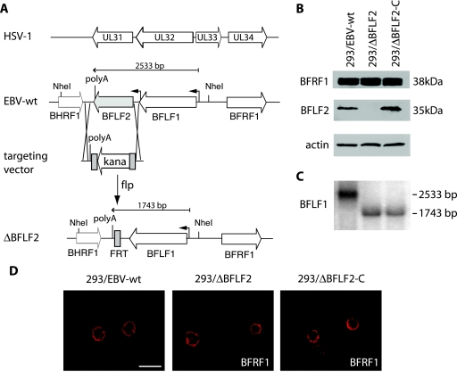
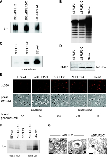
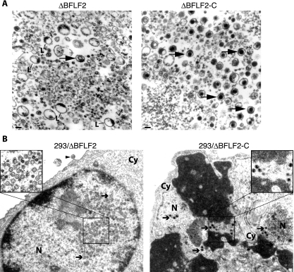
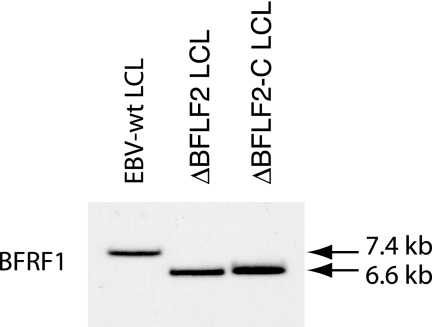
Previous genetic and biochemical studies performed with several members of the Alphaherpesvirus subfamily have shown that the UL31 and UL34 proteins are essential components of the molecular machinery that mediates the primary egress of newly assembled capsids across the nuclear membrane. Further, there is substantial evidence that BFLF2 and BFRF1, the respective positional homologs of UL31 and UL34 in the Epstein-Barr virus (EBV) genome, are also their functional homologs, i.e., that the UL31/UL34 pathway is common to distant herpesviruses. However, the low degree of protein sequence identity between UL31 and BFLF2 would argue against such a hypothesis. To further clarify this issue, we have constructed a recombinant EBV strain devoid of BFLF2 (DeltaBFLF2) and show that BFLF2 is crucial for efficient virus production but not for lytic DNA replication or B-cell transformation. This defective phenotype could be efficiently restored by trans complementation with a BFLF2 expression plasmid. Detailed analysis of replicating cells by electron microscopy revealed that, as expected, DeltaBFLF2 viruses not only failed to egress from the nucleus but also showed defective DNA packaging. Nonfunctional primary egress did not, however, impair the production and extracellular release of enveloped but empty viral particles that comprised L particles containing tegument-like structures and a few virus-like particles carrying empty capsids. The DeltaBFLF2 and DeltaUL31 phenotypes therefore only partly overlap, from which we infer that BFLF2 and UL31 have substantially diverged during evolution to fulfil related but distinct functions.


Figures

FIG. 1.
FIG. 1.
Construction of a ΔBFLF2 mutant virus. (A) Schematic drawing of the HSV-1 and EBV loci that carry positional homologs UL31/UL34 and BFLF2/BFRF1. The EBV genome is shown before and after disruption of the BFLF2 open reading frame. One FLP recombinase target (FRT) site is left between the intact BHRF1 and BFLF1 genes after excision of the KAN resistance cassette with FLP recombinase. (B) Whole protein extracts from lytically induced 293/EBV-wt, 293/ΔBFLF2, and 293/ΔBFLF2-C cells were used to assess BFRF1 (upper panel), BFLF2 (middle panel), and actin (lower panel) protein synthesis in a Western blot analysis. The BFRF1 expression level is unchanged in the 293/ΔBFLF2 mutant. (C) Northern blot analysis of lytically induced cells. Total RNAs extracted from induced 293/ΔBFLF2, 293/ΔBFLF2-C, and 293/wt-EBV cells were separated in an agarose gel and hybridized with a BFLF1-specific probe. The BFLF1 primary transcript encoded by ΔBFLF2 is smaller than its wild-type counterpart as a result of the deletion, but expression levels are similar in all three samples. (D) Immunofluorescence analysis with a confocal laser scanning microscope of BFRF1 localization in induced 293/EBV-wt, 293/ΔBFLF2, and 293/ΔBFLF2-C cells. BFRF1 localization at the perinuclear region is not altered in 293/ΔBFLF2 cells. Scale bar, 20 μm.
FIG. 2.
FIG. 2.
BFLF2 is not required for DNA replication but is important for DNA packaging. (A) Gardella gel analysis of lytically induced producer cells, followed by Southern blotting with a gp350-specific DNA probe. 293/ΔBFLF2 cells produce amounts of viral DNA similar to those produced by trans-complemented (293/ΔBFLF2-C) and 293/EBV-wt cells. L, linear DNA. (B) Southern blot analysis with a TR-specific probe confirmed the presence of newly replicated monomeric linear DNA molecules in lytically induced 293/ΔBFLF2 cells. Genomic DNA was cleaved with BamHI and separated on a 0.8% agarose gel. (C) Gardella gel analysis of equal volumes of pelleted virus supernatants from the same cell lines as in panel A shows a drastic reduction in mature virus particles in the ΔBFLF2 virus stocks. The difference in signal intensities between the ΔBFLF2-C and EBV-wt virus stocks reflects the difference in MOIs (1.5 × 10⁷ and 7 × 10⁷ genome equivalents/ml, respectively). (D) Western blot analysis of virus supernatant showing the same amount of BNRF1 tegument protein in ΔBFLF2, ΔBFLF2-C, and EBV-wt virus particles. Protein extract from 5 ml of ultracentrifuged virus supernatant was separated on a 10% sodium dodecyl sulfate-polyacrylamide gel and probed with an antiserum against BNRF1 tegument protein. (E) Detection of bound viruses on resting B cells. Cells were incubated with supernatants containing mutant or wild-type viruses either at an MOI of 10 or with equal volumes and immunostained with an antibody specific to gp350. B cells exposed to ΔBFLF2 are covered with a higher number of viral gp350-positive structures (red dots on the cell surface) than are B cells exposed to EBV-wt and ΔBFLF2-C virus particles at an equal MOI (left part of panel). Exposure of B cells with equal volumes of supernatants showed similar levels of staining (right part of panel). (F) Gardella gel analysis of virus bound to B cells. B cells incubated with the same virus stocks as in panel E were submitted to Southern blotting and hybridization with a gp350-specific DNA probe. No viral DNA could be detected after incubation of B cells with ΔBFLF2 supernatants. L, linear DNA. (G) ΔBFLF2 virus particles bound to B cells are devoid of DNA. B cells incubated with ΔBFLF2 or ΔBFLF2-C virus supernatant were analyzed by electron microscopy. Only VLP (left side) and L particles (right side) were identified at the surface and in intracellular vesicles of B cells, respectively, after incubation with ΔBFLF2 supernatants. In contrast, most particles bound after incubation with ΔBFLF2-C supernatant are mature and contain DNA. Cy, cytoplasm; Pm, plasma membrane; N, nucleus. Bars, 100 nm.
FIG. 3.
FIG. 3.
BFLF2 is required for efficient nuclear egress and DNA packaging. (A) Electron micrographs of pelleted ΔBFLF2 and ΔBFLF2-C particles. L particles dominated the picture in the ΔBFLF2 supernatant, but rare VLP could also be identified (arrow, left side). ΔBFLF2-C supernatant mainly contained mature virions (arrows, right side). Bar, 100 nm. L, L particles. (B) Electron micrograph of lytically induced 293/ΔBFLF2 cells showing empty A capsids and B capsids (arrows, left side) in the nucleus. One virus-like empty particle could be identified at the surface of these cells (arrowhead). In contrast, induced 293/ΔBFLF2-C cells contain a normal number of electron-dense C capsids (arrows, right side). Cy, cytoplasm; N, nucleus.
FIG. 4.
FIG. 4.
Southern blot analysis of LCLS obtained by infection of resting B cells with ΔBFLF2, ΔBFLF2-C, or EBV-wt supernatants. Genomic DNA was cleaved with BamHI and separated by gel electrophoresis. Hybridization with a BFRF1-specific probe detected a 6.6-kb BamHI F fragment corresponding to the deleted BFLF2-encoding gene in both 293/ΔBFLF2 and 293/ΔBFLF2-C LCLs. LCLs infected with the wild-type virus carry an intact 7.4-kb BamHI F fragment.

References

    1. Calderwood, M. A., K. Venkatesan, L. Xing, M. R. Chase, A. Vazquez, A. M. Holthaus, A. E. Ewence, N. Li, T. Hirozane-Kishikawa, D. E. Hill, M. Vidal, E. Kieff, and E. Johannsen. 2007. Epstein-Barr virus and virus human protein interaction maps. Proc. Natl. Acad. Sci. USA 1047606-7611. - PMC - PubMed
    1. Chang, Y. E., C. Van Sant, P. W. Krug, A. E. Sears, and B. Roizman. 1997. The null mutant of the UL31 gene of herpes simplex virus 1: construction and phenotype in infected cells. J. Virol. 718307-8315. - PMC - PubMed
    1. Cherepanov, P. P., and W. Wackernagel. 1995. Gene disruption in Escherichia coli: TcR and KmR cassettes with the option of Flp-catalyzed excision of the antibiotic-resistance determinant. Gene 1589-14. - PubMed
    1. Delecluse, H. J., T. Hilsendegen, D. Pich, R. Zeidler, and W. Hammerschmidt. 1998. Propagation and recovery of intact, infectious Epstein-Barr virus from prokaryotic to human cells. Proc. Natl. Acad. Sci. USA 958245-8250. - PMC - PubMed
    1. Farina, A., R. Feederle, S. Raffa, R. Gonnella, R. Santarelli, L. Frati, A. Angeloni, M. R. Torrisi, A. Faggioni, and H. J. Delecluse. 2005. BFRF1 of Epstein-Barr virus is essential for efficient primary viral envelopment and egress. J. Virol. 793703-3712. - PMC - PubMed

LinkOut - more resources