

Benefit of time-of-flight in PET: experimental and clinical results
- PMID: 18287269
- PMCID: PMC2639717
- DOI: 10.2967/jnumed.107.044834
Benefit of time-of-flight in PET: experimental and clinical results
Abstract
Significant improvements have made it possible to add the technology of time-of-flight (TOF) to improve PET, particularly for oncology applications. The goals of this work were to investigate the benefits of TOF in experimental phantoms and to determine how these benefits translate into improved performance for patient imaging.
Methods: In this study we used a fully 3-dimensional scanner with the scintillator lutetium-yttrium oxyorthosilicate and a system timing resolution of approximately 600 ps. The data are acquired in list-mode and reconstructed with a maximum-likelihood expectation maximization algorithm; the system model includes the TOF kernel and corrections for attenuation, detector normalization, randoms, and scatter. The scatter correction is an extension of the model-based single-scatter simulation to include the time domain. Phantom measurements to study the benefit of TOF include 27-cm- and 35-cm-diameter distributions with spheres ranging in size from 10 to 37 mm. To assess the benefit of TOF PET for clinical imaging, patient studies are quantitatively analyzed.
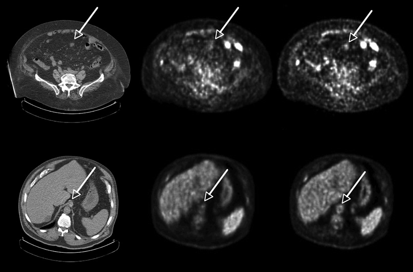
Results: The lesion phantom studies demonstrate the improved contrast of the smallest spheres with TOF compared with non-TOF and also confirm the faster convergence of contrast with TOF. These gains are evident from visual inspection of the images as well as a quantitative evaluation of contrast recovery of the spheres and noise in the background. The gains with TOF are higher for larger objects. These results correlate with patient studies in which lesions are seen more clearly and with higher uptake at comparable noise for TOF than with non-TOF.
Conclusion: TOF leads to a better contrast-versus-noise trade-off than non-TOF but one that is difficult to quantify in terms of a simple sensitivity gain improvement: A single gain factor for TOF improvement does not include the increased rate of convergence with TOF nor does it consider that TOF may converge to a different contrast than non-TOF. The experimental phantom results agree with those of prior simulations and help explain the improved image quality with TOF for patient oncology studies.
Figures


References
-
- Anger HO. Survey of Radioisotope Cameras. ISA Trans. 1966;5(4):311.
-
- Brownell GL, Burnham CA, Wilensky S, Aronow S, Kazemi H, Streider . New developments in positron scintigraphy and the application of cyclotron produced positron emitters. Vienna: 1969.
-
- Budinger TF. Instrumentation trends in nuclear medicine. Semin Nucl Med. 1977;7(4):285–297. - PubMed
-
- Ter-Pogossian MM, Ficke DC, Hood JT, Sr, Yamamoto M, Mullani NA. PETT VI: A positron emission tomograph utilizing cesium fluoride scintillation detectors. J Comput Assist Tomogr. 1982;6:125–133. - PubMed
-
- Allemand R, Gresset C, Vacher J. Potential advantages of a cesium fluoride scintillator for a time-of-flight positron camera. J Nucl Med. 1980;21(2):153–155. - PubMed
Publication types
MeSH terms
Grants and funding
LinkOut - more resources
Full Text Sources
Other Literature Sources