. 2008 Apr;146(4):1862-77.
doi: 10.1104/pp.107.113217. Epub 2008 Feb 20.

DNA-binding study identifies C-box and hybrid C/G-box or C/A-box motifs as high-affinity binding sites for STF1 and LONG HYPOCOTYL5 proteins

Affiliations

DNA-binding study identifies C-box and hybrid C/G-box or C/A-box motifs as high-affinity binding sites for STF1 and LONG HYPOCOTYL5 proteins

Young Hun Song et al. Plant Physiol. 2008 Apr.

Abstract

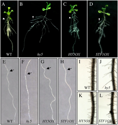
LONG HYPOCOTYL5 (HY5) is a bZIP (basic leucine zipper) transcription factor that activates photomorphogenesis and root development in Arabidopsis (Arabidopsis thaliana). Previously, STF1 (soybean [Glycine max] TGACG-motif binding factor 1), a homologous legume protein with a RING-finger motif and a bZIP domain, was reported in soybean. To investigate the role of STF1, the phenotypes of transgenic Arabidopsis plants overexpressing STF1 and HY5 were compared. In addition, the DNA-binding properties of STF1 and HY5 were extensively studied using random binding site selection and electrophoretic mobility shift assay. Overexpression of STF1 in the hy5 mutant of Arabidopsis restored wild-type photomorphogenic and root development phenotypes of short hypocotyl, accumulation of chlorophyll, and root gravitropism with partial restoration of anthocyanin accumulation. This supports that STF1 is a homolog of HY5 with a role in light and hormone signaling. The DNA-binding properties of STF1 and HY5 are shown to be similar to each other in recognizing many ACGT-containing elements with a consensus sequence motif of 5'-(G/A)(G/A) TGACGT(C/G/A)(A/T/G)-3'. The motif represents a characteristically strong preference for flanking sequence to TGACGT and a larger sequence than the sequences recognized by the G-box binding factor and TGA protein families. The finding of C-box, hybrid C/G-, and C/A-boxes as high-affinity binding sites over the G-box and parameters associated with HY5 recognition define the criteria of HY5/STF1 protein-DNA interaction in the promoter regions. This study helps to predict the precise in vivo binding sites of the HY5 protein from the vast number of putative HY5 genomic binding sites analyzed by chromatin immunoprecipitation on chip.


Figures

Figure 1.
Figure 1.
Phenotypes of the hy5 mutant and HY5 and STF1 complementation lines. A, Diagram of the STF1 protein structure. The basic DNA-binding region and Leu zipper region, casein kinase II (CKII in the image) phosphorylation sites, COP1 interaction sites to HY5, and the conserved motifs are indicated in the bottom. B, The effect of the hy5 mutation and HY5 and STF1 complementation lines on hypocotyl elongation, chlorophyll production, and anthocyanin accumulation; the phenotypes of light- and dark-grown seedlings (a and b); the hypocotyl lengths of light- and dark-grown seedlings (c and d); chlorophyll and anthocyanin levels (e and f). Hypocotyl length is the mean ± se (n ≥ 35) of 6-d-old seedlings grown in either LL or dark. The contents of chlorophyll (e) and anthocyanin (f) are from 5-d-old seedlings grown in constant white light. Lanes 1, 2, 3, and 4 denote wild type (WT in the image), hy5-Ks50 (hy5), 35SHY5/hy5-Ks50 (HY5OX), and 35SSTF1/hy5-Ks50 (STF1OX), respectively. Error bars represent the sd. [See online article for color version of this figure.]
Figure 2.
Figure 2.
Root morphologies of the wild type, the hy5 mutant, and the STF1 and HY5 overexpression lines. Wild-type Arabidopsis (Wassilewskija ecotype; A, E, and I), hy5-Ks50 (hy5; B, F, and J), 35S:HY5/hy5-Ks50 (HY5OX; C, G, and K), and 35S:STF1/hy5-Ks50 (STF1OX; D, H, and L) are shown. Plants were grown, vertically positioned, on MS agar plates supplemented with 2% Suc for 21 d (A–D). Arrowheads indicate lateral roots. Vertically grown 3-d-old seedlings were tilted to 45° and grown for 3 d to see the wavy pattern of root growth (E–H). Arrows indicate the positions of root tips at the time of the position change. Root hairs of each plant are shown (I–L). The hy5 mutants display elongated root hairs (J).
Figure 3.
Figure 3.
Comparison of the DNA-binding properties of STF1 and HY5. A, The seven ACEs and the AP-1 site used as binding-site probes. B, EMSA using different concentrations of STF1 and HY5. Increasing concentrations (100, 250, and 500 nm) of purified STF1 and HY5 protein were added to reaction mixtures containing 20,000 cpm of each binding-site probe (lanes 1–8; PA G-box, CHS-U1, Hex, as-1, nos-1, CREG/A, CREA/T, AP-1). Two probes, CHS-U1 and as-1, are very weak binding sites for both bZIP factors, which show binding only at high concentrations. C, Binding affinity of full-length STF1 and HY5 to CRE, PA G-box, and CHS-U1 probes. Radioactivity of the bands corresponding to free and bound DNA were measured from the dried gel using a Bio-Image analyzer (BAS 2500; Fuji Photo Film) and calculated as the percentage of bound versus free DNA. This experiment was performed three times with the same results. D, The effect of the N-terminal domain on STF1 binding to G-box sequences (PA G-box and CHS-U1 G-box). The HY5-like homologous domain is shown as a black bar at the top. Increasing concentrations (lanes 2–7; 0.05, 0.1, 0.2, 0.5, 1.0, and 1.73 μm) of each purified protein were added to the reaction mixtures containing PA G-box and CHS-U1 as binding site probes prior to EMSA.
Figure 4.
Figure 4.
Comparison of EMSA pattern and methylation interference of three soybean bZIP proteins: STGA1, STF1, and SGBF1. A, EMSA of three bZIP proteins using seven selected DNA sequences. The sequences of binding site probes are the same as in Figure 3A. B, Methylation interference assay using Hex as a probe. STF1 binds differently to the Hex sequence than to the two other soybean bZIP proteins. Methylation interference shows that STF1 binds a wider sequence than the GBF (SGBF1) and TGA (STGA1) proteins recognize. Both strands (upper, lower in the image) of the DNA fragment containing the cloned Hex sequence (5′-ggGTGACGTGGCca-3′) were partially methylated and incubated with in vitro generated recombinant bZIP proteins. Free (f) and protein-complexed (b) DNA fragments were separated, eluted, and, after piperidine cleavage, analyzed on a denaturing polyacryamide gel. Markers labeled G refer to the Maxam-Gilbert sequencing reactions of this DNA fragment (Maxam and Gilbert, 1980). The brackets indicate the location of protected sequences. The DNA sequence of the protected region is given below. Strong protection and weak protection by protein binding during methylation reaction are indicated as circles and triangles, respectively.
Figure 5.
Figure 5.
DNA-binding sequences selected by STF1. A, Compilation of STF1 binding sites identified by RBSS in 50 and 150 mm KCl. Binding sites were selected from a pool of oligonucleotides carrying random 13 bp flanked by a defined sequence of 26 bp on either side. DNA sequences that bound to the GST-STF1 fusion protein were selected and analyzed using DNA sequencing. Binding sites were aligned according to the consensus sequence. Nucleotides corresponding to the flanking sequences on either side of the random 13 bp are underlined. An asterisk before a clone number indicates a selected sequence containing more or less than 13 random nucleotides. In the image, “rev” refers to the reverse orientation of the individual sequence. Selected binding sites are grouped according to the bases at position −2/+2 and divided further by the base at position +3. B, Consensus sequences of STF1 binding sites derived from the base frequencies of the selected sequences at 50 and 150 mm KCl. Numbers indicate the base frequencies from the sequence data. R denotes either base G or A. V denotes bases G, A, and C. D denotes bases G, A, and T. C, The selected binding sites are arranged by C-box, C/G-box, C/A-box, and C/T-box sequences. The number in parentheses indicates the occurrence of the group.
Figure 6.
Figure 6.
EMSA analysis of selected binding sites. A, EMSA analysis of purified STF1 and HY5 proteins using various ACEs. Purified STF1 (0.1 μm) and HY5 protein was incubated with 20,000 cpm of P32-labeled binding-site probes. The 24 binding sites used as probes for the EMSA analysis are listed on the right. These sequences are arranged to allow a comparison of the binding properties of each type of ACE. The bases, which are different from the high-affinity consensus binding site, are boxed. B, EMSA analysis of three soybean bZIP proteins: STF1, STGA1, and SGBF1. Three soybean bZIP proteins were interacted with selected C-box sequences to compare the binding properties of each bZIP protein against different flanking sequences to the ACGT-motif.
Figure 7.
Figure 7.
Binding strength of STF1 and HY5 to different ACEs. A, The half-site combinations that fit the parameter of STF1 and HY5 binding. Symmetric half-sites are sequences that can be bound by protein when made symmetric (i.e. C-box), whereas dependent half-sites are sequences that cannot be bound or are weakly bound when made symmetric but that can be bound when combined with a symmetric half-site. The binding affinities to STF1 are indicated. B, The diagram of predicted binding affinity. Thick lines indicate strong or moderate binding of the half-site combinations; thin lines indicate weak binding of half-sites.
Figure 8.
Figure 8.
Regulation of anthocyanin biosynthetic genes by HY5 and the predicted HY5 binding sites in the promoter region. A, Simplified schematic representation of the biosynthesis pathway of anthocyanins and flavonols. Abbreviations of the seven enzyme designations are: CHS, Chalcone synthase; CHI, chalcone isomerase; F3H, flavonone 3-hydroxylase; DFR, dihydroflavonol 4-reductase; LDOX, leucoanthocyanidin dioxygenase; F3H, flavonone 3′-hydroxylase; FLS, flavonol synthase. B, Semiquantitative RT-PCR. mRNA abundance of genes involved in anthocyanin biosynthesis pathways in the hy5 mutation and HY5 complementation lines grown under LL. Transcript levels of CAB1 (chlorophyll a/b-binding protein 1) are shown for comparative purpose (Lee et al., 2007). ACTIN2 (Act2) mRNA level was used as an internal control. C, Bar graphs represent the relative expression levels of genes as obtained by RT-PCR (B). Band intensities were quantified using Bio-Rad's Quantity One software, and values were normalized against Act2 transcript. D, Diagram of F3H promoter fragments, including ACEs (A) as well as E- and G-boxes (G), according to Shin et al. (2007; top). Thick bars represent fragments identified by in vivo ChIP. The 12 nucleotides of the indicated elements are shown at the bottom. The types of ACEs are indicated to the right. *F3H promoter fragments are named according to Shin et al. (2007). **Prediction is based on binding parameter of STF1/HY5 binding. WB, Weak binding; NB, no binding. ***This is based on in vitro binding assay (Shin et al., 2007). The base number indicates the center of each element as counted from the translation start site (+1). E, The list of predicted HY5 binding sites in the seven anthocyanin biosynthetic genes that satisfy the binding parameter identified from this study and the fragments identified by in vivo ChIP (Shin et al., 2007). The type of ACE and the predicted binding affinity are indicated on the right. In vitro binding data of direct binding to HY5 protein is not available (NA). ACE is identified by functional analysis and in vitro nuclear protein binding study (Hartmann et al., 2005). All functionally defined ACEs satisfy the criteria of STF1/HY5 binding but predicted as weak binding sites.

References

    1. Ang LH, Chattopadhyay S, Wei N, Oyama T, Okada K, Batschauer A, Deng XW (1998) Molecular interaction between COP1 and HY5 defines a regulatory switch for light control of Arabidopsis development. Mol Cell 1 213–222 - PubMed
    1. Ang LH, Deng XW (1994) Regulatory hierarchy of photomorphogenic loci: allele-specific and light-dependent interaction between the HY5 and COP1 loci. Plant Cell 6 613–628 - PMC - PubMed
    1. Chattopadhyay S, Ang LH, Puente P, Deng XW, Wei N (1998) Arabidopsis bZIP protein HY5 directly interacts with light-responsive promoters in mediating light control of gene expression. Plant Cell 10 673–683 - PMC - PubMed
    1. Cheong YH, Park JM, Yoo CM, Bahk JD, Cho MJ, Hong JC (1994) Isolation and characterization of STGA1, a member of the TGA1 family of bZIP transcription factors from soybean. Mol Cells 4 405–412
    1. Cheong YH, Yoo CM, Park JM, Ryu GR, Goekjian VH, Nagao RT, Key JL, Cho MJ, Hong JC (1998) STF1 is a novel TGACG-binding factor with a zinc-finger motif and a bZIP domain which heterodimerizes with GBF proteins. Plant J 15 199–209 - PubMed

Publication types

MeSH terms