Comparative Study
. 2008 Feb 20;28(8):1916-28.
doi: 10.1523/JNEUROSCI.4755-07.2008.

An input-representing interneuron regulates spike timing and thereby phase switching in a motor network

Affiliations
Comparative Study

An input-representing interneuron regulates spike timing and thereby phase switching in a motor network

Kosei Sasaki et al. J Neurosci. .

Abstract

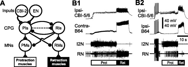
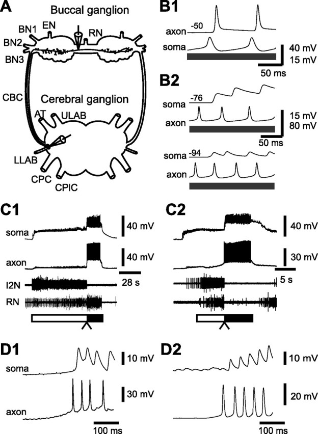
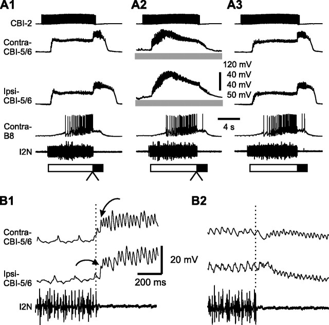
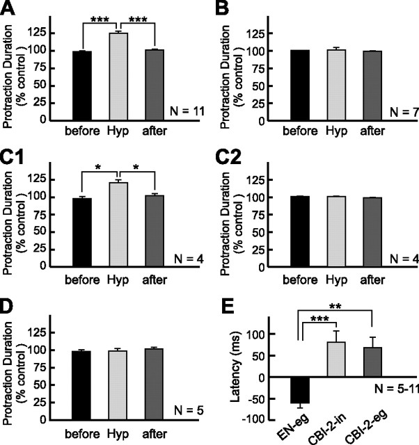
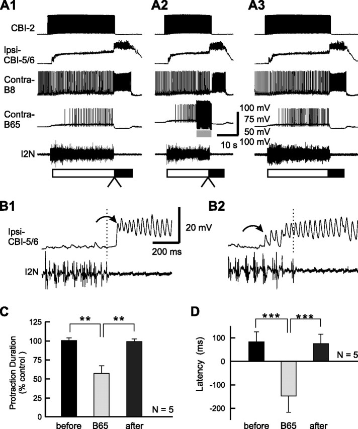
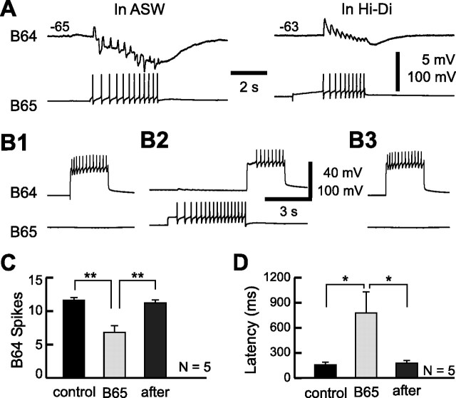
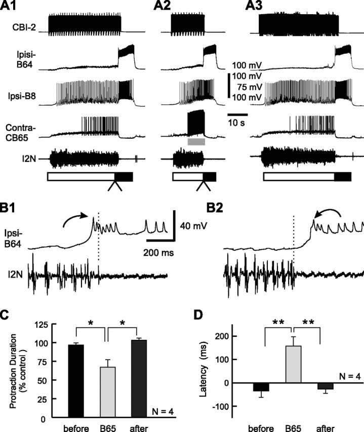
Despite the importance of spike-timing regulation in network functioning, little is known about this regulation at the cellular level. In the Aplysia feeding network, we show that interneuron B65 regulates the timing of the spike initiation of phase-switch neurons B64 and cerebral-buccal interneuron-5/6 (CBI-5/6), and thereby determines the identity of the neuron that acts as a protraction terminator. Previous work showed that B64 begins to fire before the end of protraction phase and terminates protraction in CBI-2-elicited ingestive, but not in CBI-2-elicited egestive programs, thus indicating that the spike timing and phase-switching function of B64 depend on the type of the central pattern generator (CPG)-elicited response rather than on the input used to activate the CPG. Here, we find that CBI-5/6 is a protraction terminator in egestive programs elicited by the esophageal nerve (EN), but not by CBI-2, thus indicating that, in contrast to B64, the spike timing and protraction-terminating function of CBI-5/6 depends on the input to the CPG rather than the response type. Interestingly, B65 activity also depends on the input in that B65 is highly active in EN-elicited programs, but not in CBI-2-elicited programs independent of whether the programs are ingestive or egestive. Notably, during EN-elicited egestive programs, hyperpolarization of B65 delays the onset of CBI-5/6 firing, whereas in CBI-2-elicited ingestive programs, B65 stimulation simultaneously advances CBI-5/6 firing and delays B64 firing, thereby substituting CBI-5/6 for B64 as the protraction terminator. Thus, we identified a neural mechanism that, in an input-dependent manner, regulates spike timing and thereby the functional role of specific neurons.


Figures

Figure 1.
Figure 1.
The schematic diagram of the core circuitry that mediates the protraction-retraction sequence of Aplysia feeding motor programs and activity patterns of CBI-5/6 and B64 in different types of programs. A, Two inputs, CBI-2 and EN, were used to activate the CPG. A single cycle of the Aplysia feeding motor programs consists of two phases, a radula protraction phase followed by a radula retraction phase. During protraction, PIs drive PMs, which in turn activate protraction muscles. PIs also inhibit RIs and RMs. During retraction, RIs drive RMs, which in turn activate retraction muscles. RIs also inhibit PIs and PMs. Protraction/retraction phase transitions are triggered, at least partly, through slow excitation of RIs by PIs, which, at a delay, overcomes inhibition of RIs by PIs. MNs, Motorneurons. Connections: Open triangle, excitation; filled circle, inhibition; broken line, polysynaptic connection. B, Examples of activity patterns of CBI-5/6 and B64 in ingestive motor programs elicited by CBI-2 stimulation (B1) or in egestive motor programs elicited by EN stimulation (B2). Protraction (Prot; open bar) is monitored by activity in the I2N. Retraction (Ret; filled bar) is monitored by robust activities of CBI-5/6 and B64, both of which are active throughout the retraction phase in both types of motor programs. Radula closure activity is monitored on the basis of activity in the RN, which contains the axons of the radula closer motoneurons B8. In the CBI-2-elicited ingestive program (B1), RN activity primarily occurred during the retraction phase; in the EN-elicited egestive program (B2), RN activity primarily occurred during the protraction phase. Each neuron is labeled with a prefix such as ipsilateral (Ipsi-) and contralateral (Contra-) related to the side on which CBI-2 or EN was stimulated. In subsequent figures, neurons are also labeled this way.
Figure 2.
Figure 2.
Spike propagation and activity of CBI-5/6 soma and axon. A, The schematic drawing of CBI-5/6 illustrates the sites at which our recordings were obtained. One electrode was used to impale the soma of CBI-5/6 in the cerebral ganglion (dorsal surface). The second electrode was used to impale the axon of CBI-5/6 in the buccal ganglion (rostral surface). The distance between the recording sites was ∼10 mm. B, Simultaneous recordings from the soma and the axon of CBI-5/6. B1, A somatic current injection (gray bar) elicited action potentials in the soma and these were followed by action potentials in the axon. B2, Axon-initiated spikes elicited by current injection were followed one-for-one by antidromic spikes in the soma (top). Hyperpolarization (−18 mV) of CBI-5/6 soma dramatically reduced the amplitude of antidromic spikes (bottom). Note that antidromic spikes in the soma were smaller than the axon-initiated spikes. Membrane potential of CBI-5/6 axon or soma is shown at the left (in millivolts). C, CBI-5/6 was recruited in both CBI-2- and EN-elicited programs. Simultaneous recordings from the axon and the soma of CBI-5/6 during a CBI-2-elicited ingestive program (C1) and an EN-elicited egestive program (C2). These recordings were obtained from ipsilateral CBI-5/6 to the side on which CBI-2 or EN was stimulated. The protraction and retraction phases of motor programs are marked by open and filled bars, respectively. In CBI-2-elicited programs, RN was predominantly active during retraction, indicating that the program was ingestive, whereas in EN-elicited programs, RN was predominantly active during protraction, indicating that the program was egestive. Note that in both programs, during the protraction phase, CBI-5/6 soma received excitatory synaptic inputs, but no spikes were present in the axon. However, during the retraction phase, a barrage of action potentials is present both in the axon and in the soma of CBI-5/6. D, Expanded views of the transition between the protraction and the retraction phases (C1, C2, arrowheads) in CBI-2-elicited ingestive programs (D1 is from C1) and in EN-elicited egestive programs (D2 is from C2). In both programs, spikes in the axon were followed one-for-one by antidromic spikes in the soma, indicating that when programs are generated, the spikes of CBI-5/6 originate in the axonal region and propagate toward the soma. BN, Buccal nerve; CBC, cerebral-to-buccal connective; ULAB, upper labial nerve; AT, anterior tentacular nerve; LLAB, lower labial nerve; CPC, cerebral-to-pedal connective; CPlC, cerebral-to-pleural connective.
Figure 3.
Figure 3.
Effect of CBI-5/6 hyperpolarization on the duration of the protraction phase in EN-elicited egestive programs. A1, An EN-elicited egestive program in control condition. A2, Bilateral hyperpolarization (gray bars) of ipsilateral CBI-5/6 and contralateral CBI-5/6 extended protraction duration. A3, The program after the hyperpolarization was released. B, Expanded views of the transition between the protraction and retraction phases (A1, A2, arrowheads) without CBI-5/6 hyperpolarization (B1 is from A1) and with CBI-5/6 hyperpolarization (B2 is from A2). Vertical dashed lines are drawn just above the last spike in I2N, to facilitate the visualization of the difference between the end of the protraction phase and the onset of the antidromic spikes in bilateral CBI-5/6 somata (arrows). Antidromic spikes in one CBI-5/6 appear before the end of I2N activity (B1). Synaptic inputs in CBI-5/6 during the protraction phase were unmasked during hyperpolarization, and the amplitude of antidromic spikes in CBI-5/6 was decreased, making it difficult to distinguish the first antidromic spike (B2).
Figure 4.
Figure 4.
Effect of CBI-5/6 hyperpolarization on the duration of the protraction phase in CBI-2-elicited ingestive programs. A1, A CBI-2-elicited ingestive program in control condition. A2, Bilateral hyperpolarization of CBI-5/6 had no effect on protraction duration. A3, The program after the hyperpolarization was released. B, Expanded views of the transition between the protraction and retraction phases (A1, A2, arrowheads) without CBI-5/6 hyperpolarization (B1 is from A1) and with CBI-5/6 hyperpolarization (B2 is from A2). Importantly, in CBI-2-elicited ingestive programs, antidromic spikes in CBI-5/6 began after the end of I2N activity (B1). In these experiments, CBI-2 stimulation was manually terminated after the protraction phase has ended. Note that CBI-2 stimulation continued for a brief period of time after the end of protraction and the beginning of retraction (B1, B2).
Figure 5.
Figure 5.
Effect of CBI-5/6 hyperpolarization on the duration of the protraction phase in CBI-2-elicited egestive programs. A1, A CBI-2-elicited egestive program in control condition. Before the programs were elicited by CBI-2 stimulation, four successive programs were evoked by continuous stimulation of EN (1.5 Hz). Note that, compared with CBI-2-elicited ingestive programs (Figs. 4A, 8A, and 10A), the high level of B8 activity occurred during the protraction phase. A2, Bilateral hyperpolarization of CBI-5/6 had no effect on protraction duration. A3, The program after the hyperpolarization was released. B, Expanded views of the transition between the protraction and retraction phases (A1, A2, arrowheads) without CBI-5/6 hyperpolarization (B1 is from A1) and with CBI-5/6 hyperpolarization (B2 is from A2). Note that in CBI-2-elicited egestive programs, antidromic spikes in CBI-5/6 began after the end of I2N activity (B1).
Figure 6.
Figure 6.
Grouped data of the effects of CBI-5/6 hyperpolarization. A, The normalized protraction duration, before, during (Hyp), and after CBI-5/6 hyperpolarization in EN-elicited egestive programs (see Fig. 3). B, The normalized protraction duration, before, during, and after CBI-5/6 hyperpolarization in CBI-2-elicited ingestive programs (see Fig. 4). C, The lengthening effect of CBI-5/6 hyperpolarization is not caused by variability of different preparations we used. In four preparations, we examined the effects of CBI-5/6 hyperpolarization both in EN-elicited egestive programs (C1) and in CBI-2-elicited ingestive programs (C2). Graphs in C1 and C2 represent a subset of data in A and B, respectively. D, The normalized protraction duration before, during, and after CBI-5/6 hyperpolarization in CBI-2-elicited egestive programs (see Fig. 5). E, The mean latencies of CBI-5/6 firing relative to the termination of I2N activity in EN-elicited egestive programs (EN-eg), in CBI-2-elicited ingestive programs (CBI-2-in), and in CBI-2-elicited egestive programs (CBI-2-eg). Latency of CBI-5/6 firing was calculated as the time that elapsed from the last extracellular signal peak recorded in I2N to the first peak of the antidromic spike recorded in CBI-5/6 soma. If the first antidromic spike preceded the last signal in I2N, we defined the latency of CBI-5/6 firing as negative, and if the first antidromic spike followed the last signal in I2N, we defined the latency of CBI-5/6 firing as positive. *p < 0.05; **p < 0.01; ***p < 0.001 (Bonferroni's post-test). Error bars indicate SEM.
Figure 7.
Figure 7.
Effect of B65 hyperpolarization on the latency of CBI-5/6 firing in EN-elicited egestive programs. A, Intracellular recordings from a CBI-5/6 axon and B65 in normal saline. B65 elicited one-for-one EPSPs in the CBI-5/6 axon. Note that B65-elicited EPSPs in CBI-5/6 showed summation. B1, An EN-elicited egestive program in control condition. B2, Bilateral hyperpolarization of B65 extended the protraction duration. B3, The program after the hyperpolarization was released. C, Expanded views of the transition between the protraction and retraction phases (B1, B2, arrowheads) without B65 hyperpolarization (C1 is from B1) and with B65 hyperpolarization (C2 is from B2). Note that antidromic spikes in CBI-5/6 occurred before the end of I2N activity in the program in which B65s were not hyperpolarized (C1), whereas they began after the end of I2N activity when B65s were hyperpolarized (C2). D, Grouped data showing the normalized protraction duration before, during (B65), and after B65 hyperpolarization. E, The mean latency of CBI-5/6 firing relative to the termination of I2N activity before, during (B65), and after B65 hyperpolarization. *p < 0.05 (Bonferroni's post-test). Error bars indicate SEM.
Figure 8.
Figure 8.
Effect of B65 stimulation on the latency of CBI-5/6 firing in CBI-2-elicited ingestive programs. A1, A CBI-2-elicited ingestive program in control condition. A2, Stimulation of B65 at 14 Hz during protraction terminated the phase before the protraction termination was expected to occur. A3, Recovery. B, Expanded views of the transition between the protraction and retraction phases (A1, A2, arrowheads) without B65 stimulation (B1 is from A1) and with B65 stimulation (B2 is from A2). Note that the antidromic spikes in CBI-5/6 began after the end of I2N activity in the control (B1), whereas they began before the end of I2N activity when B65 was stimulated (B2). C, Grouped data showing the normalized protraction duration before, during (B65), and after B65 stimulation. D, The mean latency of CBI-5/6 firing relative to the termination of I2N activity before, during, and after B65 stimulation. **p < 0.01; ***p < 0.001 (Bonferroni's post-test). Error bars indicate SEM.
Figure 9.
Figure 9.
B65 decreases the excitability of interneuron B64. A, B65 elicited not only the one-for-one fast EPSPs, but also a slow inhibitory response in the contralateral B64 in normal saline (left). Both PSP components persisted in Hi-Di (right). B, Stimulation of B65 reduced the number of spikes in B64 elicited by depolarizing current pulses (2.5 s duration) every 60 s. Without B65 stimulation (B1, B3), the current pulse in B64 induced 12 spikes in B64. When B65 was stimulated for 5 s before the current injection into B64, the number of B64 spikes was reduced to eight (B2). Note that compared with the control, prestimulation of B65 also delayed the onset of B64 firing relative to the onset of current injection into it. C, Grouped data showing the mean number of B64 spikes elicited by a constant current injection before, during (B65), and after B65 stimulation. D, The mean latencies between the onset of current injection into B64 and the appearance of the first action potential in B64 before, during (B65), and after B65 stimulation. *p < 0.05; **p < 0.01 (Bonferroni's post-test). Error bars indicate SEM.
Figure 10.
Figure 10.
Effect of B65 stimulation on the latency of B64 firing in CBI-2-elicited ingestive programs. A1, A CBI-2-elicited ingestive program in control condition. A2, Stimulation of B65 at 14 Hz shortened the protraction phase compared with the duration of this phase in control. A3, Recovery. B, Expanded views of the transition between the protraction and retraction phases (A1, A2, arrowheads) without B65 stimulation (B1 is from A1) and with B65 stimulation (B2 is from A2). Note that B64 began to fire spikes before the end of I2N activity in the control condition (B1), whereas B64 fired spikes after the activity in I2N ceased when B65 was stimulated (B2). C, Grouped data showing the normalized protraction duration before, during (B65), and after B65 stimulation. D, The mean latency of B64 firing relative to the termination of I2N activity before, during (B65), and after B65 stimulation. *p < 0.05; **p < 0.01 (Bonferroni's post-test). Error bars indicate SEM.
Figure 11.
Figure 11.
A schematic diagram illustrating synaptic mechanisms that an input-representing interneuron B65 uses to regulate the spike timing and the phase-switching function of CBI-5/6 and B64. Both CBI-5/6 and B64 receive fast excitatory inputs from B65, but B64 also receives prominent slow inhibitory inputs. Here, only the inhibitory connection from B65 to B64 is depicted because the mixed excitatory and inhibitory actions of B65 functionally decrease B64 excitability. In addition, both CBI-5/6 (Sasaki et al., 2007) and B64 (Hurwitz and Susswein, 1996; Kabotyanski et al., 1998) make inhibitory synaptic connections with protraction neurons including B65. A, In EN-elicited egestive programs, B65 is activated at high frequency (Proekt et al., 2007), thereby advancing the timing of CBI-5/6 firing, but delaying the timing of B64 firing (lower electrophysiological recordings). Thus, in this type of program, CBI-5/6, but not B64 functions as a protraction terminator by inhibiting protraction neurons (Sasaki et al., 2007; present study). B, In CBI-2-elicited ingestive programs, B65 is inhibited by CBI-2 (Jing and Weiss, 2005). In the absence of excitation of the CBI-5/6 axon by B65, the timing of CBI-5/6 firing may be delayed. In contrast, without B65 inhibition, B64 fires early, in part because of slow excitation from protraction interneurons, such as B63, and excitation from neuropeptides released by CBI-2 (Koh and Weiss, 2007). Thus, in this type of program, B64, but not CBI-5/6, functions as a protraction terminator by inhibiting protraction neurons.

Similar articles

Cited by

References

    1. Ayali A, Harris-Warrick RM. Monoamine control of the pacemaker kernel and cycle frequency in the lobster pyloric network. J Neurosci. 1999;19:6712–6722. - PMC - PubMed
    1. Bartos M, Nusbaum MP. Intercircuit control of motor pattern modulation by presynaptic inhibition. J Neurosci. 1997;17:2247–2256. - PMC - PubMed
    1. Berkowitz A. Spinal interneurons that are selectively activated during fictive flexion reflex. J Neurosci. 2007;27:4634–4641. - PMC - PubMed
    1. Berkowitz A, Stein PS. Activity of descending propriospinal axons in the turtle hindlimb enlargement during two forms of fictive scratching: phase analyses. J Neurosci. 1994;14:5105–5119. - PMC - PubMed
    1. Bi GQ, Poo MM. Synaptic modifications in cultured hippocampal neurons: dependence on spike timing, synaptic strength, and postsynaptic cell type. J Neurosci. 1998;18:10464–10472. - PMC - PubMed

Publication types