Comparative Study
. 2008 Feb 20;28(8):1961-9.
doi: 10.1523/JNEUROSCI.3061-07.2008.

TGFbeta-Smad2 signaling regulates the Cdh1-APC/SnoN pathway of axonal morphogenesis

Affiliations
Comparative Study

TGFbeta-Smad2 signaling regulates the Cdh1-APC/SnoN pathway of axonal morphogenesis

Judith Stegmüller et al. J Neurosci. .

Abstract

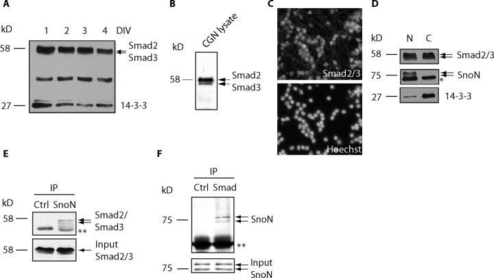
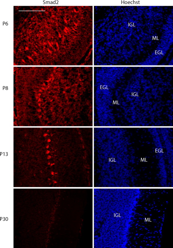
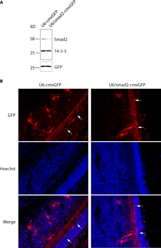
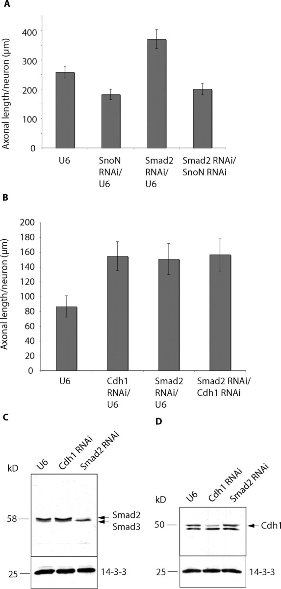
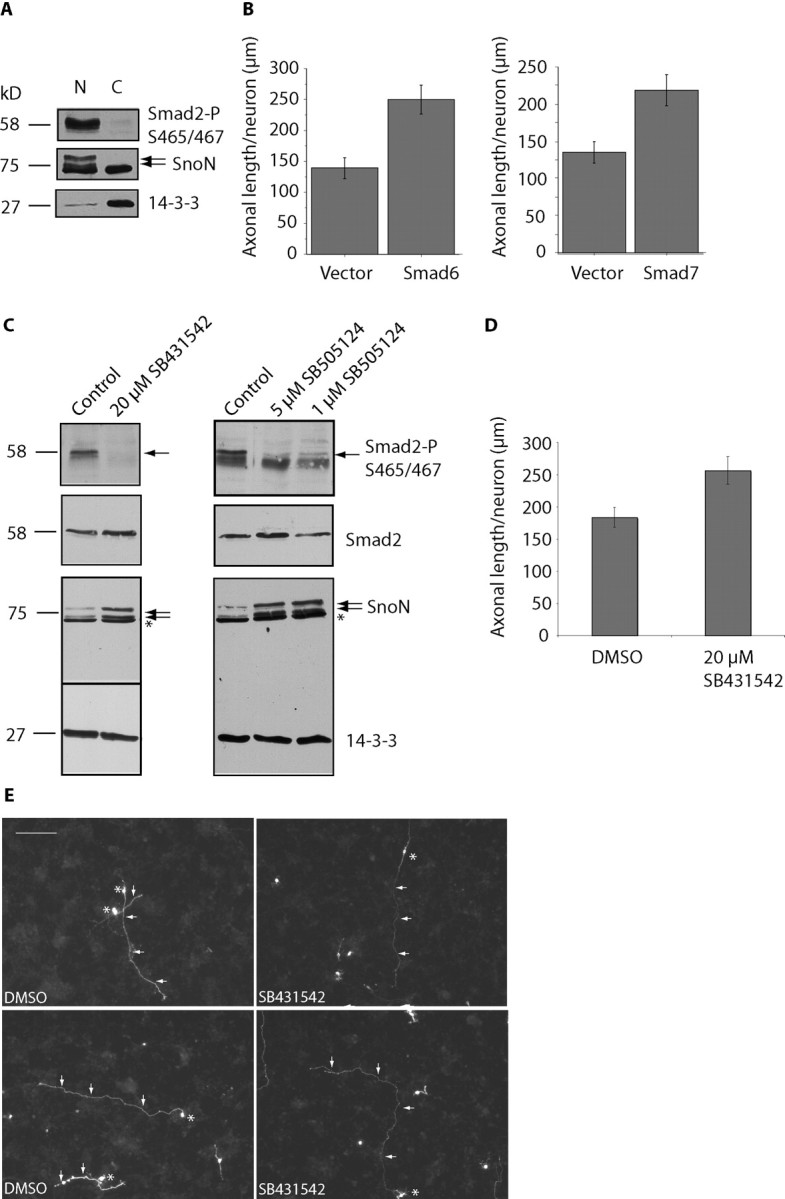
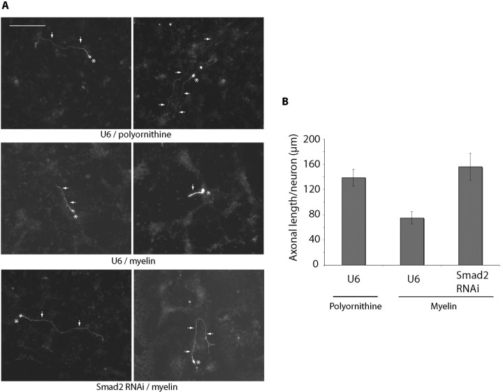
Axon growth is critical to the establishment of neuronal connectivity. The E3 ubiquitin ligase Cdh1-anaphase-promoting complex (Cdh1-APC) and its substrate the transcriptional modulator SnoN form a cell-intrinsic pathway that orchestrates axonal morphogenesis in the mammalian brain. How the Cdh1-APC/SnoN pathway is controlled in the nervous system remained unknown. Here, we report that the TGFbeta-regulated signaling protein Smad2 plays a key role in regulating the Cdh1-APC/SnoN pathway in neurons. We find that Smad2 is expressed in primary granule neurons of the developing rat cerebellar cortex. The Smad signaling pathway is basally activated in neurons. Endogenous Smad2 is phosphorylated, localized in the nucleus, and forms a physical complex with endogenous SnoN in granule neurons. Inhibition of Smad signaling by several distinct approaches, including genetic knock-down of Smad2, stimulates axonal growth. Biochemical evidence and genetic epistasis analyses reveal that Smad2 acts upstream of SnoN in a shared pathway with Cdh1-APC in the control of axonal growth. Remarkably, Smad2 knock-down also overrides the ability of adult rat myelin to inhibit axonal growth. Collectively, our findings define a novel function for Smad2 in regulation of the Cdh1-APC/SnoN cell-intrinsic pathway of axonal morphogenesis, and suggest that inhibition of Smad signaling may hold therapeutic potential in stimulating axonal growth after injury in the CNS.


Figures

Figure 1.
Figure 1.
Smad2/3 is expressed in the cerebellum and interacts with SnoN in neurons. A, Cerebellar granule neurons were lysed at the indicated DIV after plating and immunoblotted using an antibody that recognizes Smad2 and Smad3 and an antibody to 14-3-3, the latter to serve as loading control. B, Cerebellar granule neuron (CGN) lysates were separated by SDS-PAGE for an extended period of time and subjected to immunoblotting using the Smad2/3 antibody to distinguish Smad2 and Smad3. C, Cultured granule neurons (DIV2) were subjected to immunofluorescence analysis using the Smad2/3 antibody (top) and the DNA dye bisbenzimide (bottom). D, Granule neurons were subjected to subcellular fractionation. Nuclear (N) and cytoplasmic (C) fractions were immunoblotted using the Smad2/3, SnoN, and 14-3-3 antibodies. The asterisk indicates nonspecific band. A large portion of Smad2/3 is present in the nucleus. E, F, Granule neuron lysates were subjected to immunoprecipitation (IP) using the hemagglutinin control antibody and the SnoN antibody followed by immunoblotting with the Smad2/3 antibody (E) or reciprocal coimmunoprecipitation (F). Double asterisks indicate heavy chain IgGs. Endogenous SnoN and Smad2/3 form a physical complex in neurons.
Figure 2.
Figure 2.
Smad2 expression in the developing cerebellar cortex. Sagittal cerebellar sections from P6, P8, P13, and P30 were subjected to immunohistochemistry using an antibody that specifically recognizes Smad2. Scale bar, 100 μm. EGL, External granular layer; ML, molecular layer; IGL, internal granular layer. Smad2 is expressed in the postnatal cerebellum.
Figure 3.
Figure 3.
Smad2 suppresses axonal growth in cerebellar granule neurons. A, Granule neurons were transfected 8 h after plating with the Smad2 RNAi or control U6 plasmid together with the GFP expression plasmid and maintained in media supplemented with insulin. Three days after transfection, neurons were fixed and subjected to immunocytochemistry using an antibody to GFP. Images of control U6-transfected and U6/smad2-transfected neurons are shown, with asterisks indicating cell bodies and arrows indicating axons. Scale bar, 100 μm. Smad2 knock-down neurons had longer axons than control U6-transfected neurons. B, Axonal length was measured in GFP-positive transfected neurons using SPOT software. Axonal length was significantly longer in Smad2 knock-down neurons compared with control U6-transfected neurons (t test, p < 0.0001). A total of 268 neurons were measured. C, 293T cells were transfected with the Smad2 RNAi plasmid or control U6 plasmid together with Smad2-WT or Smad2-Rescue. Lysates were immunoblotted using the Smad2/3 and 14-3-3 antibodies, the latter to serve as loading control. Smad2 RNAi led to the knock-down of Smad2-WT, but failed to induce knock-down of Smad2-Rescue. D, Granule neurons transfected with the Smad2 RNAi or control U6 plasmid together with pcDNA3 vector, Smad2-WT, or Smad2-Rescue expression plasmid were analyzed as in Figure 2B. Whereas expression of Smad2-WT did not affect Smad2 RNAi-mediated increase in axonal length, expression of Smad2-Rescue reversed the Smad2 RNAi phenotype (ANOVA, p < 0.0001). A total of 297 neurons were measured.
Figure 4.
Figure 4.
In vivo Smad2 knock-down does not appear to affect parallel fiber patterning in the cerebellar cortex. A, Lysates of 293T cells transfected with the control U6-cmvGFP plasmid and the U6/smad2-cmvGFP RNAi plasmid were immunoblotted with the Smad2, 14-3-3, or GFP antibody. A bicistronic plasmid encoding a Smad2 hairpin RNA and GFP efficiently knocks down endogenous Smad2. B, The control U6-cmvGFP plasmid or U6/smad2-cmvGFP RNAi plasmid together with a Bcl-Xl expression plasmid were injected into the cerebella of P3 rat pups. Pups were subjected to electroporation and killed 5 d later. Coronal cerebellar sections were analyzed by immunohistochemistry using a GFP antibody. Arrows indicate parallel fibers. No difference was observed in parallel fiber patterning.
Figure 5.
Figure 5.
Smad2 acts in the Cdh1-APC/SnoN pathway of axonal morphogenesis. A, Neurons transfected with the control U6, U6/smad2, or U6/snon RNAi plasmid or both U6/smad2 and U6/snon plasmids were maintained in media supplemented with 10% calf serum and membrane-depolarizing concentrations of KCl and analyzed as in Figure 3B. Although Smad2 RNAi increased and SnoN RNA; reduced axonal length, simultaneous knock-down of Smad2 and SnoN reduced axonal growth (ANOVA, p = 0.0633). A total of 438 neurons were measured. B, Neurons transfected with the control U6, U6/cdh1, or U6/smad2 RNAi plasmid or both U6/cdh1 and U6/smad2 plasmids were placed in media supplemented with insulin and analyzed as in Figure 3B. Knock-down of Cdh1 and Smad2 individually increased axonal length compared with control neurons (ANOVA, p < 0.001), but no additive effect was detected after simultaneous knock-down of Cdh1 and Smad2. A total of 429 neurons were measured. C, D, Granule neurons were transfected using an electroporation-based nucleofection method with the U6 control, U6/cdh1 RNAi, or U6/smad2 RNAi plasmid. Lysates were analyzed 5 d later by immunoblotting using the Smad2/3 (C), Cdh1 (D), or 14-3-3 antibody. 14-3-3 served as loading control. The Smad2 RNAi and Cdh1 RNAi plasmids led to specific knock-down of endogenous Smad2 and Cdh1, respectively, in granule neurons.
Figure 6.
Figure 6.
TGFβ-Smad signaling suppresses axonal growth. A, Granule neurons were subjected to subcellular fractionation and analyzed with immunoblotting using an antibody that recognizes Smad2 specifically when phosphorylated at serines 465 and 467 as well as the SnoN and 14-3-3 antibodies. Phosphorylated Smad2 is localized in the nucleus in granule neurons. B, Neurons transfected with the control pCMV5 vector, Smad6, or Smad7 expression plasmids were placed in media supplemented with 10% calf serum and analyzed as in Figure 3B. Expression of Smad6 or Smad7 significantly increased axonal length (t test, p < 0.001). A total of 247 and 305 neurons were measured, respectively. C, Lysates of granule neurons exposed to SB431542 and SB505124 at the indicated concentrations for 48 h were immunoblotted with the SnoN, phosphoS465/467-Smad2, Smad2/3, and 14-3-3 antibodies. Exposure of neurons to small molecule inhibitors of TGFβ receptors reduced Smad2 phosphorylation and increased SnoN levels in neurons. Asterisks indicate nonspecific band. D, Granule neurons transfected with the GFP expression plasmid at DIV0 and placed in media supplemented with 10% calf serum. Neurons were treated with SB431542 or its vehicle (DMSO) starting at DIV1 for 48 h and analyzed as in Figure 3B. Treatment with SB431542 significantly increased axonal length (t test, p < 0.01). A total of 170 neurons were measured. E, Images of control vehicle- and SB431542-treated neurons are shown, with asterisks indicating cell bodies and arrows indicating axons. Scale bar, 100 μm. Neurons that were treated with SB431542 have longer axons than vehicle-treated neurons.
Figure 7.
Figure 7.
Smad2 knock-down overrides myelin inhibition of axonal growth. A, Images of neurons plated on polyornithine- or myelin-coated coverslips (13.3 μg/ml) and transfected with the control U6 or U6/smad2 RNAi plasmid were analyzed at DIV3 as in Figure 3A. Asterisks and arrows indicate cell bodies and axons, respectively. Scale bar, 100 μm. B, Quantitation of neurons in A analyzed as in Figure 3B. Axonal length is significantly reduced in control U6-transfected neurons on myelin compared with polyornithine substrate (ANOVA, p < 0.001). Axonal length is significantly increased in Smad2 knock-down neurons compared with control U6-transfected neurons on myelin (ANOVA, p < 0.0005). A total 130 of neurons were measured.

Similar articles

Cited by

References

    1. Angley C, Kumar M, Dinsio KJ, Hall AK, Siegel RE. Signaling by bone morphogenetic proteins and Smad1 modulates the postnatal differentiation of cerebellar cells. J Neurosci. 2003;23:260–268. - PMC - PubMed
    1. Attisano L, Wrana JL. Smads as transcriptional co-modulators. Curr Opin Cell Biol. 2000;12:235–243. - PubMed
    1. Bashir T, Dorrello NV, Amador V, Guardavaccaro D, Pagano M. Control of the SCF(Skp2-Cks1) ubiquitin ligase by the APC/C(Cdh1) ubiquitin ligase. Nature. 2004;428:190–193. - PubMed
    1. Bernard DJ. Both SMAD2 and SMAD3 mediate activin-stimulated expression of the follicle-stimulating hormone beta subunit in mouse gonadotrope cells. Mol Endocrinol. 2004;18:606–623. - PubMed
    1. Bonni S, Wang HR, Causing CG, Kavsak P, Stroschein SL, Luo K, Wrana JL. TGF-beta induces assembly of a Smad2-Smurf2 ubiquitin ligase complex that targets SnoN for degradation. Nat Cell Biol. 2001;3:587–595. - PubMed

Publication types

MeSH terms

LinkOut - more resources