. 2008 May;19(5):1942-51.
doi: 10.1091/mbc.e08-02-0110. Epub 2008 Feb 20.

Protein networks supporting AP-3 function in targeting lysosomal membrane proteins

Affiliations

Protein networks supporting AP-3 function in targeting lysosomal membrane proteins

Thorsten Baust et al. Mol Biol Cell. 2008 May.

Abstract

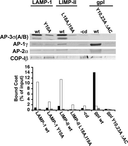
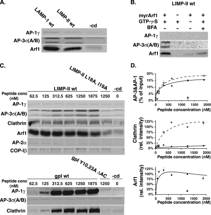
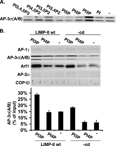
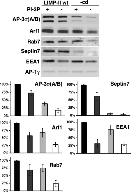
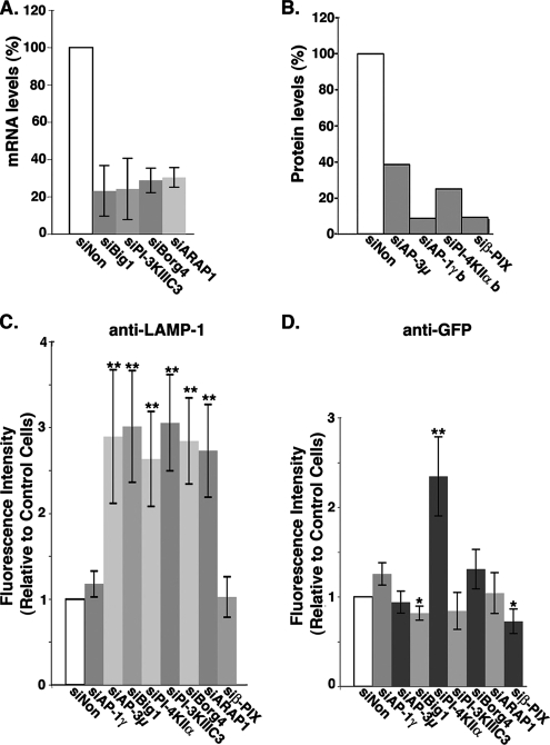
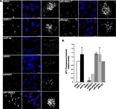
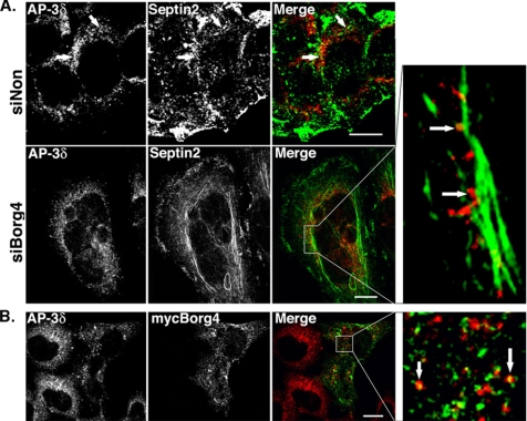
The AP-3 adaptor complex targets selected transmembrane proteins to lysosomes and lysosome-related organelles. We reconstituted its preferred interaction with liposomes containing the ADP ribosylation factor (ARF)-1 guanosine triphosphatase (GTPase), specific cargo tails, and phosphatidylinositol-3 phosphate, and then we performed a proteomic screen to identify new proteins supporting its sorting function. We identified approximately 30 proteins belonging to three networks regulating either AP-3 coat assembly or septin polymerization or Rab7-dependent lysosomal transport. RNA interference shows that, among these proteins, the ARF-1 exchange factor brefeldin A-inhibited exchange factor 1, the ARF-1 GTPase-activating protein 1, the Cdc42-interacting Cdc42 effector protein 4, an effector of septin-polymerizing GTPases, and the phosphatidylinositol-3 kinase IIIC3 are key components regulating the targeting of lysosomal membrane proteins to lysosomes in vivo. This analysis reveals that these proteins, together with AP-3, play an essential role in protein sorting at early endosomes, thereby regulating the integrity of these organelles.


Figures

Figure 1.
Figure 1.
AP-3 recruitment requires intact sorting signals. Liposomes with cytoplasmic domains (wild type or mutants) of LAMP-1 or LIMP-II, or gpI, or no cytoplasmic domain (−cd) were incubated with cytosol, and GTPγS. AP-1, AP-3, AP-2, and COP-I bound to liposomes were detected and quantified after SDS-PAGE and Western blotting by using specific antibodies against AP-1γ (black bars), AP-3σA/B (white bars), AP-2α (dark gray bars), and COP-Iβ (light gray bars).
Figure 2.
Figure 2.
AP-3 recruitment is ARF-1 dependent. (A) Liposomes with LAMP-1, LIMP-II, and no cytoplasmic domain (−cd) were incubated with cytosol and GTPγS. AP-1γ, AP-3σA/B, and ARF-1 bound to liposomes were detected after SDS-PAGE and Western blotting by using specific antibodies. (B) Liposomes with LIMP-II cytoplasmic domains were incubated with ARF-depleted cytosol (3 mg/ml) and supplemented or not with recombinant, myristoylated ARF-1 (30 μg/ml) with or without GTPγS and with or without brefeldin A (100 μg/ml). AP-1γ, AP-3σA/B, and ARF-1 bound to liposomes were detected after SDS-PAGE and Western blotting. (C) Top, liposomes with increasing amounts of LIMP-II wt (♦), LIMP-II L18A, I19A (■), or no cytoplasmic domain (−cd) (♦) were incubated with cytosol and GTPγS. Bottom, liposomes with increasing amounts of gpI (♦), gpI Y10, 23A, DAC (■), or no cytoplasmic domain (−cd) (♦) were incubated with cytosol in the presence of GTPγS. After incubation, AP-1γ, AP-2α, AP-3σA/B, clathrin, and COP-Iβ bound to liposomes were detected after SDS-PAGE and Western blotting. (D) Quantification of C. The clathrin signal was quantified and normalized to the values obtained with gpI-containing liposomes.
Figure 3.
Figure 3.
PI-3P increases AP-3 recruitment. (A) Liposomes with LIMP-II tails containing different or no PIPs were incubated with cytosol (3 mg/ml), GTPγS, and phosphatase inhibitors. AP-3σA/B bound to liposomes was detected after SDS-PAGE and Western blotting. (B) Liposomes with or without LIMP-II tails and either PI-3P or PI-4P or no PIP were incubated with cytosol (3 mg/ml), GTPγS, and phosphatase inhibitors. AP-1γ, AP-2α, AP-3σA/B, COP-Iβ, and ARF-1 bound to liposomes were detected after SDS-PAGE and Western blotting. The AP-3σA/B signal was quantified.
Figure 4.
Figure 4.
Requirements for stabilization of AP-3, septins, and Rab7 on liposomes. Liposomes with or without LIMP-II and with or without PI-3P were incubated with cytosol and GTPγS. After incubation, AP-3σA/B, AP-1γ, ARF-1, Rab7, and Septin 7 were analyzed by Western blotting by using specific antibodies. The recruitment of these markers was quantified (black bar, LIMP-II and PI-3P; dark gray bar, LIMP-II no PI-3P; light gray bar, PI-3P and no LIMP-II; and white bar, no LIMP-II and no PI-3P).
Figure 5.
Figure 5.
LAMP-1 lysosomal targeting in siRNA-treated HeLa cells. Cells were transfected for 72 h with siRNAs against the indicated molecules or with control nontargeting siRNAs (siNon). (A) mRNA levels after siRNA-mediated interference with siBig1, siBorg4, siARAP1, and siPI-3KIII C3 were evaluated by QPCR, and data were plotted relative to those in control siNon-treated cells. (B) siRNA-mediated knockdowns of AP-1, AP-3, PI-4K IIα, and β-PIX were evaluated by Western blots (also see Supplemental Figure S4) and plotted relative to control siNon-treated cells. (C and D) The uptakes of anti LAMP-1 (C) or anti-GFP antibodies (D) after the knockdown of the indicated molecules were analyzed by fluorescence confocal microscopy. Fluorescence levels were normalized, and data were plotted relative to those in control siNon-treated cells. Bars indicate average values ± SD (n = 4 independent experiments). **p < 0.005 and *p < 0.02 (analysis of variance single factor analysis).
Figure 6.
Figure 6.
AP-3 localization in siRNA-treated cells. (A) Cells transfected for 72 h with siRNAs against the indicated molecules or with control nontargeting siRNAs (siNon) were then fixed, permeabilized, and labeled with antibodies against AP-3δ (red). The cells were analyzed by fluorescence confocal microscopy. Right, AP-3δ signal at higher magnifications. Bars, 10 μm. (B) Total cellular intensity of the AP-3δ signal was quantified for each condition, and the averages were plotted relative to control (value = 1) and to the siAP-3 (value = 0). n = 2 independent experiments performed in triplicates with 100 cells/condition.
Figure 7.
Figure 7.
Localization of AP-3δ in siBorg4 and myc-Borg4-treated cells. (A) HeLa cells were transfected with either control siNon or siBorg4, double labeled with antibodies against AP-3δ (red) and septin2 (green). (B) HeLa cells expressing myc-Borg4 were fixed, permeabilized, and labeled with antibodies against AP-3δ (red) and myc (green). Cells were then fixed and analyzed by fluorescence confocal microscopy. The arrows indicate AP-3δ–positive structures that overlap with septin2 (A) or myc-Borg4 (Figure 7B) are aligned along septin2-positive filaments. Bars, 10 μm.

References

    1. Aniento F., Emans N., Griffiths G., Gruenberg J. Cytoplasmic dynein-dependent vesicular transport from early to late endosomes. J. Cell Biol. 1993;123:1373–1387. - PMC - PubMed
    1. Barral Y., Mermall V., Mooseker M. S., Snyder M. Compartmentalization of the cell cortex by septins is required for maintenance of cell polarity in yeast. Mol. Cell. 2000;5:841–851. - PubMed
    1. Baust T., Czupalla C., Krause E., Bourel-Bonnet L., Hoflack B. Proteomic analysis of adaptor protein 1A coats selectively assembled on liposomes. Proc. Natl. Acad. Sci. USA. 2006;103:3159–3164. - PMC - PubMed
    1. Bonifacino J. S., Glick B. S. The mechanisms of vesicle budding and fusion. Cell. 2004;116:153–166. - PubMed
    1. Bonifacino J. S., Traub L. M. Signals for sorting of transmembrane proteins to endosomes and lysosomes. Annu. Rev. Biochem. 2003;72:395–447. - PubMed

Publication types

MeSH terms

Substances