. 2008 May;19(5):2147-53.
doi: 10.1091/mbc.e07-12-1269. Epub 2008 Feb 20.

GEF-H1 couples nocodazole-induced microtubule disassembly to cell contractility via RhoA

Affiliations

GEF-H1 couples nocodazole-induced microtubule disassembly to cell contractility via RhoA

Yuan-Chen Chang et al. Mol Biol Cell. 2008 May.

Abstract

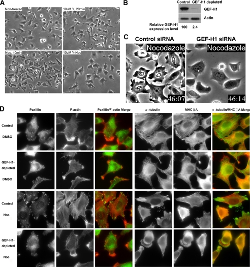
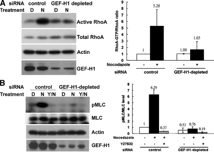
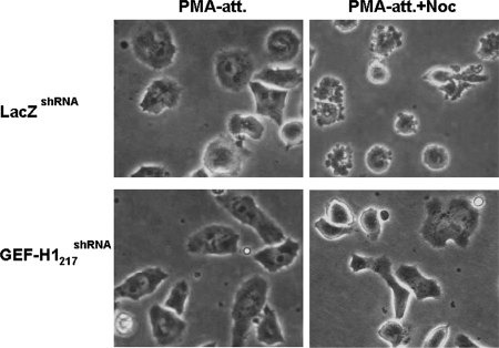
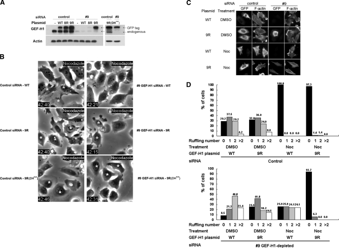
The RhoA GTPase plays a vital role in assembly of contractile actin-myosin filaments (stress fibers) and of associated focal adhesion complexes of adherent monolayer cells in culture. GEF-H1 is a microtubule-associated guanine nucleotide exchange factor that activates RhoA upon release from microtubules. The overexpression of GEF-H1 deficient in microtubule binding or treatment of HeLa cells with nocodazole to induce microtubule depolymerization results in Rho-dependent actin stress fiber formation and contractile cell morphology. However, whether GEF-H1 is required and sufficient to mediate nocodazole-induced contractility remains unclear. We establish here that siRNA-mediated depletion of GEF-H1 in HeLa cells prevents nocodazole-induced cell contraction. Furthermore, the nocodazole-induced activation of RhoA and Rho-associated kinase (ROCK) that mediates phosphorylation of myosin regulatory light chain (MLC) is impaired in GEF-H1-depleted cells. Conversely, RhoA activation and contractility are rescued by reintroduction of siRNA-resistant GEF-H1. Our studies reveal a critical role for a GEF-H1/RhoA/ROCK/MLC signaling pathway in mediating nocodazole-induced cell contractility.


Figures

Figure 1.
Figure 1.
GEF-H1 depletion impairs nocodazole-induced contractility. (A) HeLa cells were pretreated with or without 10 μM of ROCK inhibitor Y27632 (Y) for 20 min and then treated with or without 10 μM nocodazole (Noc) for 40 min, as indicated. Representative phase-contrast images of the treated cells are shown. (B) HeLa cells treated with GEF-H1–specific siRNA pool or control siRNA for 72 h were harvested for Western blotting using anti-GEF-H1 antibody. Actin is shown as a loading control. (C) siRNA transfected cells as described in B were treated with nocodazole and imaged by time-lapse video phase-contrast microscopy. Note the morphological differences in the two cell populations: The control cells exhibited retraction of the cell edges and overall cell shrinkage. In contrast, the GEF-H1–depleted cells remained well spread and showed rounded edges with membrane ruffles (see Videos 1 and 2). (D) Control or GEF-H1–specific siRNA-transfected cells were treated with DMSO or nocodazole for 40 min and then fixed for immunostaining against α-tubulin/MHCIIA or paxillin/F-actin, as in Materials and Methods. Results shown are representative of three independent experiments.
Figure 2.
Figure 2.
Depletion of GEF-H1 impairs nocodazole-induced RhoA/pMLC activation. (A) HeLa cells transfected with GEF-H1–specific siRNA pool or control siRNA were treated with DMSO (D) or 10 μM nocodazole (N) for 40 min. Cells were then harvested to quantify endogenous RhoA activity by GST-RBD pulldown assay. RhoA in GTP-bound form and in total lysates were analyzed by Western blotting with anti-RhoA antibody. By densitometric scanning, the intensity ratio of RhoA in GTP-bound form to total RhoA in lysate of control siRNA transfection with DMSO treatment was set to 1, and the relative ratio of active RhoA for each condition is shown in the right panel. Results are presented as means ± SD of three individual experiments. (B) HeLa cells transfected with siRNA as described above were pretreated with or without ROCK inhibitor Y27632 (Y) and then treated with DMSO (D) or 10 μM nocodazole (N) for 40 min, as indicated. Cells were then lysed in lysis buffer containing Ser/Thr phosphatase inhibitors and analyzed by Western blotting against pMLC, MLC, GEF-H1, and actin, as in Materials and Methods. By densitometric scanning, the intensity ratio of pMLC to total MLC in lysate of control siRNA transfection with DMSO treatment was set to 1, and the relative ratio of pMLC for each condition is shown in the right panel. Results are presented as means ± SD of three individual experiments.
Figure 3.
Figure 3.
GEF-H1 depletion abolishes nocodazole-induced contraction in PMA-induced differentiated D2 cells. Two stable shRNA-expressing erythroblastoma D2 cell lines, control LacZshRNA, and GEF-H1–depleted GEF-H1217shRNA were treated with 32 nM PMA in serum-free RPMI medium for 2 h to induce adhesion and differentiation. Cells were then treated with or without 3.3 μM nocodazole for 1.5 h. Representative PMA-induced attached (PMA-att) and nocodazole-treated adherent (PMA-att+Noc) D2 cells were observed by phase-contrast microscopy and are shown as indicated.
Figure 4.
Figure 4.
Expression of siRNA-resistant GEF-H1 restores nocodazole-induced contractility. (A) HeLa cells treated with individual GEF-H1–specific siRNA oligo 9 or control siRNA oligo were then transfected with or without each EGFP-GEF-H1 construct as indicated. 8R, 9R, and 9R(DHmut) represent GEF-H1 constructs individually resistant to GEF-H1–specific siRNA oligo 8 and 9 (see Materials and Methods.). Cells were lysed and analyzed by Western blotting using anti-GEF-H1 antibody. Note that the transfected GEF-H1 migrated slower because of the attached EGFP tag. (B) HeLa cells transfected with siRNA and EGFP-GEF-H1 plasmid as indicated were treated with 10 μM nocodazole for 40 min and imaged by phase-contrast and fluorescence microscopy. EGFP-expressing cells are indicated by asterisks. (C and D) Cells transfected with siRNA and EGFP-GEF-H1 construct were treated with DMSO or 10 μM nocodazole for 40 min then fixed and stained for GFP and F-actin. Representative fluorescent images are shown in C to show the distribution of membrane ruffles and/or stress fibers. Quantitative analysis of ruffle number in each transfected cell was derived from two independent transfection and double-staining experiments with at least 50 cells counted for each condition. The percent of cells exhibiting the indicated number of ruffles for each siRNA/EGFP-GEF-H1 combination is shown in D.

Similar articles

Cited by

References

    1. Aijaz S., D'Atri F., Citi S., Balda M. S., Matter K. Binding of GEF-H1 to the tight junction-associated adaptor cingulin results in inhibition of Rho signaling and G1/S phase transition. Dev. Cell. 2005;8:777–786. - PubMed
    1. Amano M., Chihara K., Kimura K., Fukata Y., Nakamura N., Matsuura Y., Kaibuchi K. Formation of actin stress fibers and focal adhesions enhanced by Rho-kinase. Science. 1997;275:1308–1311. - PubMed
    1. Amano M., Ito M., Kimura K., Fukata Y., Chihara K., Nakano T., Matsuura Y., Kaibuchi K. Phosphorylation and activation of myosin by Rho-associated kinase (Rho-kinase) J. Biol. Chem. 1996;271:20246–20249. - PubMed
    1. Birkenfeld J., Nalbant P., Bohl B. P., Pertz O., Hahn K. M., Bokoch G. M. GEF-H1 modulates localized RhoA activation during cytokinesis under the control of mitotic kinases. Dev. Cell. 2007;12:699–712. - PMC - PubMed
    1. Birukova A. A., Adyshev D., Gorshkov B., Bokoch G. M., Birukov K. G., Verin A. A. GEF-H1 is involved in agonist-induced human pulmonary endothelial barrier dysfunction. Am. J. Physiol. Lung Cell Mol. Physiol. 2005;290:540–548. - PubMed

Publication types

MeSH terms