. 2008 May;19(5):2039-50.
doi: 10.1091/mbc.e07-10-1048. Epub 2008 Feb 20.

Organization of the pre-autophagosomal structure responsible for autophagosome formation

Affiliations

Organization of the pre-autophagosomal structure responsible for autophagosome formation

Tomoko Kawamata et al. Mol Biol Cell. 2008 May.

Abstract

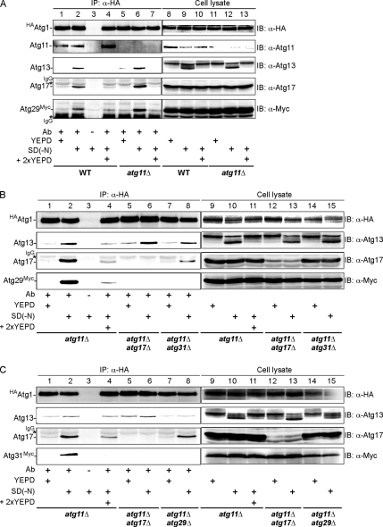
Autophagy induced by nutrient depletion is involved in survival during starvation conditions. In addition to starvation-induced autophagy, the yeast Saccharomyces cerevisiae also has a constitutive autophagy-like system, the Cvt pathway. Among 31 autophagy-related (Atg) proteins, the function of Atg17, Atg29, and Atg31 is required specifically for autophagy. In this study, we investigated the role of autophagy-specific (i.e., non-Cvt) proteins under autophagy-inducing conditions. For this purpose, we used atg11Delta cells in which the Cvt pathway is abrogated. The autophagy-unique proteins are required for the localization of Atg proteins to the pre-autophagosomal structure (PAS), the putative site for autophagosome formation, under starvation condition. It is likely that these Atg proteins function as a ternary complex, because Atg29 and Atg31 bind to Atg17. The Atg1 kinase complex (Atg1-Atg13) is also essential for recruitment of Atg proteins to the PAS. The assembly of Atg proteins to the PAS is observed only under autophagy-inducing conditions, indicating that this structure is specifically involved in autophagosome formation. Our results suggest that Atg1 complex and the autophagy-unique Atg proteins cooperatively organize the PAS in response to starvation signals.


Figures

Figure 1.
Figure 1.
Atg29 interaction with Atg17 is essential for autophagy. (A) The Atg17 binding site in Atg29. The indicated segments of Atg29 were fused to the Gal4 DNA-binding domain (BD). Binding to Atg17 fused to the Gal4-activating domain was evaluated by growth on −Ade plates for 3 d. (B) The Atg29 binding sites in Atg17. The indicated segments of Atg17 were fused to the Gal4 (transcriptional) activation domain (AD). Binding to Atg29 was tested by growth on −His plates containing 3 mM 3-AT. Atg17 interacts with Atg29 via coiled-coil domain 2. (C) Mutant Atg29s (Atg29W54R, Atg29P64H, Atg29F67L, or Atg29R71G) did not associate with Atg17. Two-hybrid assays were carried out as described in A. (D) Mutant Atg29s failed to interact with Atg17 in vivo. atg29Δ (TMK4) cells carrying the indicated myc-tagged ATG29 plasmids (wild-type and mutants) were grown in YEPD, and then they were treated with 0.2 μg/ml rapamycin for 3 h. Cell extracts were immunoprecipitated with anti-myc antibody. Coimmunoprecipitated proteins were analyzed by immunoblotting with anti-myc (top) or anti-Atg17 antibodies (middle). The bottom row indicates the amount of Atg17 in the total lysates. (E) The binding of Atg29 and Atg17 is essential for autophagy. Wild-type (BY4741 + pTN3) or atg29Δ (TMK181 + pTN3) cells carrying pTM108 (Atg29), pTM109 (Atg29F67L), pTM110 (Atg29R71G), or pRS313 (empty vector) were cultured in SCD, and then they were transferred to SD(−N) for 4 h, lysed, and assayed for ALP activity. The bars represent the SD of three independent experiments. (F) Atg17 is required for proper Atg29 localization to the PAS. WT (TMK190) or atg17Δ (TMK214) cells were grown in SD medium containing casamino acid supplemented with adenine, tryptophan, and uracil, and then they were treated with 0.2 μg/ml rapamycin. Growing (0 h) and rapamycin-treated (3 h) cells were observed by fluorescence microscopy. The percentages of cells with fluorescent dot signals at the PAS were determined. Bar, 5 μm. (G) Atg29 is not required for proper Atg17 localization to the PAS. WT (TMK576) or atg29Δ (TMK600) cells were tested as in F.
Figure 2.
Figure 2.
Deletion of ATG29 in atg11Δ cells affects PAS localization of Atg proteins. Localization of GFP-tagged Atg17, Atg29, and Atg31 was analyzed in the indicated atg mutant cells. Similarly, localization of Atg2 and Atg5 was observed. Rapamycin-treated (3 h) cells were observed by fluorescence microscopy. The percentages of cells with fluorescent dot signals at the PAS were determined. Bar, 5 μm.
Figure 3.
Figure 3.
The PAS localization of Atg17 or Atg29 does not depend on Atg2, Atg9, or Atg14 in atg11Δ cells. Localization of Atg17-GFP and Atg29-GFP in the indicated atg mutants (treated with rapamycin for 3 h) was observed by fluorescence microscopy. The percentages of cells with fluorescent dot signals at the PAS were determined. Bars, 5 μm.
Figure 4.
Figure 4.
All of five proteins (Atg1, Atg13, Atg17, Atg29, and Atg31) play important role in their localization at the Atg11-independent PAS. (A) In atg11Δ cells, both Atg1 and Atg13 maintain Atg17-GFP, Atg29-GFP, and Atg31-GFP at the PAS. Rapamycin-treated (3 h) cells were observed by fluorescence microscopy. (B) Localization of Atg1-GFP and Atg13-GFP in the indicated atg cells was examined as described in A. Bars, 5 μm.
Figure 5.
Figure 5.
Atg17–Atg29 and Atg1–Atg13 interactions are indispensable for their recruitment to the PAS. (A) Atg29 mutant (Atg29F67L and Atg29R71G) is defective for the ATG11-independent PAS localization of Atg1, Atg13, or Atg17. atg11Δatg29Δ cells expressing GFP-tagged Atg1, Atg13, or Atg17 were transformed with a plasmid expressing either Atg29wild-type, Atg29F67L, or Atg29F71G. Cells were treated with rapamycin for 3 h before observation. (B) Atg131-448 does not recruit Atg29 to the PAS in atg11Δ cells. atg11Δatg13Δ cells expressing Atg29-GFP were transformed with a plasmid expressing either Atg13wild type or Atg131-448. Cells were observed after rapamycin treatment for 3 h. (C) Atg1 kinase activity is not essential for the PAS targeting of Atg17 and Atg29 in atg11Δ. atg11Δatg1Δ cells expressing Atg17-GFP or Atg29-GFP were transformed with a plasmid expressing either wild-type Atg1 or Atg1 kinase-deficient mutant (Atg1D211A or Atg1K54A). Cells were observed after rapamycin-treatment for 3 h. Bars, 5 μm.
Figure 6.
Figure 6.
Assembly of Atg proteins to the PAS is regulated in response to nutrient conditions. (A) Recruitment of Atg17 or Atg29 to the PAS is induced by starvation in atg11Δ cells. Atg29-GFP and Atg17-GFP were observed in wild-type or atg11Δ cells under both growth and rapamycin-treated (3 h) conditions. (B) Atg29 localization in response to nutrient conditions in wild-type or in atg11Δ cells. Cells grown in SD medium containing casamino acid supplemented with adenine, tryptophan, and uracil were shifted to SD(−N). Nitrogen-starved cells (−N, 2 h) were supplied with 2× SD medium containing casamino acid supplemented with adenine, tryptophan, and uracil, and then they were incubated for 10 min. (C) PAS recruitment of Atg8 in response to starvation. atg11Δatg8Δ cells (YYK865) harboring the CFP-ATG8 (pRS314) were observed as described in B. CFP-Atg8 and Atg29-YFP were colocalized in starved cells. (Noted that CFP signal was observed in the vacuole indicating the induction of autophagy.) (D) Accumulation of Atg17 and Atg29 to the PAS in atg1D211Aatg11Δ is starvation specific. The localization of Atg17-GFP and Atg29-GFP was analyzed as described in B. (E) Comparison of autophagic activity of wild-type cells and atg11Δ cells in response to cellular nutrient condition. Wild-type (OND88) or atg11Δ (YYK762) cells genomically expressing Pho8Δ60 protein were cultured in YEPD, and then they were shifted to SD(−N) at time 0. After SD(−N) for 4 h, nitrogen-starved cells were supplied with nutrients as described in B. After 1 or 2 h, cells were corrected and subjected to ALP assay. The bars represent the SD of three independent experiments. Bars (A–D), 5 μm.
Figure 7.
Figure 7.
Association of Atg1, Atg13, Atg17, Atg29, and Atg31 is regulated by nutrient condition. (A) Interaction of Atg1 with Atg13, 17, and Atg29 is enhanced by starvation. Wild-type (YYK850) or atg11Δ (YYK868) cells grown in YEPD (lanes 1, 5, 8, and 11) were transferred to SD(−N) for 2 h (lanes 2, 3, 6, 9, and 12). Starvation was terminated by adding 2× YEPD for 10 min (lanes 4, 7 10, and 13). HA-Atg1 was immunoprecipitated by anti-HA ascite (Ab), and immunocomplex (lanes 1–7) was analyzed by immunoblot with the indicated antibodies. Total cell extracts also are shown (lanes 8–13). Note that all Atg proteins were chromosomally expressed under their own promoter. (B) Atg17 and Atg31 are required for starvation-induced Atg1–Atg29 association. atg11Δ (YYK868), atg11Δ atg17Δ (YYK870) and atg11Δatg31Δ (YYK872) cells were analyzed as described in A. (C) Atg1–Atg31 association is disrupted in atg11Δatg17Δ and atg11Δatg29Δ cells. The experiment described in A was carried out using atg11Δatg29Δ (YYK874) harboring plasmid pTM4 (atg11Δ), atg11Δatg17Δ atg29Δ (YYK878) harboring plasmid pTM4 (atg11Δatg17Δ), or atg11Δatg29Δ (YYK874).

References

    1. Baba M., Takeshige K., Baba N., Ohsumi Y. Ultrastructural analysis of the autophagic process in yeast: detection of autophagosomes and their characterization. J. Cell Biol. 1994;124:903–913. - PMC - PubMed
    1. Cheong H., Yorimitsu T., Reggiori F., Legakis J. E., Wang C. W., Klionsky D. J. Atg17 regulates the magnitude of the autophagic response. Mol. Biol. Cell. 2005;16:3438–3453. - PMC - PubMed
    1. Giaever G., et al. Functional profiling of the Saccharomyces cerevisiae genome. Nature. 2002;418:387–391. - PubMed
    1. Goldstein A. L., McCusker J. H. Three new dominant drug resistance cassettes for gene disruption in Saccharomyces cerevisiae. Yeast. 1999;15:1541–1553. - PubMed
    1. James P., Halladay J., Craig E. A. Genomic libraries and a host strain designed for highly efficient two-hybrid selection in yeast. Genetics. 1996;144:1425–1436. - PMC - PubMed

Publication types

Substances