. 2008 May;19(5):2059-68.
doi: 10.1091/mbc.e07-09-0902. Epub 2008 Feb 20.

Rab8 regulates basolateral secretory, but not recycling, traffic at the recycling endosome

Affiliations

Rab8 regulates basolateral secretory, but not recycling, traffic at the recycling endosome

Lauren Henry et al. Mol Biol Cell. 2008 May.

Abstract

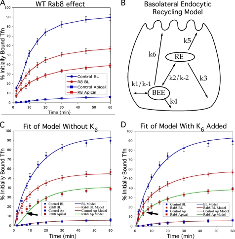
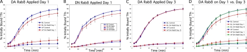
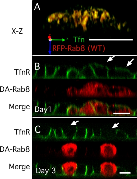
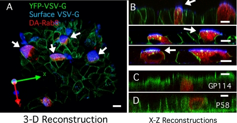
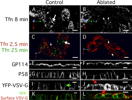
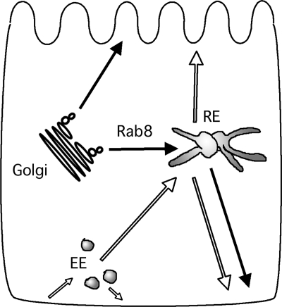
Rab8 is a monomeric GTPase that regulates the delivery of newly synthesized proteins to the basolateral surface in polarized epithelial cells. Recent publications have demonstrated that basolateral proteins interacting with the mu1-B clathrin adapter subunit pass through the recycling endosome (RE) en route from the TGN to the plasma membrane. Because Rab8 interacts with these basolateral proteins, these findings raise the question of whether Rab8 acts before, at, or after the RE. We find that Rab8 overexpression during the formation of polarity in MDCK cells, disrupts polarization of the cell, explaining how Rab8 mutants can disrupt basolateral endocytic and secretory traffic. However, once cells are polarized, Rab8 mutants cause mis-sorting of newly synthesized basolateral proteins such as VSV-G to the apical surface, but do not cause mis-sorting of membrane proteins already at the cell surface or in the endocytic recycling pathway. Enzymatic ablation of the RE also prevents traffic from the TGN from reaching the RE and similarly results in mis-sorting of newly synthesized VSV-G. We conclude that Rab8 regulates biosynthetic traffic through REs to the plasma membrane, but not trafficking of endocytic cargo through the RE. The data are consistent with a model in which Rab8 functions in regulating the delivery of TGN-derived cargo to REs.


Figures

Figure 1.
Figure 1.
Rab8 overexpression before polarization causes mis-sorting of Tfn traffic by disrupting cell polarity. (A) Effect of Rab8 overexpression. 125I-Tfn bound to the basolateral surface of MDCKT cells on ice was internalized at 37°C, and label released into the apical and basolateral media was measured at the times indicated. Blue circles, recycling into the basolateral medium in control cells; blue squares, transcytosis to the apical medium in control cells; red circles, recycling into the basolateral medium in cells overexpressing Rab8; and red squares, transcytosis into the apical medium in cells overexpressing Rab8. (B) Cartoon of possible Tfn recycling pathways, whose rate constants were used in the mathematical model. The rapid recycling pathway includes k1 and k4. The long recycling pathway includes k1 and k3. k6 indicates hypothetical mis-sorting pathway that would occur only in cells that are not properly polarized so that the apical and basolateral surfaces are functionally equivalent. BEE, basolateral early endosomes. (C) Fit of mathematical model to data in A using rate constants in Table 1, assuming k6 = 0. Arrow indicates area of poor fit. Data points are as in A; blue line models recycling in control cells; brown line models transcytosis in control cells, red line models recycling in cells overexpressing Rab8; and green line models transcytosis in cells overexpressing Rab8. (D) Fit of mathematical model to data in A when loss of cell polarity is assumed. Data points and lines are as in C. Error bars, SD for experimental data. n = 9 for all data points.
Figure 2.
Figure 2.
Dominant-active (DA) and -negative (DN) Rab8 mutants cause mis-sorting of Tfn traffic when applied before, but not after, cell polarity is established. All panels compare recycling (circles) and transcytosis (squares) of 125I-Tfn in control cells (blue) and Rab8 overexpression mutants (red) after 4 d of polarization. (A) Lentiviral DA-Rab8 application 24 h after cell plating results in significant mis-sorting to the apical surface. (B) Lentiviral DN-Rab8 application 24 h after cell plating results in significant mis-sorting to the apical surface. (C) Lentiviral DA-Rab8 application 3 d after cell plating does not result in mis-sorting. (D) Overlay of results from control cells in A with those obtained for cells infected with DA-Rab8 in A and C, to directly compare the effects of infection after 24 h (red) versus 3 d (green).
Figure 3.
Figure 3.
DA-Rab8 causes mis-sorting of TfnR when applied before, but not after, cell polarity is established. (A) X-Z reconstruction of an MDCKT monolayer infected with wild-type RFP-Rab8 (red) 24 h after plating and labeled with Alexa-488 Tfn internalized for 25 min (green, marks the RE). (B) X-Z reconstruction of MDCKT monolayer infected with DA-RFP-Rab8 (red) 24 h after plating and surface labeled on ice with Alexa-488 Tfn (green) on day 4. Arrows indicate mis-sorted TfnR. (C) MDCKT cells infected with DA-Rab8 (red) 3 d after plating and surface labeled on ice with Alexa-488 Tfn (green) on day 4 (green). Arrows indicate cells that express DA-Rab8 but did not mis-sort TfnR. Bar, 10 μm.
Figure 4.
Figure 4.
DA-Rab8 Rab8 applied 3 d after plating causes mis-sorting of VSV-G. (A) 3D reconstruction from a z-stack of confocal images spanning the full height of the cells. The image is rotated as shown by the axes at the lower left. GFP-VSV-G (green)–expressing MDCKT cells were infected with lentiviral DA-Rab8 at low MOI (red). Cells were kept at the nonpermissive temperature until 4 d after plating and then shifted to the permissive temperature as described in the text. The apical surface of live cells was labeled with an antibody against the VSV-G ectodomain (blue). All cells expressing DA-Rab8 (arrows) mis-sorted VSV-G to the apical surface. A few cells not expressing DA-Rab8 also mis-sorted VSV-G to the apical surface (top left) (B) Three X-Z reconstructions of confocal line-scan z-stacks from other fields of MDCKT cells labeled as in A, with arrows indicating mis-sorted VSV-G. (C) X-Z reconstruction of MDCKT cells expressing DA-Rab8 (red) as in A and labeled for apical marker GP114 (green). (D) X-Z reconstruction of MDCKT cells expressing DA-Rab8 (red) as in A and labeled for basolateral marker P58 (green). Bars, 10 μm.
Figure 5.
Figure 5.
Ablation of the RE causes mis-sorting of newly synthesized VSV-G. All panels show MDCKT cells (4 d after plating) that have internalized Tfn-HRP into the RE. (A, C, E, G, I, and K) Control cells treated with DAB but not H2O2; (B, D, F, H, J, and L) cells treated with DAB and H2O2. (A and B) Basolaterally applied Alexa-546 Tfn internalized into MDCKT cells for 8 min labels early (small arrow) and recycling (large arrow) endosomes. REs are not visible in RE-ablated cells. (C and D) Basolaterally applied Alexa-546 Tfn internalized into control MDCKT cells for 25 min labels RE (large arrows). Small peripheral early endosomes are visible in RE-ablated cells (small arrows). (E and F) In MDCKT cells labeled for Gp-114, both control and RE-ablated cells are labeled apically. (G and H) In MDCKT cells labeled for p58, both control and RE-ablated cells are labeled basolaterally. (I and J) MDCKT cells expressing VSV-G tsO45 released at the permissive temperature for 2 h after RE ablation. Green arrow in I indicates normal basolateral distribution of VSV-G. Red arrow in J indicates putative apical VSV-G in RE-ablated cells. (K and L) MDCKT cells as in I and J containing cellular GFP-VSV-G (green) and with surface VSV-G labeled by anti-VSV-G ectodomain antibody (red). Red arrow indicates mis-sorted apical VSV-G in ablated cells. Bars, 10 μm.
Figure 6.
Figure 6.
Cartoon of trafficking pathways that pass through the RE. Solid arrows represent secretory pathways. Open arrows represent recycling pathways. EE, early endosome; RE, recycling endosome.

Similar articles

Cited by

References

    1. Anderson E., Maday S., Sfakianos J., Hull M., Winckler B., Sheff D., Folsch H., Mellman I. Transcytosis of NgCAM in epithelial cells reflects differential signal recognition on the endocytic and secretory pathways. J. Cell Biol. 2005;170:595–605. - PMC - PubMed
    1. Ang A. L., Folsch H., Koivisto U. M., Pypaert M., Mellman I. The Rab8 GTPase selectively regulates AP-1B-dependent basolateral transport in polarized Madin-Darby canine kidney cells. J. Cell Biol. 2003;163:339–350. - PMC - PubMed
    1. Ang A. L., Taguchi T., Francis S., Folsch H., Murrells L. J., Pypaert M., Warren G., Mellman I. Recycling endosomes can serve as intermediates during transport from the Golgi to the plasma membrane of MDCK cells. J. Cell Biol. 2004;167:531–543. - PMC - PubMed
    1. Bomsel M., Prydz K., Parton R. G., Gruenberg J., Simons K. Endocytosis in filter-grown Madin-Darby canine kidney cells. J. Cell Biol. 1989;109:3243–3258. - PMC - PubMed
    1. Breitfeld P. P., Harris J. M., Mostov K. E. Postendocytotic sorting of the ligand for the polymeric immunoglobulin receptor in Madin-Darby canine kidney cells. J. Cell Biol. 1989;109:475–486. - PMC - PubMed

Publication types