. 2008 May;19(5):2101-12.
doi: 10.1091/mbc.e07-09-0897. Epub 2008 Feb 20.

Cholesterol loss enhances TrkB signaling in hippocampal neurons aging in vitro

Affiliations

Cholesterol loss enhances TrkB signaling in hippocampal neurons aging in vitro

Mauricio G Martin et al. Mol Biol Cell. 2008 May.

Abstract

Binding of the neurotrophin brain-derived neurotrophic factor (BDNF) to the TrkB receptor is a major survival mechanism during embryonic development. In the aged brain, however, BDNF levels are low, suggesting that if TrkB is to play a role in survival at this stage additional mechanisms must have developed. We here show that TrkB activity is most robust in the hippocampus of 21-d-old BDNF-knockout mice as well as in old, wild-type, and BDNF heterozygous animals. Moreover, robust TrkB activity is evident in old but not young hippocampal neurons differentiating in vitro in the absence of any exogenous neurotrophin and also in neurons from BDNF -/- embryos. Age-associated increase in TrkB activity correlated with a mild yet progressive loss of cholesterol. This, in turn, correlated with increased expression of the cholesterol catabolic enzyme cholesterol 24-hydroxylase. Direct cause-effect, cholesterol loss-high TrkB activity was demonstrated by pharmacological means and by manipulating the levels of cholesterol 24-hydroxylase. Because reduced levels of cholesterol and increased expression of choleseterol-24-hydroxylase were also observed in the hippocampus of aged mice, changes in cellular cholesterol content may be used to modulate receptor activity strength in vivo, autonomously or as a way to complement the natural decay of neurotrophin production.


Figures
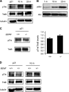
Figure 1.
Figure 1.
TrkB signaling robustness in vivo is independent of the levels of BDNF. (A) Western blot of hippocampal membranes from postnatal day 21 (P21), postnatal month 10 (10 m) and postnatal month 20 (20 m), with antibodies against active (pTrk) and total (TrkB) Trk receptors. The active form of the receptor increases dramatically with age, reaching highest levels in 20-mo-old mice. (B) A similar curve was observed for the downstream target Akt, suggesting that the levels of receptor phosphorylation lead to activation of survival effectors. (C) Western blot of hippocampal membranes from 21-d-old BDNF knockout (BDNF −/−) and wild-type animals (BDNF +/+) animals, with antibodies against active (pTrk) and total (TrkB) receptor. Note that the absence of ligand (BDNF −/−) does not affect the levels of receptor expression nor activity (compare with BDNF +/+). The relative amount of pTrk/TrkB present in KO mice respect to the wild-type case is shown on the right. (D) Western blot of hippocampal membranes from post natal day 21 (P21) and postnatal month 10 (10 m) wild-type (BDNF +/+) and BDNF heterozygous mice (+/−). Levels of the active receptor (pTrk) increase with age, in both types of animals.
Figure 2.
Figure 2.
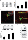
TrkB activity increases during in hippocampal neurons in vitro. (A) Immunoblotting (IB) of membranes from hippocampal neurons maintained for 10, 15, and 26 d in vitro (DIV). Increased receptor activity (pTrk) occurs with days in vitro, despite equal levels of receptor expression. Protein loading was assessed with anti-TrkB (TrkB) antibody. Quantification of blots from different experiments (n = 5) shows the increase of the pTrk/TrkB levels in aged cells. (B) Immunofluorescence microscopy analysis of phospho-TrkB of the surface of young and old cells. Distribution revealed more and more peripherally distributed structures in old neurons as compared with young neurons. This analysis reveals a high degree of colocalization between active Trk (green) and the ganglioside GT1b (red) in the aged neurons (insets). (C) Immunoblot analysis of Akt activity in cellular extracts from 10, 15, and 26 DIV neurons. Akt activity is low in 10 DIV neurons in comparison with 15 and 26 DIV neurons. This age-dependent increase in activity is not due to differences in the amount Akt protein expression. Quantitative analysis is shown on the right. (D) Effect of addition of the Trk inhibitor K252a on Trk phosphorylation and Akt activity. Addition of the inhibitor to 26-d-old cells (+, treated; −, untreated) result in the great reduction of Trk and Akt phosphorylation after 24-h treatment. These results suggest that the high levels of Trk activity found in old cells promote survival.
Figure 3.
Figure 3.
TrkB activity in fully differentiated hippocampal neurons in vitro is ligand-independent. (A) Phase-contrast images of hippocampal neurons from BDNF +/+ and BDNF −/− embryos, maintained in culture for 1, 4, and 15 DIV: cell number and degree of morphological differentiation is similar. (B) Immunoblotting of membranes from hippocampal and cortical neurons, from BDNF −/− and BDNF +/+ embryos, maintained for 15 DIV: the intensity of phosphorylated TrkB signal in the −/− cells is comparable to that of +/+ cells. Protein loading was assessed with anti-TrkB (TrkB) antibody, and the relative amount of pTrk/TrkB present in −/− cells with respect to the wild-type case is shown below. (C) Neurons from wild-type rat embryos maintained in vitro for 10 d (10 DIV) were treated for 30 min with the medium of neurons maintained in vitro for 26 d (26 DIV medium): this treatment does not elicit any TrkB activation in these cells, despite abundant levels of receptor. (D) Western blot analysis of TrkB activation of 15 DIV neurons incubated or not with a cocktail of neurotrophin-blocking antibodies (Anti-NT Abs), starting on day 10 in vitro (see Materials and Methods). Note that the antibody mix did not perturb the increase in TrkB signal, whether for total Trk (TrkB) or its phosphorylated form (pTrk). (E) Control of antibody-mix efficacy as neurotrophin-blocking TrkB activation. Neurons kept in vitro for 10 DIV neurons were incubated with 100 mg/ml BDNF; effect on TrkB activity (pTrkB) was measured 30 min later. Treatment triggers intense TrkB activity (pTrkB; BDNF +) in nonneurotrophin antibody-mix–incubated neurons (Anti-NT Abs, −); TrkB activity is absent in cells coincubated with the antibody mix (BDNF +/Anti-NT Abs, +).
Figure 4.
Figure 4.
Moderate still continuous loss of cholesterol from the plasma membrane of differentiated hippocampal neurons in vitro determines TrkB DRM partitioning. (A) Colorimetric measurement of plasma membrane cholesterol from hippocampal neurons kept for 10, 15, and 26 DIV. Mean ± SD values (nmol/mg protein, n = 4) were 0.61 ± 0.029, 0.55 ± 0.047, and 0.43 ± 0.023 in 10, 15, and 26 DIV neurons, respectively. (B) TLC analysis of the membrane cholesterol content in DRMs of 10, 15, and 26 DIV neurons; cholesterol is clearly and progressively reduced in DRMs from 15 and 26 DIV neurons. Percentages of cholesterol reduction in DRMs (fractions 4 and 5) and non-DRMs (fractions 6–8) were measured by densitometry and normalized for sphingomyelin values of different sucrose fractions (n = 2). In DRMs, cholesterol was decreased to 43.29% in 15 DIV cells, and to 25.92% in 26 DIV cells. The migration of the cholesterol standard is indicated (cholesterol). (C) Western blotting with anti-total Trk, anti-phospho Trk, and anti-prion protein (PrPc) antibodies, of 4°C detergent-extracted, sucrose-gradient centrifuged membranes from 10 and 26 DIV hippocampal neurons in vitro. Note that receptor activity is exclusively present in the DRM domains (fractions 4 and 5) of 26 DIV neurons. The PrPc lane demonstrates that the presence in fractions 4 and 5 truly reflect DRM partitioning. (D) Immunoprecipitations of TrkB from the DRMs and non-DRMs of 10 and 26 DIV neurons, probed with antibodies against phospho Trk and the anti-p85 subunit of PI3K. Note that the downstream survival effector of TrkB is precipitated exclusively from the DRM domains of 26 DIV neurons. (E) Control for correct separation between DRMs and non-DRMs fractions. Lysates utilized in C were hybridized with antibodies against the non-DRM protein Transferrin receptor (IB: TfR) or the DRM protein PrPc (IB: PrPc).
Figure 5.
Figure 5.
(A) Sucrose gradient membrane fractions from cold detergent-solubilized plasma membranes from 10, 15, and 26 DIV hippocampal neurons followed by immunodetection for the protein BACE 1. Note that BACE1 segregation to DRMs progressively decreases with time. Percentages of BACE1 distribution in DRMs (fractions 4 and 5, □) and nonDRMs (fractions 6–8, ■) were measured by densitometry and reported in the graphics to the right of the Western lot (mean ± SD, n = 4). In exact numbers, BACE 1 was 35.3 ± 5.8% in the DRMs of 10 DIV neurons, decreasing to 25.3 ± 4.1% in 15 DIV neurons and further decreasing to 15.8 ± 6.4% in senescent cells. (B) Sucrose gradient fractions of cold detergent-solubilized plasma membranes from 10 and 26 DIV neurons followed by immunodetection for the protein Fyn. Percentages of Fyn distribution in DRMs (fractions 4 and 5, □) and non-DRMs (fractions 6–8, ■) were measured by densitometry and reported in the graphics on the right (mean ± SD, n = 4). In 10 DIV neurons 34.75 ± 2.8% of Fyn is in DRMs, increasing to 58.9 ± 8.6% in 26 DIV neurons.
Figure 6.
Figure 6.
Pharmacological-induced changes in cholesterol levels in young neurons modulate pTrk activity. (A) Western blot of hippocampal membranes from 10, 15, and 26 d in vitro (DIV) control neurons as well as from 10 d in vitro (10 DIV) neurons with cholesterol levels reduced to the values of untreated neurons 26 DIV neurons (see Figure 2A; ↓Chol) and 10 DIV neurons with cholesterol levels replenished after reduction, to reach 10 DIV levels (↑Chol). Note how cholesterol reduction in the 10 DIV cells triggers intense TrkB activity, whereas its replenishment to their natural values makes receptor activity disappear. (B) Sucrose gradient fractions of cold detergent-solubilized plasma membranes from 10 in vitro (DIV) with control (ctrl), pharmacological reduction of membrane cholesterol (↓ Chol, 25% less than in control cells) or with cholesterol levels replenished after reduction to control levels (↑Chol). The 25% decrease in membrane cholesterol induces the displacement of Flotillin-1 away from DRMs (↓Chol), yet replenishment restores DRM-preferential partitioning (↑Chol, compare with ↓Chol). Percentages of Flotillin-1 distribution in DRMs (fractions 4 and 5, □) and nonDRMs (fractions 6–8, ■) were measured by densitometry (graphics on the right, mean ± SD, n = 4). Flotillin 1 was 31.3 ± 8% in the DRM domains of 10 DIV neurons. Cholesterol depletion reduced Flotillin-1 in DRMs to 14 ± 3.0%; cholesterol replenishment of the latter cells increased Flotillin 1 in DRMs to 50.4 ± 8.0%.
Figure 7.
Figure 7.
Differentiation-occurring increase in cholesterol-24-hydorxylase is sufficient and necessary for TrkB activation. (A) Compared with 10 DIV neurons, and after normalization for actin mRNA, mRNA expression levels of cholesterol-24-hydroxylase (CYP46) increase at 26 DIV. Variations related to 10 DIV (value of 1) are indicated. (B) Immunofluorescence analysis of the expression levels of cholesterol-24-hydroxylase in hippocampal neurons maintained for 10 and 26 DIV. Antibody concentration and image exposure were identical for all three time points. Higher reactivity occurs in the 26 DIV neurons, consistent with the higher expression of the messenger (see A). (C) Hippocampal neurons in suspension were transfected either with the plasmid expressing cyp46a1 (p46+) or scrambled vector (pLL3.7) or with the plasmid expressing the cyp46a1-siRNA (pSi46). TrkB activity was measured at 7 DIV or 10 DIV. Note how the increased expression of the cholesterol-24-hydroxylase (CYP46) resulted in high levels of active TrKB in these cells, and reduction of the cholesterol 24-hydroxylase by siRNA resulted in lower levels of Trk phosphorylation. The amount of pTrk/tubulin either in pSi46 or p46+ transfected cells respect to the control is shown. Quantification of the Western blottings performed using anti-cholesterol-24-hydroxylase antibody reveals the efficacy of the knockdown strategy. (D) Immunofluorescence microscopy with anti phospho-Trk antibody (p-Trk) reveals that reduction in cholesterol-24-hydroxylase results in reduced levels of active Trk (IF:pTrk). The efficacy of the knockdown strategy was analyzed with anti-cholesterol-24-hydroxylase antibody reveals (see IF: CYP46).
Figure 8.
Figure 8.
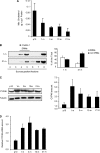
In vivo reduction of membrane cholesterol, changes in membrane DRM partitioning characteristics and increased levels of cholesterol-24-hydroxylase. (A) Colorimetric analysis of membrane cholesterol levels in hippocampal membranes at different postnatal times: 10 d (p10), 1 mo (1 m), 3 mo (3 m), 10 mo (10 m), and 21 mo (21 m). Cholesterol is most abundant in the membranes of early postnatal hippocampus (p10) undergoing significant loss with time, reaching the lowest levels at month 21 (21 m). (B) Western blot analysis with an antibody against the canonical DRM protein Flotillin 1 in detergent extracted, sucrose gradient separated, DRM and non-DRM fractions from 1- and 21-mo-old mice hippocampi. Note the clear DRM partitioning in the membranes from young animals and the more homogeneous distribution in the old animals. (C) Western blotting (left) and quantification (right) of the expression levels of cholesterol-24-hydroxylase in hippocampal extracts from 10 d (p10) and 1-, 3-, 10-, and 21-mo-old mice. CYP46 levels were normalized for tubulin. (D) Real-time PCR analysis of expression levels of cholesterol-24-hydroxylase. Total RNA was obtained from hippocampus from 10 d (p10) and 1-, 3-, 10-, and 21-mo-old mice. As shown in C and D, time-associated increased expression of the enzyme parallels both loss of cholesterol (see above, A) as well as the increased partitioning of TrkB in DRMs in this structure (see Figure 3C).

Similar articles

Cited by

References

    1. Abad-Rodriguez J., et al. Neuronal membrane cholesterol loss enhances amyloid peptide generation. J. Cell Biol. 2004;167:953–960. - PMC - PubMed
    1. Agerman K., Hjerling-Leffler J., Blanchard M. P., Scarfone E., Canlon B., Nosrat C., Ernfors P. BDNF gene replacement reveals multiple mechanisms for establishing neurotrophin specificity during sensory nervous system development. Development. 2003;130:1479–1491. - PubMed
    1. Bligh E. G., Dyer W. J. A rapid method of total lipid extraction and purification. Can. J. Biochem. Physiol. 1959;37:911–917. - PubMed
    1. Blum R., Konnerth A. Neurotrophin-mediated rapid signaling in the central nervous system: mechanisms and functions. Physiology. 2005;20:70–78. - PubMed
    1. Borroni B., Archetti S., Agosti C., Akkawi N., Brambilla C., Caimi L., Caltagirone C., Di Luca M., Padovani A. Intronic CYP46 polymorphism along with ApoE genotype in sporadic Alzheimer disease: from risk factors to disease modulators. Neurobiol. Aging. 2004;25:747–751. - PubMed

Publication types

MeSH terms