. 2008 May;19(5):1848-61.
doi: 10.1091/mbc.e07-08-0815. Epub 2008 Feb 20.

FBXO25-associated nuclear domains: a novel subnuclear structure

Affiliations

FBXO25-associated nuclear domains: a novel subnuclear structure

Adriana O Manfiolli et al. Mol Biol Cell. 2008 May.

Abstract

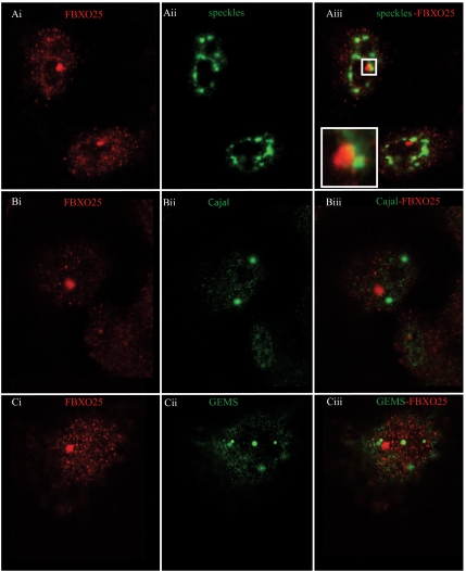
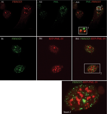
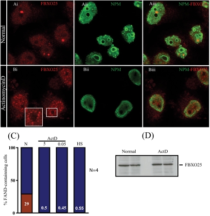
Skp1, Cul1, Rbx1, and the FBXO25 protein form a functional ubiquitin ligase complex. Here, we investigate the cellular distribution of FBXO25 and its colocalization with some nuclear proteins by using immunochemical and biochemical approaches. FBXO25 was monitored with affinity-purified antibodies raised against the recombinant fragment spanning residues 2-62 of the FBXO25 sequence. FBXO25 protein was expressed in all mouse tissues tested except striated muscle, as indicated by immunoblot analysis. Confocal analysis revealed that the endogenous FBXO25 was partially concentrated in a novel dot-like nuclear domain that is distinct from clastosomes and other well-characterized structures. These nuclear compartments contain a high concentration of ubiquitin conjugates and at least two other components of the ubiquitin-proteasome system: 20S proteasome and Skp1. We propose to name these compartments FBXO25-associated nuclear domains. Interestingly, inhibition of transcription by actinomycin D or heat-shock treatment drastically affected the nuclear organization of FBXO25-containing structures, indicating that they are dynamic compartments influenced by the transcriptional activity of the cell. Also, we present evidences that an FBXO25-dependent ubiquitin ligase activity prevents aggregation of recombinant polyglutamine-containing huntingtin protein in the nucleus of human embryonic kidney 293 cells, suggesting that this protein can be a target for the nuclear FBXO25 mediated ubiquitination.


Figures

Figure 1.
Figure 1.
Expression of FBXO25 protein in mouse tissues and cultured cell lines. Approximately 150 μg of protein from the indicated tissues (A) and cultured cells (B) lysates were subjected to SDS-PAGE, transferred onto nitrocellulose membranes, and probed with affinity-purified anti-FBXO25 antibodies (1:1500). Tissue and cell extracts were prepared as described in Materials and Methods. The specificity of the anti-FBXO25 antibodies (C) were ascertained by probing twin blots prepared from SDS-PAGE loaded with two separate sets of cell lysates from HEK293H cells and HEK293H cells stably transfected with GST-FBXO25 (HEK293HFB25-WT-1), with antibodies in the absence and presence of 6 μg/ml recombinant FBXO25 N-terminal fragment. Note that the FBXO25 fragment fully blocked the antibody reaction with both the endogenous and GST-fusioned protein. Ponceau-S staining showed protein loading (middle) and anti-GST antibodies labeling showed the expression of GST-FBXO25 protein in HEK293HFB25-WT-1 cells (bottom).
Figure 2.
Figure 2.
Mammalian cell nuclei contain domains highly enriched in FBXO25. Affinity-purified antibodies directed against FBXO25 were used to perform immunofluorescence on a variety of mammalian cell types. These included HeLa (A), COS-7 (B), and HEK293H (C). Note that anti-FBXO25 antibodies label both nucleus and cytoplasm, staining of the nucleoplasm is more intense. Observe the presence of bright dot-like structure within the nucleoplasm. DAPI was used to stain nuclei and images were taken by confocal microscopy. Bars, 5 μm.
Figure 3.
Figure 3.
Nuclear association of FBXO25. (A) Biochemical partitioning. Whole-cells extracts of controls HeLa cells or cells treated were prepared under the indicated conditions (see Materials and Methods) and analyzed by immunoblotting using anti-FBXO25 antibodies. (B) HeLa cells (T) were fractionated into nucleoplasm (N) and insoluble nuclear pellet (NP) as described in Materials and Methods. Proteins were separated by SDS-PAGE and the corresponding blotted nitrocellulose membranes were probed with anti-FBXO25, anti-nucleophosmin (NPM), and anti-β-tubulin antibodies. (C) An overlay of the differential interference contrast (DIC) microscopy image of HeLa cells and the corresponding FBXO25 immunostaining image generated by affinity-purified anti-FBXO25 antibodies is shown in Ci. Confocal analysis of cells stained for FBXO25 and nuclei/nucleoli with the affinity-purified anti-FBXO25 antibodies and anti-B23 nucleophosmin (anti-NPM) antibodies, respectively, is shown in Cii. FBXO25 is predominantly confined to the nucleus, outside nucleoli structures.
Figure 4.
Figure 4.
Localization of overexpressed tagged-FBXO25 in HEK293H cells. (A) HEK293H cells transfected with EGFP/HA-FBXO25-FLAG were fixed and immunostained with anti-NPM antibodies. (B) Western blotting analysis of transiently expressed EGFP-FBXO25 protein. Approximately 50 μg of protein from the nontransfected (NT) and transfected cells (T) lysates were subjected to SDS-PAGE, transferred onto nitrocellulose membranes and probed with monoclonal anti-GFP antibodies.
Figure 5.
Figure 5.
FBXO25 protein is found in distinct subnuclear domain. Confocal microscopy of cells double stained with affinity-purified anti-FBXO25 antibodies and antibodies against SC35, which label splicing speckles (Ai–Aiii), or against p80-coilin, which label Cajal bodies (Bi–Biii), or against SMN, which label GEMS (Ci–Ciii). The proteins labeled in each panel are indicated in the top left and right of the panel in the relevant color.
Figure 6.
Figure 6.
FBXO25 protein is organized in subnuclear structures distinct from PML-IV clastosomes in HeLa cells. (A) Confocal analysis of cells labeled with the FBXO25 and PML antibodies. PML bodies were labeled with monoclonal antibodies that react against of all isoforms of PML proteins (Ai–Aiii). (B) HeLa cells were transiently transfected with RFP-PML isoform IV and immunostained with the FBXO25 antibodies to detect sites of colocalization. FANDs did not colocalize with a large ring-like structure of PML-IV clastosomes (Bi–Biii).
Figure 7.
Figure 7.
(A) Double labeling confocal experiments for the detection of FBXO25 and proteasomes in HeLa cells. (A) Proteasomes are accumulated in domains resembling clastosomes (Aiii, inset). (B) Proteasomes show a diffuse nucleoplasmic pattern. (C) Density and colocalization of FANDs and clastosomes in nuclei of HeLa cells as determined by counting 100 cells in each of four microscopic-slides. (D) Confocal analysis of cells double-labeled with antibodies against FBXO25 and Skp1 (Ci–Ciii). The labeled proteins in each panel are indicated in the top left and right of each panel in the respective color.
Figure 8.
Figure 8.
Inhibition of RNA polymerases using actinomycin D disrupts FBXO25-associated nuclear domains. Confocal microscopy of labeled FBXO25 in untreated and ActD-treated (5 μg/ml for 2 h) HeLa cells are shown. Without ActD treatment, FANDs were found in the nucleoplasm (Ai–Aiii). In the presence of ActD, the majority of endogenous (Bi–Biii) FANDs disappeared (B). Note that FBXO25 occurs in perinucleolar structures in cells treated with ActD (Bi, inset). (C) Proportion of cells containing FANDs was estimated. After treatment with ActD, the proportion of cells containing FANDs (red) significantly differs from the control population. Four separate experiments were performed, and ∼100 cells were analyzed for drug treatment. CR, untreated; 0.5, ActD, 0.5 μg/ml; ActD 0.05, 0.05 μg/ml; and HS, heat shock. (D) Western blotting analysis of FBXO25 from protein extracts of cells at time points as shown in A. ActD does not alter FBXO25 subcellular levels.
Figure 9.
Figure 9.
Distribution of FANDs throughout cell division. (A) Three separate experiments were performed, and ∼100 cells were analyzed for each mitotic phase. The percentages of cells containing FANDs are indicated (red). (B) Western blotting of FBXO25 protein levels during the cell cycle. HeLa cell lysates were prepared by harvested cells in RIPA buffer at the indicated time points after release from the thymidine arrest. RIPA cell extracts (60 μg/lane) were processed for protein blots. The membrane was stripped and reprobed with an anti-β-actin antibodies.
Figure 10.
Figure 10.
FANDs are dispersed throughout the nucleoplasm during mitosis. HeLa cells released from a double thymidine block at the G1/S were immunolabeled using antibodies to the FBXO25 (red), costained with α-tubulin (green) and DAPI (blue) to identify the mitotic phase. Merged images are shown in the last column.
Figure 11.
Figure 11.
FANDs are major focal sites of ubiquitination but not transcription. (A) Ubiquitin-conjugates were labeled with antibody FK2, specific for conjugated ubiquitin but not for free ubiquitin (Fujimuro et al., 1994). The localization of FBXO25 (red) and ubiquitin-conjugates (green) are shown. (B) The transcription sites in HeLa cells were labeled using in vivo Br-UTP incorporation, followed by detection with anti-BrdUTP. No labeling was observed in control labeling in the absence of Br-UTP (data not shown). The localization of FBXO25 (green) and BrRNA (red) are shown.
Figure 12.
Figure 12.
FBXO25 prevents aggregation of polyQ-containing proteins. (A) HeLa cells transfected with EGFP-httEx1-103Q were fixed and labeled with anti-FBXO25 antibodies. Images were taken by confocal microscopy, and the labeled proteins are indicated on top of each panel with the respective color. (B) Slot-blot (S.B) filter retardation assays of polyQ aggregates formed in HEK293T cells cotransfected as indicated in B (top). Results shown correspond to two of four sets of independently cotransfected cells. The EGFP-httEx1-74Q protein-containing aggregates were detected with anti-GFP antibodies. (C) Western blotting (W.B) analysis of cell lysates of one set of HEK293T cells cotransfected as indicated in B. The different forms of FBXO25 protein, indicated by arrows, were revealed with ati-FBXO25 antibodies. Input: lysates of control, WT and ΔF cells. (D) Densitometric analysis of the slot-blot membranes prepared with samples of lysates from all four sets of cotransfected cells indicated in B. Results are expressed as percentage of aggregated protein in the control samples. (E) Detection of EGFP-httEx1-74Q mRNAs by RT-PCR from HEK293T cells cotransfected as indicated in E (top). Coexpression of full-length wild-type or mutant version of FBXO25 had no effect on expression of EGFP-httEx1-74Q mRNA. (F) S.B filter retardation assays of polyQ aggregates formed in HEK293T cells that were transfected with either Cul1WT or Cul1DN and EGFP-httEx1-74Q, FBXO25WT, Skp1, and Roc1. Results shown correspond to two of four sets of independently cotransfected cells. (G) W.B analysis of cell lysates of one set of HEK293T cells cotransfected as indicated in F. The different forms of FLAG-Cul1 protein, indicated by arrows, were revealed with anti-FLAG antibodies. Input, lysates of Cul1WT and Cul1DN cells. (H) Densitometric analysis of the slot-blot membranes prepared with samples of lysates from all four sets of cotransfected cells indicated in F. Results are expressed as percentage of aggregated protein in the Cul1DN.

Similar articles

Cited by

References

    1. Ang X. L., Harper J. W. Interwoven ubiquitination oscillators and control of cell cycle transitions. Oncogene. 2005;17:2860–2870. - PubMed
    1. Ardley H. C., Robinson P. A. E3 ubiquitin ligases. Essays Biochem. 2005;41:15–30. - PubMed
    1. Ausubel F. M., Brent R., Kingston R. E., Moore D. D, Seidman J. G., Smith J. A., Struhl K. Short Protocols in Molecular Biology. 3rd ed. New York: John Wiley & Sons; 1997.
    1. Bond U. Heat shock but not other stress inducers leads to the disruption of a sub-set of snRNPs and inhibition of in vitro splicing in HeLa cells. EMBO J. 1988;7:3509–3518. - PMC - PubMed
    1. Carmo-Fonseca M., Mendes-Soares L., Campos I. To be or not to be in the nucleolus. Nat. Cell Biol. 2000;2:E107–E112. - PubMed

Publication types

MeSH terms