. 2008 Apr;19(4):780-8.
doi: 10.1681/ASN.2007040420. Epub 2008 Feb 20.

The use of magnetic resonance to evaluate tissue oxygenation in renal artery stenosis

Affiliations

The use of magnetic resonance to evaluate tissue oxygenation in renal artery stenosis

Stephen C Textor et al. J Am Soc Nephrol. 2008 Apr.

Abstract

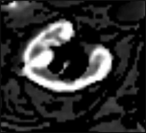
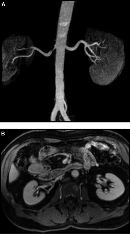
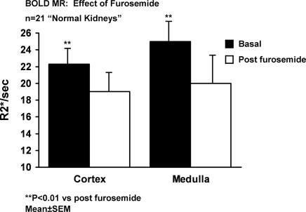
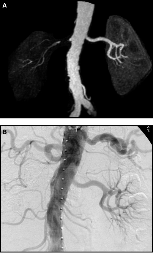
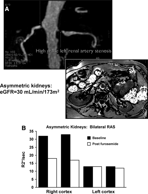
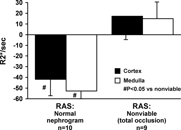
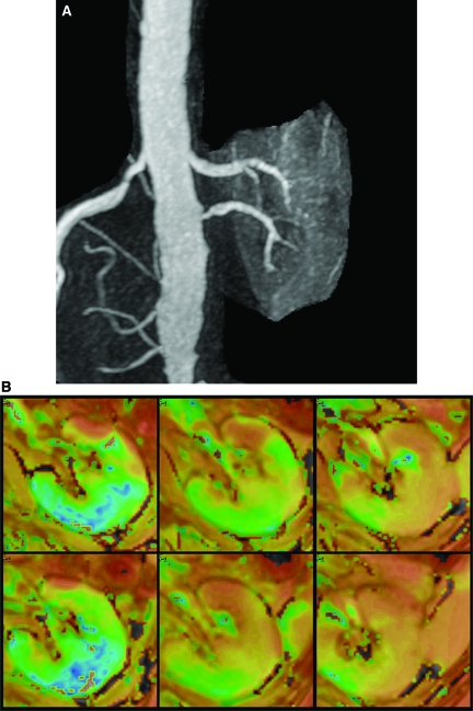
Vascular occlusive disease poses a threat to kidney viability, but whether the events leading to injury and eventual fibrosis actually entail reduced oxygenation and regional tissue ischemia is unknown. Answering this question has been difficult because of the lack of an adequate method to assess tissue oxygenation in humans. BOLD (blood oxygen-level-dependent) magnetic resonance imaging detects changes in tissue deoxyhemoglobin during maneuvers that affect oxygen consumption, therefore this technique was used to image and analyze cortical and medullary segments of 50 kidneys in 25 subjects undergoing magnetic resonance (MR) angiography to diagnose renal artery stenosis (RAS). Magnetic rate of relaxation (R2*) positively correlates with deoxyhemoglobin levels and was therefore used as a surrogate measure of tissue oxygenation. Furosemide was administered to examine the effect of inhibiting energy-dependent electrolyte transport on tissue oxygenation in subjects with renovascular disease. In 21 kidneys with normal nephrograms, administration of furosemide led to a 20% decrease in medullary R2* (P < 0.01) and an 11.2% decrease in cortical R2*. In normal-size kidneys downstream of high-grade renal arterial stenoses, R2* was elevated at baseline, but fell after furosemide. In contrast, atrophic kidneys beyond totally occluded renal arteries demonstrated low levels of R2* that did not change after furosemide. In kidneys with multiple arteries, localized renal artery stenoses produced focal elevations of R2*, suggesting areas of deoxyhemoglobin accumulation. These results suggest that BOLD MR coupled with a method to suppress tubular oxygen consumption can be used to evaluate regional tissue oxygenation in the human kidney affected by vascular occlusive disease.


Figures

Figure 1.
Figure 1.
Parametric BOLD (blood oxygen-level-dependent) image of the left kidney. The image represents a map of T2 values calculated from fitting voxel signal intensities from each echo time (TE) value of the multiecho gradient echo sequence to an exponential function. The bright intensity in the cortex defines a region of interest as contrasted to darker medullary segments.
Figure 2.
Figure 2.
Magnetic resonance (MR) angiogram (A) demonstrating focal stenosis of the proximal right renal artery. Despite the stenosis and poststenotic dilation, filtration and kidney volume (B) were preserved in this kidney. This is an example of a “normal” appearing kidney beyond a stenotic lesion (see text). The left kidney in this patient was considered a “normal” kidney.
Figure 3.
Figure 3.
BOLD MR measurements (R2*, s−1) in cortex and medullary segments in 21 “normal” appearing kidneys before and after intravenous furosemide. Relative reduction in cortical segments (11.2 ± 2%) was less than that observed in medullary regions (20 ± 2%) (P < 0.05). Enhanced reductions in R2* after furosemide in medullary segments is consistent with relatively greater accumulation of deoxyhemoglobin related to oxygen consumption due to chloride and sodium transport in the thick ascending limb of Henle (see text).
Figure 4.
Figure 4.
MR angiogram (A) demonstrating near total occlusion to the right kidney with minimal filtration. Conventional intra-arterial contrast angiography (B) (one week later) confirmed total occlusion and nonfunction of this kidney. This is an example of “nonviable” kidney as summarized in Table 2.
Figure 5.
Figure 5.
MR angiogram in a patient with bilateral renal arterial stenosis (A), more severe on the left, on the basis of poststenotic dilation and reduced parenchymal volume. BOLD imaging demonstrated low levels of R2* both before and after furosemide (B). The right kidney had normal volume with higher baseline R2* with a large fall in R2* after administration of furosemide. These data suggest higher deoxyhemoglobin levels in the right kidney with exaggerated furosemide-suppressible oxygen consumption.
Figure 6.
Figure 6.
Changes in BOLD MR measurements (Delta R2*/s) before and after furosemide (FSOC, furosemide-suppressible oxygen consumption) in patients with atherosclerotic renal artery stenosis (RAS) with “normal” kidneys as compared with “nonviable” kidneys with total occlusion (Table 2). Nonfunctioning kidneys demonstrated no R2* response after intravenous furosemide, whereas poststenotic kidneys otherwise had consistent falls in R2* after furosemide, particularly in medullary regions.
Figure 7.
Figure 7.
(A) Gadolinium-enhanced MR angiogram demonstrating a left kidney supplied by two renal arteries, one of which (inferior) has a high grade stenosis. (B) Parametric map of T2* (1/R2*) in the left kidney supplied by two renal arteries, one of which has high-grade stenosis. Top row are prefurosemide images from the upper pole, mid pole, and lower pole regions, respectively, and bottom row images are after furosemide administration. T2* intensity differs between regions supplied by stenotic and nonstenotic renal arteries.

References

    1. Garovic V, Textor SC: Renovascular hypertension and ischemic nephropathy. Circulation 112: 1362–1374, 2005 - PubMed
    1. Nielsen K, Rehling M, Henriksen JH: Renal vein oxygen saturation in renal artery stenosis. Clin Physiol 12: 179–184, 1992 - PubMed
    1. Textor SC, Lerman LO: Renal artery disease: Pathophysiology. In: Vascular Medicine: A Companion to Braunwald's Heart Disease, edited by Creager MA, Dzau VJ, Loscalzo J, Philadelphia, Saunders-Elsevier, 2006, pp 323–334
    1. Pohl MA: Renal artery stenosis, renal vascular hypertension and ischemic nephropathy. In: Diseases of the Kidney, Sixth Ed., edited by Schrier RW, Gottschalk CW, Boston, Little, Brown and Company, 1997, pp 1367–1423
    1. Alcazar JM, Rodicio JL: Ischemic Nephropathy: Clinical characteristics and treatment. Am J Kidney Dis 36: 883–893, 2000 - PubMed

Publication types