. 2008 Apr;15(4):697-706.
doi: 10.1128/CVI.00045-08. Epub 2008 Feb 20.

Dose-dependent protection against or exacerbation of disease by a polylactide glycolide microparticle-adsorbed, alphavirus-based measles virus DNA vaccine in rhesus macaques

Affiliations

Dose-dependent protection against or exacerbation of disease by a polylactide glycolide microparticle-adsorbed, alphavirus-based measles virus DNA vaccine in rhesus macaques

Chien-Hsiung Pan et al. Clin Vaccine Immunol. 2008 Apr.

Abstract

Measles remains an important cause of vaccine-preventable child mortality. Development of a low-cost, heat-stable vaccine for infants under the age of 6 months could improve measles control by facilitating delivery at the time of other vaccines and by closing a window of susceptibility prior to immunization at 9 months of age. DNA vaccines hold promise for development, but achieving protective levels of antibody has been difficult and there is an incomplete understanding of protective immunity. In the current study, we evaluated the use of a layered alphavirus DNA/RNA vector encoding measles virus H (SINCP-H) adsorbed onto polylactide glycolide (PLG) microparticles. In mice, antibody and T-cell responses to PLG-formulated DNA were substantially improved compared to those to naked DNA. Rhesus macaques received two doses of PLG/SINCP-H delivered either intramuscularly (0.5 mg) or intradermally (0.5 or 0.1 mg). Antibody and T-cell responses were induced but not sustained. On challenge, the intramuscularly vaccinated monkeys did not develop rashes and had lower viremias than vector-treated control monkeys. Monkeys vaccinated with the same dose intradermally developed rashes and viremia. Monkeys vaccinated intradermally with the low dose developed more severe rashes, with histopathologic evidence of syncytia and intense dermal and epidermal inflammation, eosinophilia, and higher viremia compared to vector-treated control monkeys. Protection after challenge correlated with gamma interferon-producing T cells and with early production of high-avidity antibody that bound wild-type H protein. We conclude that PLG/SINCP-H is most efficacious when delivered intramuscularly but does not provide an advantage over standard DNA vaccines for protection against measles.


Figures

FIG. 1.
FIG. 1.
Immune responses of mice to PLG/SINCP-H, SINCP-H, and SINCP. BALB/c mice (n = 5/group) were immunized with 10 μg or 1 μg PLG/SINCP-H, 10 μg naked SINCP-H, or 10 μg naked SINCP and were boosted with the same vaccines and doses 4 weeks later (arrow). (A) MV-specific IgG in serum was assayed by EIA. (B) Production of IFN-γ and IL-4 by splenocytes in response to MV H peptides was measured by ELISPOT assay at 8 weeks after immunization. Values are plotted as means, with error bars indicating standard deviations.
FIG. 2.
FIG. 2.
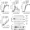
Immune responses of rhesus macaques to different doses and routes of inoculation of PLG/SINCP-H. Pairs of rhesus macaques were immunized with 0.5 mg (high dose) of PLG/SINCP-H either i.d. or i.m., 0.1 mg (low dose) of PLG/SINCP-H i.d., or 0.5 mg of PLG/SINCP vector as a control. All monkeys were boosted 11 weeks later using the same vaccine dose and route (arrows). (A) MV-specific neutralizing antibody was measured by plaque reduction on Vero cells. The generally accepted protective level of neutralizing antibody (120 mIU/ml) is shown with a solid line. (B) MV-specific IgG was measured by EIA. (C) The H-specific IFN-γ response was measured by ELISPOT. Error bars indicate standard deviations. *, P < 0.05.
FIG. 3.
FIG. 3.
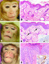
Clinical disease and skin rash histopathology. All monkeys were challenged approximately 1 year after immunization and monitored for development of a rash. (A to C) SINCP-immunized vector control monkeys (e.g., monkey 11P) developed a typical rash (A), while monkeys given the low dose of PLG/SINCP-H i.d. (12P and 14P) developed severe rashes (B and C). (D to E) Skin biopsies were taken and sections stained with hematoxylin and eosin; skin without a rash (D), skin with a typical rash (E), and skin with a severe rash (F) are shown. Insets show the results of immunohistochemical staining for MV N protein (brown color) in skin with a typical rash (E) and with a severe rash (F). Bars, 100 μm. Arrows, inflammatory infiltrates; arrowhead, MV-positive cell.
FIG. 4.
FIG. 4.
Viremia after challenge. (A and B) Viremia was measured by cocultivation of PBMCs with B95-8 cells (A) and by flow cytometry analysis of PBMCs positive for N protein (B). (C) The types of cells infected were analyzed by gating on cells stained with antibody to CD3 for T cells or antibody to CD20 for B cells. Error bars indicate standard deviations.
FIG. 5.
FIG. 5.
Numbers of lymphocytes and eosinophils in peripheral blood after challenge. Blood was analyzed for white blood cell count and for the numbers of lymphocytes (A) and eosinophils (B) in circulation after challenge. The mean percent change with standard deviation for each group relative to counts before challenge is shown.
FIG. 6.
FIG. 6.
Antibody responses after challenge. (A) Neutralizing antibody titers were measured by plaque neutralization assay. (B and C) MV-specific IgM (B) and IgG (C) were measured by EIA. The changes in OD from baseline at 1:200 (IgM) and 1:400 (IgG) dilutions of monkey plasma are shown. The mean and standard deviation for each group are shown. (D) MV-specific IgG avidity was measured by ammonium thiocyanate disruption of antibody binding in the EIA assay. The avidity index is the thiocyanate concentration required to remove 50% of the bound IgG. (E) The capacity to immunoprecipitate the native MV Moraten and Bilthoven H proteins was measured by RIPA.
FIG. 7.
FIG. 7.
T-cell responses after challenge. IFN-γ responses to stimulation by peptide pools of MV H protein (A) or F protein (B) were assayed by ELISPOT. The number of SFC per million PBMCs is shown. The mean and standard deviation for two monkeys in each pair are indicated.

Similar articles

Cited by

References

    1. Albrecht, P., F. A. Ennis, E. J. Saltzman, and S. Krugman. 1977. Persistence of maternal antibody in infants beyond 12 months: mechanism of measles vaccine failure. J. Pediatr. 91:715-718. - PubMed
    1. Albrecht, P., K. Herrmann, and G. R. Burns. 1981. Role of virus strain in conventional and enhanced measles plaque neutralization test. J. Virol. Methods 3:251-260. - PubMed
    1. Auwaerter, P. G., P. A. Rota, W. R. Elkins, R. J. Adams, T. DeLozier, Y. Shi, W. J. Bellini, B. R. Murphy, and D. E. Griffin. 1999. Measles virus infection in rhesus macaques: altered immune responses and comparison of the virulence of six different virus strains. J. Infect. Dis. 180:950-958. - PubMed
    1. Babaniyi, O. A., D. B. Parakoyi, B. A. Aiyedun, and M. A. Bello. 1995. Loss of maternally-acquired measles antibody during infancy in Ilorin, Nigeria. J. Trop. Pediatr. 41:115-117. - PubMed
    1. Black, F. L. 1989. Measles active and passive immunity in a worldwide perspective. Prog. Med. Virol. 36:1-33. - PubMed

Publication types

MeSH terms

LinkOut - more resources