Comparative Study
. 2008 Feb 1;7(3):335-47.
doi: 10.4161/cc.7.3.5234. Epub 2007 Nov 1.

Integrin affinity modulation in angiogenesis

Affiliations
Comparative Study

Integrin affinity modulation in angiogenesis

Ganapati H Mahabeleshwar et al. Cell Cycle. .

Abstract

Integrins, transmembrane glycoprotein receptors, play vital roles in pathological angiogenesis, but their precise regulatory functions are not completely understood and remain controversial. This study aims to assess the regulatory functions of individual beta subunits of endothelial integrins in angiogenic responses induced by vascular endothelial growth factor (VEGF). Inhibition of expression of beta(1), beta(3), or beta(5) integrins in endothelial cells resulted in down regulation of EC adhesion and migration on the primary ligand for the corresponding integrin receptor, while no effects on the recognition of other ligands were detected. Although inhibition of expression of each subunit substantially affected capillary growth stimulated by VEGF, the loss of beta(3) integrin was the most inhibitory.


Figures

Figure 1
Figure 1
Knockdowns of integrin beta subunits on endothelial cells by specific siRNAs. HUVECs were transfected with control siRNA or integrin-specific siRNA and cell lysates were analyzed for expression of β1 (A), β3 (B) or β5 (C) integrin subunits using specific antibody. Densitometry analysis was performed and results are shown in bar graphs (lower). (D and E) Cell surface expression of β1 (D), β3 (E) or β5 (F) integrin subunits in endothelial cells was assessed by FACS analysis. Cells were fixed with 3% paraformaldehyde, stained with primary antibody against the corresponding integrin and with secondary antibody labeled with Alexa Fluor 488. The mean fluorescence intensity was determined; the value obtained using control cells was assigned 100%. Asterisks indicate significant difference over control (p < 0.0038).
Figure 2
Figure 2
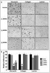
Specificity of integrins in reorganization on distinct ECM ligands. (A and B) HUVECs were transfected with control siRNA or siRNA specific for β1, β3 or β5 integrin. Wells of microtiter plates were coated with vitronectin, collagen or laminin-1 and were incubated overnight at 4°C. siRNA-transfected EC were harvested and resuspended in serum-free media at 5 × 10⁵ cells/ml. The cell suspension (100 μL) was plated on the microtiter wells coated with integrin ligand. After incubation at 37°C for 45 min, wells were gently washed three times with DMEM and photographs were taken (A). The numbers of attached cells per field were counted and untransfected cells adherent to the individual ECM ligands were assigned a value of 100% (B). Asterisks indicate significant difference over control (p < 0.0046).
Figure 3
Figure 3
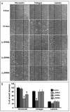
Vitronectin (αvβ3) and collagen (α5β1) receptors regulate endothelial cell migration. (A and B) HUVECs were transfected with control siRNA or siRNA specific for β1, β3 or β5 integrin. These cells were grown to confluence on 12-well plates precoated with individual integrin ligands. Cells were serum starved then wounded across the cell monolayer by scraping away a swath of cells. Wells were rinsed twice with sterile PBS and further cultured in DMEM medium containing 2% FBS. Sites were photographed immediately after wounding (zero hour) and 12 h later using a phase contrast microscope (A). Images were acquired using a Leica DMIRB phase contrast microscope, 5X objective, and a Micromax RTE/CCD-1300-V-HS camera. The mean wound area recovery by nontransfected endothelial cells on vitronectin for 12 hours was designated as 100% and the relative % of wound recovery for siRNA-transfected EC were determined (B). Asterisks indicate significant difference over control (p < 0.0058).
Figure 4
Figure 4
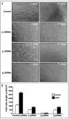
β3 integrin regulates endothelial cell morphogenesis in vitro. (A and B) HUVECs were transfected with control siRNA or siRNA specific for β1, β3 or β5 integrin. Cells were transferred to Matrigel coated plates and further incubated at 37°C for 8 h with or without 20 ng/mL VEGF. Endothelial capillary tubes formed in Matrigel were observed using an inverted phase contrast microscope and photographs were taken (A). Mean length of tubes from five random fields were measured using Image-Pro software (B).
Figure 5
Figure 5
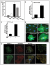
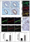
Activated αvβ3 integrin co-localizes with VEGFR-2 on endothelial cells. To evaluate growth factor-induced activation of αvβ3 integrin, semiconfluent, serum starved HUVECs were induced with VEGF-A165 (A), NIH-3T3 cells were induced with bFGF (Fig. 5B) and PC-3 cells were induced with EGF (Fig. 5C). These cells were further incubated with WOW-1 Fab and goat anti-mouse IgG labeled with Alexa Fluor 488. Fixed cells were then analyzed by flow cytometry. (D) HUVECs were grown on -gelatin-coated glass coverslips. These cells were serum starved and induced with VEGF-A165, VEGF-DΔNΔC or MnCl2 in the presence of WOW-1 Fab. Cells were washed and further incubated with goat anti-mouse IgG labeled with Alexa Fluor 488. Cells were fixed, observed via fluorescence microscopy and photographs were taken. (E) αvβ3 integrin and VEGFR-2 co-localize on endothelial cells. HUVECs were serum-starved overnight then stimulated with 20 ng/mL VEGF for 5 minutes. These cells were stained with WOW-1 and anti-VEGFR-2, followed by the incubation with goat anti-mouse IgG labeled with Alexa Fluor 488 and goat anti-rabbit IgG conjugated with Alexa Fluor 594. Without a stimulatory signal (upper), very little co-localization of β3 integrin and VEGFR-2 was observed. Upon VEGF stimulation (lower), the affinity of αvβ3 increases and co-localizes with VEGFR-2.
Figure 6
Figure 6
αvβ3 integrin activation level serve as a marker of tumor angiogenesis. (A) Activated αvβ3 integrin co-localizes with VEGFR-2 on endothelial cells of proliferating blood vessels. Parallel prostate tumor tissue sections were cut and stained for WOW-1 (activated αvβ3 integrin), CD31 (endothelial cell marker) and VEGFR-2. Blood vessels (revealed by CD31 staining) were positively stained for both WOW-1 and VEGFR-2, indicating the co-localization of activated αvβ3 with VEGFR-2 in tumor vasculature. (B) Frozen parallel prostate tumor sections were stained for activated αvβ3 integrin (WOW-1 Fab) and VEGFR-2. Tissue sections were analyzed with a confocal microscope and photographs were taken. (C and D) Association of VEGFR-2 with the active form of αvβ3 integrin on endothelial cells of angiogenic blood vessels. Robust angiogenesis was induced by left femoral artery ligation as described in Experimental Procedures and immunohistochemical analyses were performed. Ischemia-induced angiogenic blood vessels show intense staining for the activated form integrin as stained by WOW-1 Fab and endothelial cells on the blood vessels using anti-CD-31 and VEGFR-2 antibody (C and D). (E-G) Normal prostate tissue and prostate tumor sections were stained for CD-31 and WOW-1 (E). Vascular density was increased at least 6-fold in prostate tumors compared to normal prostate tissue (F). Vascular density was positively correlated with the density of WOW-1-positive vasculature in the two tissue samples (G). Asterisks indicate significant difference over normal tissue.
Figure 7
Figure 7
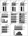
VEGF-induced VEGFR-2 phosphorylation is subordinate to αvβ3 integrin activation status. Effect of integrin knockdown on VEGFR-2 expression was evaluated by transfecting HUVECs with siRNA specific for (A) β1, (B) β3 or (C) β5 integrin. Cell lysates were analyzed for expression of VEGFR-2. Densitometry analysis was performed and results are shown as bar graphs (lower). (D) αvβ3 integrin activation dependent phosphorylation of VEGFR-2. HUVECs were transfected with β1, β3 or β5 integrin-specific siRNA and induced with 20 ng/mL VEGF. Cell lysates were analyzed for phosphorylation of VEGFR-2 using specific antibody. Densitometry analysis was performed and results are shown as bar graphs (lower D). (E-G) αvβ3 integrin and VEGF receptors interactions. HUVECs were stimulated with 20 ng/ml VEGF for 5 min. Cell lysate were subjected to immunoprecipitation with anti-β3 integrin antibody and immunoblotted with anti-VEGFR-1, anti-VEGFR-2 and anti-VEGFR-3 antibodies separately (E). HUVEC cell lysates were used as positive control (lane 1). Endothelial cells from wild type and β3 knockout mice were lysed and subjected to immunoprecipitation using anti-VEGFR-2 antibody and immunoblotted with anti-β1 integrin, anti-β3 integrin, anti-β5 integrin and anti-β6 integrin antibody (F). HUVECs were incubated with 1mM EDTA (lane 3) or 800 nM of SU1498 (lane 4), a potent VEGFR-2 specific inhibitor. These cells were stimulated with VEGF in presence of WOW-1 Fab fragments and lysed. Cell lysates subjected to immunoprecipitation with anti-VEGFR-2 antibody and immunoblotted with anti-β3 integrin or anti-His-Probe (recognizes WOW-1) antibody (G). (H) HUVECs were incubated with αvβ3 integrin-activating antibody (Libs-1, AP-7.3, CRC-54) or β3 integrin blocking antibody. These cells were induced with VEGF for 5 min and cell lysates were analyzed for phosphorylation of VEGFR-2 using specific antibody. Densitometry analysis was performed and results are shown as bar graphs (lower H).

Similar articles

Cited by

References

    1. Plow EF, Haas TA, Zhang L, Loftus J, Smith JW. Ligand binding to integrins. J Biol Chem. 2000;275:21785–8. - PubMed
    1. Arnaout MA, Goodman SL, Xiong JP. Coming to grips with integrin binding to ligands. Curr Opin Cell Biol. 2002;14:641–51. - PubMed
    1. Echarri A, Del Pozo MA. Caveolae internalization regulates integrin-dependent signaling pathways. Cell Cycle. 2006;5:2179–82. - PubMed
    1. Fang K, Fu W, Beardsley AR, Sun X, Lisanti MP, Liu J. Overexpression of caveolin-1 inhibits endothelial cell proliferation by arresting the cell cycle at G0/G1 phase. Cell Cycle. 2007;6:199–204. - PubMed
    1. Ruoslahti E. Integrin signaling and matrix assembly. Tumour Biol. 1996;17:117–24. - PubMed

Publication types

LinkOut - more resources