. 2008:2008:142082.
doi: 10.1155/2008/142082.

PPAR Signaling in Placental Development and Function

Affiliations

PPAR Signaling in Placental Development and Function

Yaacov Barak et al. PPAR Res. 2008.

Abstract

With the major attention to the pivotal roles of PPARs in diverse aspects of energy metabolism, the essential functions of PPARgamma and PPARbeta/delta in placental development came as a surprise and were often considered a nuisance en route to their genetic analysis. However, these findings provided an opportune entrée into placental biology. Genetic and pharmacological studies, primarily of knockout animal models and cell culture, uncovered networks of PPARgamma and PPARdelta, their heterodimeric RXR partners, associated transcriptional coactivators, and target genes, that regulate various aspects of placental development and function. These studies furnish both specific information about trophoblasts and the placenta and potential hints about the functions of PPARs in other tissues and cell types. They reveal that the remarkable versatility of PPARs extends beyond the orchestration of metabolism to the regulation of cellular differentiation, tissue development, and trophoblast-specific functions. This information and its implications are the subject of this review.


Figures

Figure 1
Figure 1
Trophoblast lineages in the developing mouse placenta. Shown from left to right are a blastocyst (E3.5), an E6.5 embryo, and an E9.5 embryo. Respective trophoblast lineages are traced for clarity. Al: allantois; Ch: chrion; CP: chorionic plate; De: decidua; Fmb: embryo; FPC: ectoplacental cone; 1°GC: primary giant cells; 2°GC: secondary giant cells; ICM: inner cell mass; La: labyrinth; Sp: spongiotrophoblast; TF: trophectoderm. FGF4: fibroblast growth factor 4 secreted by the embryo to maintain the chorion. Blastocyst and E6.5 embryo picture courtesy of Drs. Mimi DeVries and Tom Gridley, respectively, The Jackson Laboratory.
Figure 2
Figure 2
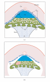
Schematic representation of the Pparg-null phenotype. (a) WT placenta. Al: allantois; Ar: maternal artery; Ch: chorion; De: decidua; FV: fetal blood vessels; La: labyrinth; MBP: maternal blood pools; Sp: spongiotrophoblast; TGC: trophoblast giant cells. (b) Pparg-null placenta. Corresponding structures are as in (a). Differences of note are marked erythrophagocytosis by spongiotrophoblast cells (red speckles), absence of fetal vessels and breakdown of the maternal blood pools in the labyrinth, and thickening of the chorion. (c,d) Ultrastructural features of WT and Pparg-null hemochorial barriers (based on [12]). See legend in (c) for identity of major features. Differences include thickening of the three trophoblast layers, near elimination of lipid droplets in layer III, and loosening of the tight adherence between the trophoblast (green) and fetal endothelium (orange).
Figure 3
Figure 3
Schematic representation of the Ppard-null phenotype. (a) WT placenta (similar to Figure 2(a)). (b) Ppard-null placenta. Hr: hemorrhage; for all other abbreviations see the legend for Figure 2. Notable differences include smaller and discontinuous giant cells, reduced size of the entire placenta and loosening of its attachment to the decidua, and sporadic severe hemorrhages at various locations in or around the placenta.

References

    1. Ornoy A, Salamon-Arnon J, Ben-Zur Z, Kohn G. Placental findings in spontaneous abortions and stillbirths. Teratology. 1981;24(3):243–252. - PubMed
    1. Krebs C, Macara LM, Leiser R, Bowman AW, Greer IA, Kingdom JCP. Intrauterine growth restriction with absent end-diastolic flow velocity in the umbilical artery is associated with maldevelopment of the placental terminal villous tree. American Journal of Obstetrics and Gynecology. 1996;175(6):1534–1542. - PubMed
    1. Zhou Y, Damsky CH, Chiu K, Roberts JM, Fisher SJ. Preeclampsia is associated with abnormal expression of adhesion molecules by invasive cytotrophoblasts. Journal of Clinical Investigation. 1993;91(3):950–960. - PMC - PubMed
    1. Cross JC. How to make a placenta: mechanisms of trophoblast cell differentiation in mice—a review. Placenta. 2005;26:S3–S9. - PubMed
    1. Simmons DG, Cross JC. Determinants of trophoblast lineage and cell subtype specification in the mouse placenta. Developmental Biology. 2005;284(1):12–24. - PubMed