Review
. 2008 Apr;466(4):791-801.
doi: 10.1007/s11999-008-0161-3. Epub 2008 Feb 21.

Imaging in the surgical management of developmental dislocation of the hip

Affiliations
Review

Imaging in the surgical management of developmental dislocation of the hip

Leslie Grissom et al. Clin Orthop Relat Res. 2008 Apr.

Abstract

Although the use of ultrasound in the diagnosis and early treatment of developmental dysplasia of the hip (DDH) has reduced the number of patients diagnosed late and decreased the number of operative procedures, surgical treatment is still needed in some patients. Late cases continue to occur as a result of missing the screening examination, being normal at initial screening and missing followup. Dysplasia may persist despite appropriate nonoperative or operative treatment. Many of these patients subsequently undergo closed or open reduction and femoral or acetabular reconstruction. Ultrasound of the hips is generally used up to 6 or 8 months of age, during which time the hips are largely cartilaginous, and radiographs after that time when bony development is more complete. Options to supplement ultrasound and radiography include arthrography, computed tomography, and magnetic resonance imaging. Several advances have been made in the imaging of DDH and its complications including acetabular labral pathology and of femoroacetabular impingement (FAI). We review imaging techniques other than ultrasound used in the management of DDH.

Level of evidence: Level V, diagnostic study. See the Guidelines for Authors for a complete description of levels of evidence.


Figures

Fig. 1
Fig. 1
A 5-year-old child was diagnosed at 15 months after open reduction and Pemberton osteotomy; the child is now asymptomatic. The figure shows postoperative deformity of the ilium and acetabular roof with an enlarged acetabulum and normally contained femoral head.
Fig. 2
Fig. 2
A lateral view of the proximal femur in a patient with decreased head neck offset demonstrating a “bump” at the head-neck junction (arrow) where the femur is impinging against the acetabular rim is shown.
Fig. 3
Fig. 3
A well centered anteroposterior radiograph of the pelvis is shown, demonstrating a crossover sign with the anterior margin of the acetabulum (dotted line) meeting the posterior margin (solid line) in the middle of the acetabulum instead of meeting at the superolateral margin of the acetabulum. This is suggestive of acetabular retroversion.
Fig. 4
Fig. 4
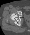
The figure shows normal hip joint width measurement demonstrated on axial computed tomography (arrows). Note wider joint anteriorly than posteriorly.
Fig. 5A–C
Fig. 5A–C
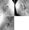
(A) A normal hip arthrogram demonstrates thin rim of contrast surrounding the femoral head and labrum covering the femoral head laterally. (B) A subluxated hip with acetabular dysplasia, pooling of contrast medially, and deflected labrum (arrow) is shown. (C) A dislocated hip with inverted labrum (arrow), narrowing of the joint capsule, and acetabular dysplasia is shown.
Fig. 6A–D
Fig. 6A–D
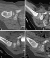
(A) A computed tomography scan shows a normally positioned hip after closed reduction with contrast surrounding the femoral head. (B) Arthrographic contrast surrounding the femoral head after successful closed reduction of the hip is demonstrated. Note the smooth arc (line) formed by the femoral metaphysis and the pubic bone anteriorly. (C) A dislocated hip is shown. Note discontinuity of the normal arc (discontinuous line) formed by the femoral metaphysis and pubic bone. Air is seen in the soft tissues from the procedure. (D) This is a mildly posteriorly displaced hip after closed reduction. F = femur; I = ischium; P = pubis.
Fig. 7A–C
Fig. 7A–C
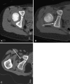
(A) The figure shows a normal acetabulum with wide radius of curvature and a smooth articular surface. (B) An example of a dysplastic acetabulum is shown: straightening and irregularity of the ischium (arrow). (C) Another example of a dysplastic acetabulum is shown: dislocated hip and fatty pulvinar centrally (arrow).
Fig. 8A–B
Fig. 8A–B
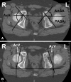
(A) A computed tomography scan for measurement of acetabular angles is shown (anterior and posterior sector angles [AASA, PASA]). The axial acetabular index (AxAI) is the sum of the two angles. (B) Acetabular anteversion (AcV) measurement is demonstrated.
Fig. 9A–C
Fig. 9A–C
(A) A three-dimensional computed tomography scan in anterior and bilateral lateral views demonstrates anterior dislocation on the right. (B) Abnormal tilting of the right acetabular roof on the lateral view (arrow) is shown. (C) Normal tilting on contralateral side.
Fig. 10A–B
Fig. 10A–B
A computed tomography scan shows the femur for femoral anteversion with cuts through the hips (A) and distal femora (B) with angle formed by lines drawn through the axis of the femoral neck and femoral condyles. There was internal rotation of the right knee but satisfactory anteversion bilaterally.
Fig. 11A–C
Fig. 11A–C
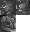
The figure shows T1-weighted magnetic resonance arthrogram images of the hip. (A) A normal hip and intact labrum (arrow) is shown. (B) A normal hip and intact labrum (arrow) is shown. (C) A torn posterior labrum in a different patient (arrow) is shown.

Similar articles

Cited by

References

    1. {'text': '', 'ref_index': 1, 'ids': [{'type': 'DOI', 'value': '10.1302/0301-620X.87B9.15928', 'is_inner': False, 'url': 'https://doi.org/10.1302/0301-620x.87b9.15928'}, {'type': 'PubMed', 'value': '16129740', 'is_inner': True, 'url': 'https://pubmed.ncbi.nlm.nih.gov/16129740/'}]}
    2. Argenson JN, Ryembault E, Fletcher X, Brassart N, Parratte S, Aubaniac JM. Three-dimensional anatomy of the hip in osteoarthritis after developmental dysplasia. J Bone Joint Surg Br. 2005;87:1192–1196. - PubMed
    1. None
    2. Bowen JR, Kotzias-Neto A. Developmental Dysplasia of the Hip. Brooklandville MD. Data Trace Publishing Co 2006;31–32:70–77.
    1. None
    2. Bowen JR, Kruse R. Complications in the treatment of developmental dysplasia of the hip. In: Epps CH Jr, Bowen JR, eds. Complications in Pediatric Orthopaedic Surgery, 3rd ed. Philadelphia, PA: JB Lippincott; 1994.
    1. {'text': '', 'ref_index': 1, 'ids': [{'type': 'PubMed', 'value': '2644290', 'is_inner': True, 'url': 'https://pubmed.ncbi.nlm.nih.gov/2644290/'}]}
    2. Clarke NM, Clegg J, Al-Chalabi AN. Ultrasound screening of hips at risk for CDH. Failure to reduce the incidence of late cases. J Bone Joint Surg Br. 1989;71:9–12. - PubMed
    1. {'text': '', 'ref_index': 1, 'ids': [{'type': 'DOI', 'value': '10.1097/01.blo.0000193511.91643.2a', 'is_inner': False, 'url': 'https://doi.org/10.1097/01.blo.0000193511.91643.2a'}, {'type': 'PubMed', 'value': '16331000', 'is_inner': True, 'url': 'https://pubmed.ncbi.nlm.nih.gov/16331000/'}]}
    2. Clohisy MD, Keeney JA, Schoenecker PL. Preliminary assessment and treatment guidelines for hip disorders in young adults. Clin Orthop Relat Res. 2005;441:163–179. - PubMed

MeSH terms