

Case Reports
doi: 10.1007/s11999-008-0159-x.
Epub 2008 Feb 21.
A 56-year-old woman with a right arm mass
Affiliations
- PMID: 18288548
- PMCID: PMC2565013
- DOI: 10.1007/s11999-008-0159-x
Item in Clipboard
Case Reports
A 56-year-old woman with a right arm mass
Clin Orthop Relat Res.
2008 Nov.
No abstract available
Figures
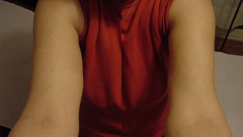
A 56-year-old woman presented with a soft, oval, deep palpable mass on the medial side of her right arm with a maximum diameter of 4 cm.

An anteroposterior plain radiograph of the right humerus shows round calcifications on the medial side of the right middle arm.

Ultrasonography of the right arm shows a 40- × 21-mm well-defined oval mass with heterogeneous internal echogenicity and moderate vascularity. The mass is closely related to the median nerve (arrow) that shows flattening, distortion of architecture, and altered echogenicity.

MR images show a fusiform lesion with well-circumscribed borders at the posteromedial aspect of the biceps, along the expected course of the median nerve. The lesion is heterogeneous and isointense or slightly hypointense to muscle on (A) T1-weighted axial and (B) T1-weighted sagittal images. On postcontrast (C) T1-weighted axial and (D) T1-weighted sagittal images, the mass enhances in fairly uniform fashion.

(A) Histologic sections of the biopsy specimen show a proliferative vascular lesion composed of thin-walled vascular channels in a focally hyalinized stroma with areas of hemorrhage. The vascular channels are lined by endothelial cells without nuclear atypia (Stain, hematoxylin and eosin; original magnification, ×400). (B) CD34 immunoperoxidase stain highlights the vascular nature of the lesion (Original magnification, ×100). (C) The thrombosed cavernous lesion developed papillary endothelial hyperplasia as an exuberant response to organizing thrombus. Embolism material was surrounded by foreign body giant cell reaction (Stain, hematoxylin and eosin; original magnification, ×100).

(A) MR and (B) conventional angiograms show a well-vascularized lesion with discrete margins. The arterial supply appears to originate from the brachial artery and venous drainage is through small branches of the brachial and basilic veins. Small defects in the contrast column correlate with the phleboliths identified on the other imaging studies.

(A) Surgical exploration of the middle arm revealed a dark-red fusiform tumor that distended the trunk of the median nerve. (B) The perineurium was incised, and the tumor was resected through intraneural microdissection.
Similar articles
-
[Intraneural hemangioma of the ulnar nerve].Nervenarzt. 2015 Feb;86(2):197-201. doi: 10.1007/s00115-014-4169-5. Nervenarzt. 2015. PMID: 25575631 German. No abstract available.
-
Intrinsic hemangiomas of the peripheral nerves report of a case and review of the literature.Conn Med. 1998 Apr;62(4):209-13. Conn Med. 1998. PMID: 9611417 Review.
-
Intramuscular thrombosed arteriovenous hemangioma of the upper right arm mimicking a neuroma of the ulnar nerve: case report.Neurosurgery. 2004 Mar;54(3):770-1; discusion 771-2. doi: 10.1227/01.neu.0000109540.92261.44. Neurosurgery. 2004. PMID: 15028157
-
Intraneural hemangioma of digital nerve diagnosed with MR imaging.Skeletal Radiol. 2007 Feb;36(2):157-60. doi: 10.1007/s00256-006-0094-4. Epub 2006 Mar 22. Skeletal Radiol. 2007. PMID: 16552604
-
Intrinsic haemangioma of the median nerve: report of a case and review of the literature.Microsurgery. 2008;28(2):89-90. doi: 10.1002/micr.20456. Microsurgery. 2008. PMID: 18220250 Review.
Cited by
-
A 3D-printed model of axillary cavernous hemangioma.J Vasc Surg Cases Innov Tech. 2025 Jun 18;11(5):101884. doi: 10.1016/j.jvscit.2025.101884. eCollection 2025 Oct. J Vasc Surg Cases Innov Tech. 2025. PMID: 40686578 Free PMC article.
-
Intraneural cavernous hemangioma: a rare case of extrafascicular left ulnar nerve tumor.Am J Blood Res. 2021 Feb 15;11(1):72-76. eCollection 2021. Am J Blood Res. 2021. PMID: 33796392 Free PMC article.
-
Chronic excruciating forearm pain in a child with intra-neural hemangioma: A challenging case report.Int J Surg Case Rep. 2021 Dec;89:106561. doi: 10.1016/j.ijscr.2021.106561. Epub 2021 Nov 3. Int J Surg Case Rep. 2021. PMID: 34864264 Free PMC article.
-
[Intraneural hemangioma of the ulnar nerve].Nervenarzt. 2015 Feb;86(2):197-201. doi: 10.1007/s00115-014-4169-5. Nervenarzt. 2015. PMID: 25575631 German. No abstract available.
References
-
- {'text': '', 'ref_index': 1, 'ids': [{'type': 'DOI', 'value': '10.1053/crad.1999.0343', 'is_inner': False, 'url': 'https://doi.org/10.1053/crad.1999.0343'}, {'type': 'PubMed', 'value': '10708612', 'is_inner': True, 'url': 'https://pubmed.ncbi.nlm.nih.gov/10708612/'}]}
- Arya S, Nagarkatti DG, Dudhat SB, Nadkarni KS, Joshi MS, Shinde SR. Soft tissue sarcomas: ultrasonographic evaluation of local recurrences. Clin Radiol. 2000;55:193–197. - PubMed
-
- {'text': '', 'ref_index': 1, 'ids': [{'type': 'DOI', 'value': '10.1097/00006123-198910000-00024', 'is_inner': False, 'url': 'https://doi.org/10.1097/00006123-198910000-00024'}, {'type': 'PubMed', 'value': '2677823', 'is_inner': True, 'url': 'https://pubmed.ncbi.nlm.nih.gov/2677823/'}]}
- Bilge T, Kaya A, Alatli M, Bilge S, Alatli C. Hemangioma of the peroneal nerve: case report and review of the literature. Neurosurgery. 1989;25:649–652. - PubMed
-
- {'text': '', 'ref_index': 1, 'ids': [{'type': 'DOI', 'value': '10.3171/foc.2007.22.6.19', 'is_inner': False, 'url': 'https://doi.org/10.3171/foc.2007.22.6.19'}, {'type': 'PubMed', 'value': '17613209', 'is_inner': True, 'url': 'https://pubmed.ncbi.nlm.nih.gov/17613209/'}]}
- Châtillon CE, Guiot MC, Jacques L. Lipomatous, vascular, and chondromatous benign tumors of the peripheral nerves. Neurosurg Focus. 2007;22:E18. - PubMed
-
- {'text': '', 'ref_index': 1, 'ids': [{'type': 'PubMed', 'value': '10433461', 'is_inner': True, 'url': 'https://pubmed.ncbi.nlm.nih.gov/10433461/'}]}
- Chesser TJ, Geraghty JM, Clarke AM. Intraneural synovial sarcoma of the median nerve. J Hand Surg Br. 1999;24:373–375. - PubMed
-
- {'text': '', 'ref_index': 1, 'ids': [{'type': 'PubMed', 'value': '3608310', 'is_inner': True, 'url': 'https://pubmed.ncbi.nlm.nih.gov/3608310/'}]}
- Chiao HC, Marks KE, Bauer TW, Pflanze W. Intraneural lipoma of the sciatic nerve. Clin Orthop Relat Res. 1987;221:267–271. - PubMed
Publication types
MeSH terms
LinkOut - more resources
Full Text Sources
Medical
