Comparative Study
. 2008 Apr;466(4):782-90.
doi: 10.1007/s11999-008-0141-7. Epub 2008 Feb 21.

Femoral morphology differs between deficient and excessive acetabular coverage

Affiliations
Comparative Study

Femoral morphology differs between deficient and excessive acetabular coverage

S D Steppacher et al. Clin Orthop Relat Res. 2008 Apr.

Abstract

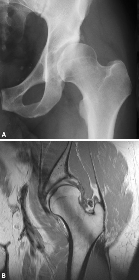
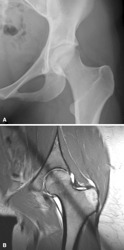
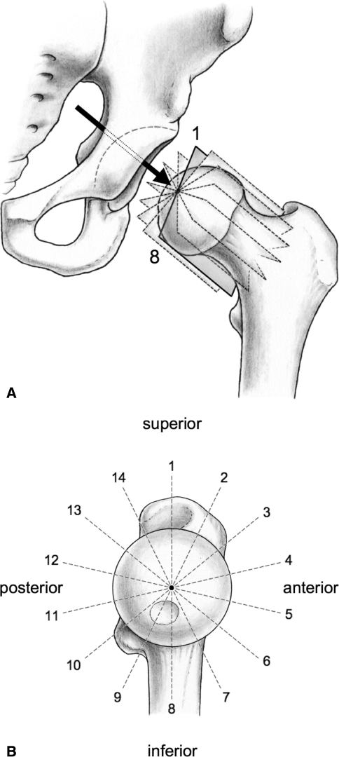
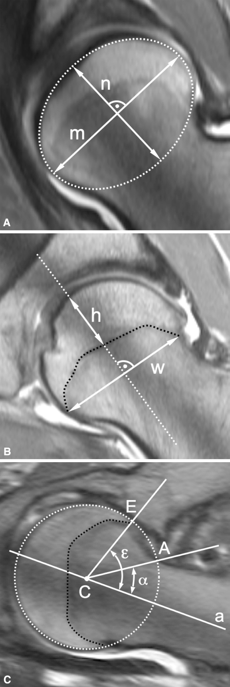
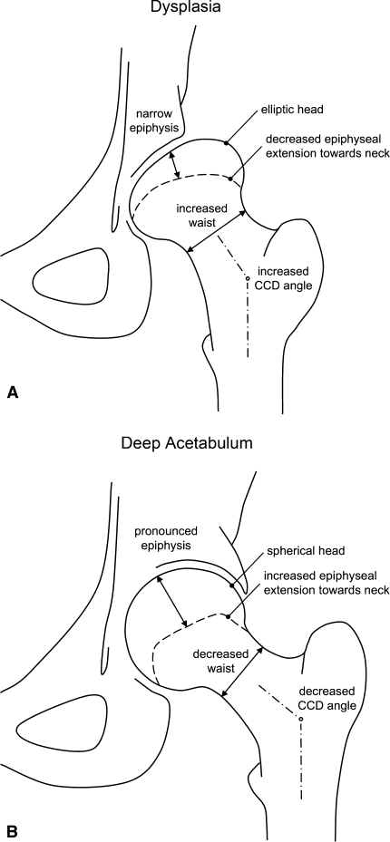
Structural deformities of the femoral head occurring during skeletal development (eg, Legg-Calvé-Perthes disease) are associated with individual shapes of the acetabulum but it is unclear whether differences in acetabular shape are associated with differences in proximal femoral shape. We questioned whether the amount of acetabular coverage influences femoral morphology. We retrospectively compared the proximal femoral anatomy of 50 selected patients (50 hips) with developmental dysplasia of the hip (lateral center-edge angle [LCE] < or = 25 degrees ; acetabular index > or = 14 degrees ) with 45 selected patients (50 hips) with a deep acetabulum (LCE > or = 39 degrees ). Using MRI arthrography we measured head sphericity, epiphyseal shape, epiphyseal extension, and femoral head-neck offset. A deep acetabulum was associated with a more spherical head shape, increased epiphyseal height with a pronounced extension of the epiphysis towards the femoral neck, and an increased offset. In contrast, dysplastic hips showed an elliptical femoral head, decreased epiphyseal height with a less pronounced extension of the epiphysis, and decreased head-neck offset. Hips with different acetabular coverage are associated with different proximal femoral anatomy. A nonspherical head in dysplastic hips could lead to joint incongruity after an acetabular reorientation procedure.

Level of evidence: Level IV, retrospective comparative study. See the Guidelines for Authors for a complete description of levels of evidence.


Figures

Fig. 1A–B
Fig. 1A–B
A left hip from a 48-year-old male patient in the dysplastic group (Group I). (A) On the conventional AP pelvic radiograph the AI is 30° and the LCE is 3°. (B) On the corresponding coronal slice of the arthro-MRI decreased coverage is apparent, as is a compensatory thickened labrum.
Fig. 2A–B
Fig. 2A–B
A left hip from a 23-year-old female patient in the deep acetabulum group (Group II). On the (A) conventional AP pelvic radiograph the AI is 4° and the LCE is 42°. (B) On the corresponding coronal slice of the arthro-MRI the femoral head has excessive acetabular coverage.
Fig. 3A–B
Fig. 3A–B
(A) MRI arthrography of the left hip with seven slices perpendicular to the neck axis was chosen for accurate and comparable measurements of the femoral head morphology and the epiphyseal extent. The arrow indicates the view direction for the scout view. (B) Each slice on the scout view is obtained by rotating clockwise in 25.7° steps around the neck axis. These slices provide 14 positions for measuring the alpha and epiphyseal angles.
Fig. 4A–C
Fig. 4A–C
These radial arthro-MRI slices show the construction of the measured parameters. (A) The head sphericity is the ratio of the minor axis (n) to the major axis (m) of the ellipse. (B) The epiphyseal index is the ratio of the epiphyseal height (h) to epiphyseal weight (w). (C) The alpha angle (α) is formed by the neck axis (a) and a line through the center of the head [C] and the offset-point [A]. The epiphyseal angle (ε) is formed in a similar way with a line through the center of the head [C] and the point [E] where the epiphysis meets the articular surface.
Fig. 5A–B
Fig. 5A–B
The schematic results of the proximal femoral morphology for (A) the dysplasia group (Group I) and (B) the deep acetabulum group (Group II) are shown.

References

    1. Anda S, Terjesen T, Kvistad KA, Svenningsen S. Acetabular angles and femoral anteversion in dysplastic hips in adults: CT investigation. J Comput Assist Tomogr. 1991;15:115–120. - DOI - PubMed
    1. Argenson JN, Ryembault E, Flecher X, Brassart N, Parratte S, Aubaniac JM. Three-dimensional anatomy of the hip in osteoarthritis after developmental dysplasia. J Bone Joint Surg Br. 2005;87:1192–1196. - DOI - PubMed
    1. Crowe JF, Mani VJ, Ranawat CS. Total hip replacement in congenital dislocation and dysplasia of the hip. J Bone Joint Surg Am. 1979;61:15–23. - PubMed
    1. Domzalski ME, Glutting J, Bowen JR, Littleton AG. Lateral acetabular growth stimulation following a labral support procedure in Legg-Calve-Perthes disease. J Bone Joint Surg Am. 2006;88:1458–1466. - DOI - PubMed
    1. Dora C, Bühler M, Stover MD, Mahomed MN, Ganz R. Morphologic characteristics of acetabular dysplasia in proximal femoral focal deficiency. J Pediatr Orthop B. 2004;13:81–87. - DOI - PubMed

Publication types