

The morphologic variations of low and high hip dislocation
- PMID: 18288552
- PMCID: PMC2504667
- DOI: 10.1007/s11999-008-0131-9
The morphologic variations of low and high hip dislocation
Abstract
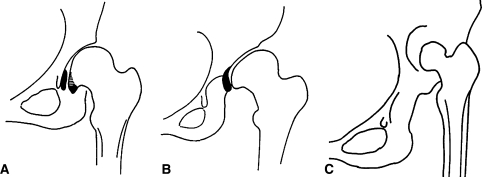
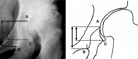
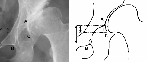
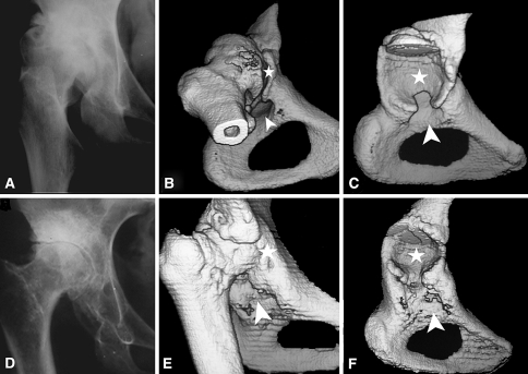
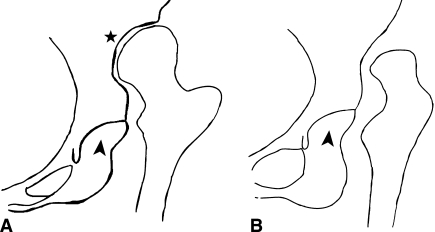
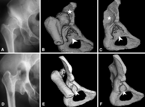
Three different types of congenital hip disease in adults have been distinguished based upon the position of the femoral head relative to the acetabulum and the underlying pathoanatomy of the joint: (1) dysplasia; (2) low dislocation; and (3) high dislocation. To facilitate classification of borderline or ambiguous cases, we studied the morphologic variations of low and high dislocation as observed on the radiographs of 101 hips with low and 74 hips with high dislocation. In low dislocation, 54 hips (53.5%) had extended coverage of the true acetabulum (Type B1) and 47 hips (46.5%) had limited coverage (Type B2). Among the cases with high dislocation, a false acetabulum with an adjacent femoral head occurred in 46 hips (62.2%) (Type C1), and the femoral head was floating within the gluteal muscles in 28 hips (37.8%) (Type C2). The kappa value for interobserver agreement between two raters who made radiographic measurements was 0.963, and for intraobserver agreement between the two evaluations of the same observer it was 0.946 and 0.971, respectively. The two types of low and high dislocation were associated with high intra- and interobserver agreement. Whether these distinctions have clinical utility requires further validation.
Level of evidence: Level III, diagnostic study. See the Guidelines for Authors for a complete description of levels of evidence.
Figures
References
-
- None
- Chung SMK. Hip Disorders in Infants and Children. Philadelphia, PA: Lea & Febiger;1981.
-
- {'text': '', 'ref_index': 1, 'ids': [{'type': 'PubMed', 'value': '365863', 'is_inner': True, 'url': 'https://pubmed.ncbi.nlm.nih.gov/365863/'}]}
- Crowe JF, Mani VJ, Ranawat CS. Total hip replacement in congenital dislocation and dysplasia of the hip. J Bone Joint Surg Am. 1979;61:15–23. - PubMed
-
- {'text': '', 'ref_index': 1, 'ids': [{'type': 'DOI', 'value': '10.1007/s00256-005-0061-5', 'is_inner': False, 'url': 'https://doi.org/10.1007/s00256-005-0061-5'}, {'type': 'PubMed', 'value': '16534641', 'is_inner': True, 'url': 'https://pubmed.ncbi.nlm.nih.gov/16534641/'}]}
- Decking R, Brunner A, Decking J, Puhl W, Gunther KP. Reliability of the Crowe und Hartofilakidis classifications used in the assessment of the adult dysplastic hip. Skeletal Radiol. 2006;35:282–287. - PubMed
-
- None
- Eftekhar NS. Principles of Total Hip Arthroplasty. St Louis, MO: CV Mosby; 1978.
-
- None
- Fleiss JL. Statistical Methods for Rates and Proportions. 2nd ed. New York, NY: John Wiley & Sons; 1981:218.
Publication types
MeSH terms
LinkOut - more resources
Full Text Sources
Research Materials
Miscellaneous
