

Review
doi: 10.2174/156802608783378891.
Immobilization of heparin: approaches and applications
Affiliations
- PMID: 18289079
- PMCID: PMC4117378
- DOI: 10.2174/156802608783378891
Item in Clipboard
Review
Immobilization of heparin: approaches and applications
Curr Top Med Chem.
2008.
Abstract
Heparin, an anticoagulant, has been used in many forms to treat various diseases. These forms include soluble heparin and heparin immobilized to supporting matrices by physical adsorption, by covalent chemical methods and by photochemical attachment. These immobilization methods often require the use of spacers or linkers. This review examines and compares various techniques that have been used for the immobilization of heparin as well as applications of these immobilized heparins. In the applications reviewed, immobilized heparin is compared with soluble heparin for efficient and versatile use in each of the various applications.
Figures

Structure of glycosaminoglycans (X = H or SO3−; Y = Ac or SO3−).
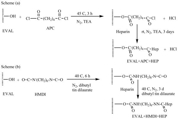
Schemes for the immobilization of heparin onto the EVAL polymer using (a) APC and (b) HMDI as linkers.

Protocols for making biocompatible vinyl surfaces with longer spacers such as TEPA and pHEMA.

Immobilization of heparin ionically through DED (DEDQ = quarternized DED).

A comparison of the bioefficiency of covalently (dotted line) and ionically formed surfaces (solid line).

Formation of PU-AB by oxygen plasma glow discharge and graft polymerization.

Functional group introduction and heparin immobilization on PU-AB.

Scheme showing the reaction protocol for the preparation of heparinized PU surfaces.

The effect of CNBr conc. on the amount of heparin immobilized (heparin concentration = 2 mg/ml, 25 °C, pH 7.4) (solid circle: p(HEMA), solid square: p(HEMA-AA), empty circle: p(HEMA-DMAEMA), empty square: p(HEMA-MMA).
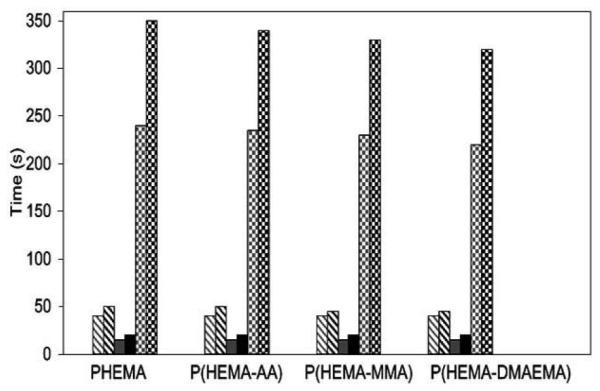
CT, APTT, PT data of various microsphere surfaces (grey: without heparin immobilized, black: with heparin immobilized; diagonal: APTT, solid: PT, checker board: CT).
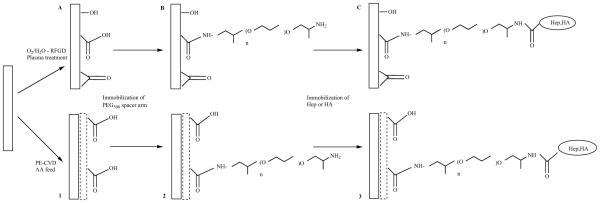
Immobilization of heparin and HA onto poly(ethylene) through O2/H2O RFGD plasma treatment (steps A,B,C) or PE-CVD AA RFGD poly(ethylene) (steps 1,2,3).

Thrombin time measurements of various surface modified PE substrates.

Schematic representation of the preparation of heparinized p(DAPAAm)-b-(PDMAAm)-block-graft-copolymer (a) Dithiocarbamate-derivatized PST film; (b) p(DMAPAAm) graft polymer; (c) heparin; and (d) p(DMAAm) graft polymer.

Schematic representation of PTFE surfaces.

Overall process of the immobilization of insulin and /or heparin on PET.
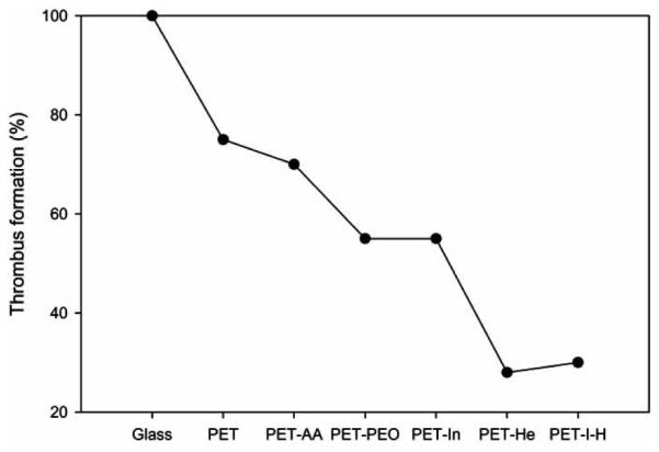
The amount of thrombus formed on PETs with various surface modifications after 30 min incubation (The value corresponding to glass was taken as 100%).
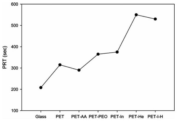
Plasma recalcification time as a function of surface-modified PETs.

APTT as a function of surface modifications.

Amount of platelets adhered on surface modified PETs with the initial platelet concentration as 100. (Circle - 60 min; diamond - 30 min incubation time).

Serotonin released from platelets on PETs with various surface modifications (solid square: 30 min without imipramine; solid circle: 30 min with imipramine; empty square: 60 min without imipramine; solid circle: 60 min with imipramine).
Heparinized PHEMA hydrogel.

Schematic representation of the various approaches of heparin immobilization on biological surfaces (a) tissue without heparinization (b) tissue with ionically bound heparin (c) tissue with covalently bound heparin via multi point attachment (d) tissue with covalently bound heparin via end point attachment.

Scheme for the heparinization of PAN through two approaches.

Heparinization of PPY through PEGMA and CC.

Heparinization of polysulfone with/without chitosan linkers.

Covalent immobilization of heparin on PLGA surface by EDC/NHS coupling.

Covalent attachment of chitosan/heparin complex to polyacrylonitrile membrane

Plasma Protein Adsorption on the surface modified PAN membranes (square: HPF; circle: HSA).

Activated partial thromboplastin time values of celluloseheparin composite in comparison with some other heparinized polymeric surfaces.
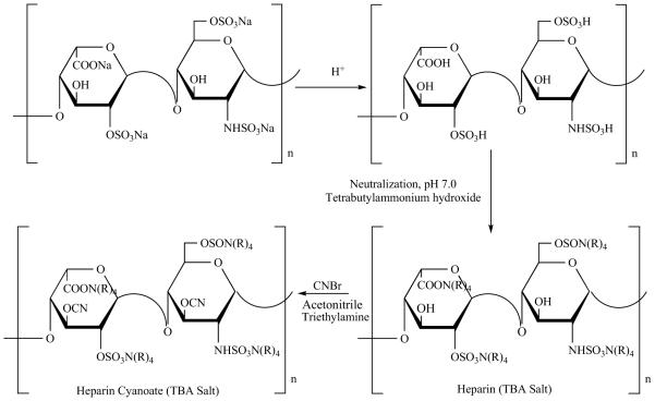
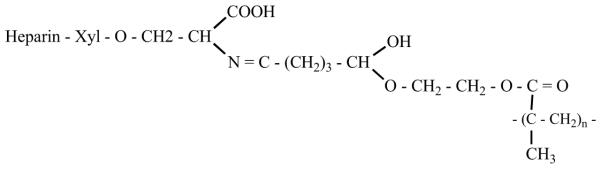
Schematic representation of the activation of heparin towards free amino groups.

APTT measurements of modified CNTs (crossed bar: No CNTs; empty bar: Pristine CNTs; black bar: PEI-CNTs; grey bar: Hep-PEI-CNTs).
Similar articles
-
Modified heparin inhibits P-selectin-mediated cell adhesion of human colon carcinoma cells to immobilized platelets under dynamic flow conditions.J Biol Chem. 2004 Jul 9;279(28):29202-10. doi: 10.1074/jbc.M312951200. Epub 2004 May 7. J Biol Chem. 2004. PMID: 15133030
-
Synthesis and evaluation of heparin immobilized "side-on" to polystyrene microspheres coated with end-group activated polyethylene oxide.Int J Biol Macromol. 2010 Aug 1;47(2):98-103. doi: 10.1016/j.ijbiomac.2010.05.015. Epub 2010 May 26. Int J Biol Macromol. 2010. PMID: 20621768
-
New methods for surface modification and covalent attachment of heparin.Med Device Technol. 1998 Jan-Feb;9(1):24-7. Med Device Technol. 1998. PMID: 10176141
-
[Review of heparin immobilization technique].Sheng Wu Yi Xue Gong Cheng Xue Za Zhi. 2007 Apr;24(2):466-9. Sheng Wu Yi Xue Gong Cheng Xue Za Zhi. 2007. PMID: 17591284 Review. Chinese.
-
Heparin-Mimicking Polymers: Synthesis and Biological Applications.Biomacromolecules. 2016 Nov 14;17(11):3417-3440. doi: 10.1021/acs.biomac.6b01147. Epub 2016 Oct 14. Biomacromolecules. 2016. PMID: 27739666 Free PMC article. Review.
Cited by
-
Grafting Strategies of Oxidation-Prone Coiled-Coil Peptides for Protein Capture in Bioassays: Impact of Orientation and the Oxidation State.ACS Omega. 2023 Jul 28;8(31):28301-28313. doi: 10.1021/acsomega.3c02172. eCollection 2023 Aug 8. ACS Omega. 2023. PMID: 37576632 Free PMC article.
-
Heparinized Polyurethane Surface Via a One-Step Photografting Method.Molecules. 2019 Feb 20;24(4):758. doi: 10.3390/molecules24040758. Molecules. 2019. PMID: 30791534 Free PMC article.
-
Xanthan-Based Materials as a Platform for Heparin Delivery.Molecules. 2023 Mar 18;28(6):2757. doi: 10.3390/molecules28062757. Molecules. 2023. PMID: 36985729 Free PMC article.
-
Ex vivo evaluation of blood coagulation on endothelial glycocalyx-inspired surfaces using thromboelastography.In Vitro Model. 2021 Oct 29;1(1):59-71. doi: 10.1007/s44164-021-00001-w. eCollection 2022 Feb. In Vitro Model. 2021. PMID: 39872977 Free PMC article.
-
Advanced strategies to thwart foreign body response to implantable devices.Bioeng Transl Med. 2022 Mar 2;7(3):e10300. doi: 10.1002/btm2.10300. eCollection 2022 Sep. Bioeng Transl Med. 2022. PMID: 36176611 Free PMC article. Review.
References
-
- Rydberg E, Westfall MJ, Nicholas RA. Low Molecular weight heparin in preventing and treating DVT. Am. Fam. Physician. 1999:1607. - PubMed
-
- Sanchez J, Elgue G, Riesenfeld J, Olsson P. Studies of adsorption, activation, and inhibition of factor XII on immobilized heparin. Thromb. Res. 1998;89:41–50. - PubMed
-
- Jackson RL, Busch SJ, Cardin ADW. Physiol. Rev. 1991;71:481–539. - PubMed
-
- Danishefsky I, Tzeng F, Ahrens M, Klien S. Thromb. Res. 1976;8:131–140. - PubMed
-
- Langer R, Tirrell DA. Designing materials for biology and medicine. Nature. 2004;428:487–492. - PubMed
Publication types
MeSH terms
Substances
Grants and funding
LinkOut - more resources
Full Text Sources
Other Literature Sources
Medical
