. 2008 Jun 15;412(3):579-88.
doi: 10.1042/BJ20080324.

Phosphorylation of eIF2alpha in response to 26S proteasome inhibition is mediated by the haem-regulated inhibitor (HRI) kinase

Affiliations

Phosphorylation of eIF2alpha in response to 26S proteasome inhibition is mediated by the haem-regulated inhibitor (HRI) kinase

Azmi Yerlikaya et al. Biochem J. .

Erratum in

  • Biochem J. 2008 Nov 1;415(3):483

Abstract

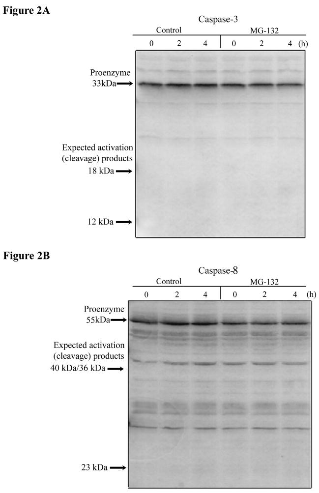
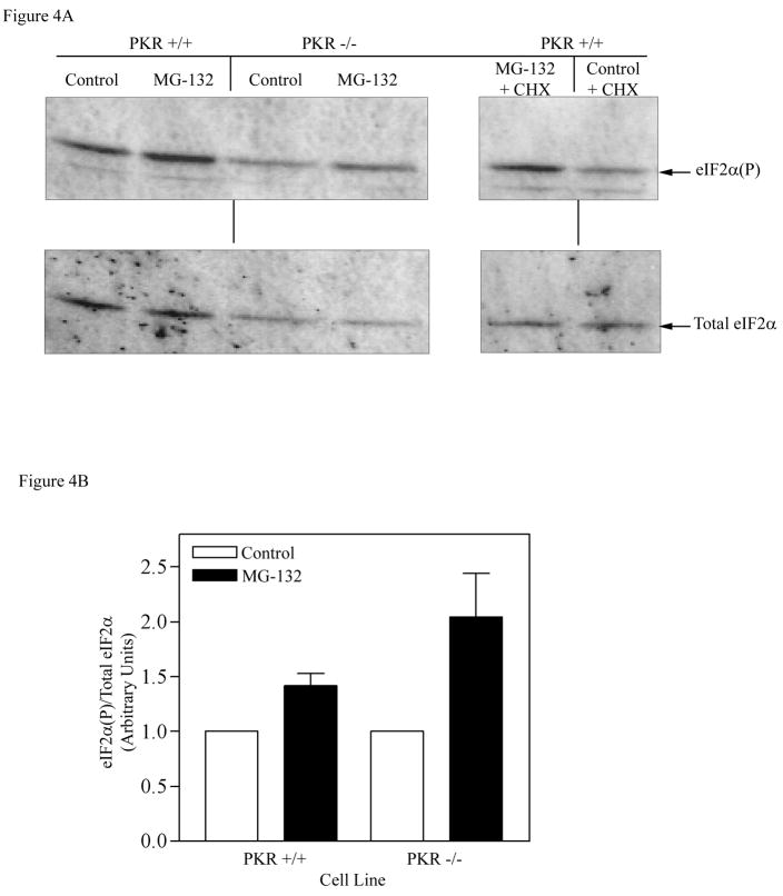
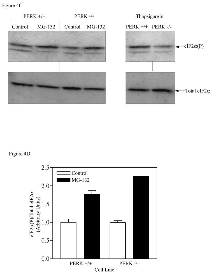
The present study demonstrates that even brief inhibition of degradation by the 26S proteasome inhibits global protein synthesis, mediated through increased phosphorylation of eIF2alpha (eukaryotic translational initiation factor 2alpha) by the HRI (haem-regulated inhibitor) kinase. Exposure of COS-7 cells to the proteasome inhibitor MG-132 (the proteasome inhibitor carbobenzoxy-L-leucyl-L-leucyl-leucinal) for 4 h resulted in a 55-60% decrease in protein synthesis rate compared with control cells. This repression of protein synthesis after treatment with MG-132 is not due to induction of apoptosis, which is known to occur after longer periods of 26S inhibition. Instead, we observed a significantly increased phosphorylation of eIF2alpha, which is known to repress global protein synthesis. In three MEF (mouse embryonic fibroblast) knockout cell lines lacking one of the four kinases known to phosphorylate eIF2alpha, increased phosphorylation of eIF2alpha still occurred after inhibition of the 26S proteasome. These three cell lines included a deletion of the PKR (double-stranded-RNA-dependent protein kinase); a deletion of the PERK (PKR-like endoplasmic reticulum resident kinase); or a deletion of the GCN2 (positive general control of transcription-2) kinase, indicating that none of these kinases was primarily responsible for the observed phosphorylation of eIF2alpha. In contrast, in a fourth MEF knockout cell line, HRI(-/-) cells lacking the HRI kinase failed to increase eIF2alpha phosphorylation upon proteasome inhibitor treatment (MG-132 or various doses of Bortezomib), indicating that the HRI kinase is the primary kinase activated by brief treatment of MEFs with 26S proteasome inhibitors.


Figures

Figure 1
Figure 1. Effect of the 26S proteasome inhibitor on protein synthesis in COS-7 cells
To determine the protein synthesis rate after MG132 treatment (50 μM) or 0.1% DSMO for 4 h, cells were labeled by addition of either 0.5 μCi/ml [3H]proline or 0.4 μCi/ml [3H]methionine to the complete media for 20 min. Cells were washed extensively with PBS, and with 10% TCA for 10 min to remove unincorporated radioactive precursor. Cells were washed with 10% TCA twice more for 5 min each. After a final wash with absolute MeOH, cells were lysed in 0.3N NaOH, 1% SDS for 30 min at room temperature. Aliquots were counted by liquid scintillation counting in 10 ml BCS. Values are means±SEM (n = 4 in [3H]proline experiment; n = 3 in [3H]methionine experiment). The p values significantly different in comparison with the control are indicated (Student’s t-test). The SEM is small and not seen in the MG132-treated group in [3H]methionine incorporation experiment (plotted on the right Y-axis).
Figure 2
Figure 2. Loss of protein synthesis activity after inhibition of the 26S proteasome is not due to activation of caspases
A) Analysis of caspase 3 and B) caspase 8 activation. After treatment of COS-7 cells with MG132 (50 μM) or 0.1% DMSO, equal amounts of protein (50 μg) from cells lysates were resolved by electrophoresis on a 12.5% polyacrylamide gel, followed by transfer to PVDF membrane at 50V at room temperature for 45 min. The membranes were probed with a caspase 3 polyclonal antibody (1:2,000 diluted) overnight at 4°C. The membrane was then washed extensively with TBS-T and incubated with alkaline phosphatase-conjugated anti-rabbit IgG (1:4,000 diluted). The membrane was developed with ECF detection reagent (Amersham), and visualized with a FluorImager. The same membrane was stripped and reprobed with a caspase 8 polyclonal antibody (1:800 diluted), and developed as described above.
Figure 3
Figure 3. Effect of the proteasome inhibitor on eIF-4G cleavage and phosphorylation of eIF2α in COS-7 cells
A) COS-7 cells were treated with MG132 (50 μM) for times indicated. Equal amounts of protein (50 μg) were separated on 7.5% SDS-PAGE gel, transferred to PVDF membrane, and detected with an eIF-4G polyclonal antibody as described in Experimental Procedures. B) To detect phosphorylated eIF2α and total eIF2α, equal amounts of protein (50μg) were separated on 12.5% SDS-PAGE gel, followed by Western blot analysis as described in Experimental Procedures. C) The results were quantitated by ImageQuant software, and the data is expressed as the level of phosphorylated eIF2α divided by the total level of eIF2α, with time point 0 set as one. Values are means±SEM (n = 2). The error bars are small and not visible for some data points.
Figure 4
Figure 4. Effect of the proteasome inhibitor on eIF2α phosphorylation in PKR KO and PERK KO cells
A) Wild type and PKR KO cells were treated with MG132 (50 μM) or 0.1% DMSO for 4 h. Equal amounts of protein (30 μg) were separated on 12.5% SDS-PAGE gel, followed by Western blot analysis as described in Experimental Procedures. B) The results were quantitated by ImageQuant software, and expressed as the amount of phosphorylated eIF2α divided by the total eIF2α level, with control (DMSO treated) set as 1. Cells were also treated with MG132 + CHX to determine whether complete inhibition of protein synthesis lowers the increase in eIF2α phosphorylation (right panel). C) Wild type and PERK KO cells were similarly treated with MG132 (50 μM) or 0.1% DMSO for 4 h and the total and phosphorylated eIF2α was determined by Western blot analysis. Wild type and PERK-KO cells were also treated with thapsigargin (1 μM) for 1 h, an agent known to activate PERK. D) Quantitation of the membrane shown in Fig. 4C (left panel). Each bar in Fig. 4B and D is the mean±SEM of an experiment run in duplicate. The error bars are small and not visible in certain bars.
Figure 5
Figure 5. Analysis of eIF2α phosphorylation in GCN2 KO and HRI KO cells
Cells were treated with MG132 as in Figure 4, and eIF2α phosphorylation was determined by Western blotting. A). Induction of eIF2α phosphorylation in GCN2 KO cells following MG132 treatment. Total and phosphorylated eIF2α were determined by Western blot analysis after treatment of wild type (GCN2+/+) and GCN2 knock out cells (GCN2−/−) with 50 μM MG132 for 4 h. B). Quantitation of the level of eIF2α phosphorylation seen in Panel A. The data are plotted as means±SEM (n = 3), normalized to the amount of total eIF2α. C). Phosphorylation of eIF2α in HRI KO cells. The total and phosphorylated eIF2α was determined by Western blot analysis after treatment of wild type (HRI+/+) and HRI knock out cells (HRI−/−) with 50 μM MG132 for 4 h. D). Quantitation of the level of eIF2α phosphorylation seen in Panel C. The data are plotted as means±SEM (n = 2).
Figure 6
Figure 6. Effect of Bortezomib on eIF2α phosphorylation in HRI wild type and HRI knock out cells
A) Cells were treated for 4 h with the various concentrations of Bortezomib indicated above the lanes, and eIF2α phosphorylation level was determined by Western blot analysis. B) Quantitation of the level of eIF2α phosphorylation in HRI wild type and HRI knock out cells. The experiment shown in Panel 6A was carried out in triplicate and the data shown in Panel 6B is the mean±SD (n = 3).

References

    1. Hershey JW. Translational control in mammalian cells. Annu Rev Biochem. 1991;60:717–755. - PubMed
    1. Kimball SR. Eukaryotic initiation factor eIF2. Int J Biochem Cell Biol. 1999;31:25–29. - PubMed
    1. Satoh S, Hijikata M, Handa H, Shimotohno K. Caspase-mediated cleavage of eukaryotic translation initiation factor subunit 2alpha. Biochem J. 1999;342:65–70. - PMC - PubMed
    1. Pain VM. Initiation of protein synthesis in eukaryotic cells. Eur J Biochem. 1996;236:747–771. - PubMed
    1. Marissen WE, Guo Y, Thomas AA, Matts RL, Lloyd RE. Identification of caspase 3-mediated cleavage and functional alteration of eukaryotic initiation factor 2alpha in apoptosis. J Biol Chem. 2000;275:9314–9323. - PubMed

Publication types