

Bile and pancreatic juice exclusion activates acinar stress kinases and exacerbates gallstone pancreatitis
- PMID: 18291265
- PMCID: PMC2278165
- DOI: 10.1016/j.surg.2007.06.004
Bile and pancreatic juice exclusion activates acinar stress kinases and exacerbates gallstone pancreatitis
Abstract
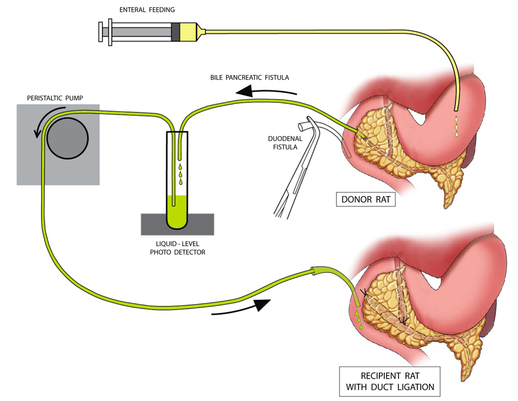
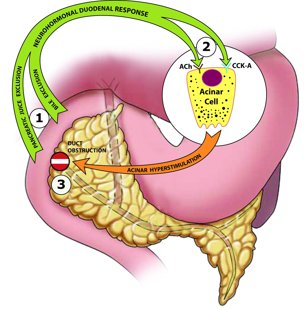
Bile and pancreatic juice exclusion from gut activates acinar stress kinases and exacerbates gallstone pancreatitis as evidenced by the ameliorating effects of replacement therapy in an experimental model of duct ligation-induced acute pancreatitis. In the early stages of gallstone pancreatitis, bile-pancreatic juice cannot enter the gut. Enteral exclusion worsens pancreatitis by causing feedback hyperstimulation of the exocrine pancreas that activates acinar cell stress kinases. Investigations using a unique surgical model, the Donor Rat Model, showed that duodenal replacement of bile-pancreatic juice in rats with duct ligation attenuates pancreatic stress kinase activation, reduces pancreatic cytokine production, and ameliorates pancreatic morphologic changes. These findings suggest that exclusion-induced acinar hyperstimulation, in the presence of duct obstruction, exacerbates acute pancreatitis via stress kinase activation. Although acinar hyperstimulation has often been implicated in the pathogenesis of acute pancreatitis, the lack of supporting evidence remains a conspicuous void. The proposed hypothesis draws on fresh evidence to present a new paradigm that reexamines the role of exocrine pancreatic hyperstimulation in gallstone pancreatitis pathogenesis.
Figures
References
-
- Samuel I, Toriumi Y, Wilcockson DP, Turkelson CM, Solomon TE, Joehl RJ. Bile and pancreatic juice replacement ameliorates early ligation- induced acute pancreatitis in rats. Am. J. Surg. 1995;169(4):391–399. - PubMed
-
- Samuel I, Zaheer S, Zaheer A. Bile-pancreatic juice exclusion increases p38MAPK activation and TNF-alpha production in ligation-induced acute pancreatitis in rats. Pancreatology. 2005;5(1):20–26. - PubMed
-
- Samuel I, Joehl RJ. Bile-pancreatic juice replacement, not cholinergic and cholecystokinin-receptor blockade, reverses acinar cell hyperstimulation after bile-pancreatic duct ligation. Am. J. Surg. 1996;171(1):207–211. - PubMed
-
- Cameron JL, Mehigan D, Zuidema GD. Evaluation of atropine in acute pancreatitis. Surg Gynecol Obstet. 1979;148:206–208. - PubMed
Publication types
MeSH terms
Substances
Grants and funding
LinkOut - more resources
Full Text Sources
Medical
