. 2008 Apr 1;316(1):110-23.
doi: 10.1016/j.ydbio.2008.01.016. Epub 2008 Jan 26.

Combinatorial signaling by the Frizzled/PCP and Egfr pathways during planar cell polarity establishment in the Drosophila eye

Affiliations

Combinatorial signaling by the Frizzled/PCP and Egfr pathways during planar cell polarity establishment in the Drosophila eye

Ursula Weber et al. Dev Biol. .

Abstract

Frizzled (Fz)/PCP signaling regulates planar, vectorial orientation of cells or groups of cells within whole tissues. Although Fz/PCP signaling has been analyzed in several contexts, little is known about nuclear events acting downstream of Fz/PCP signaling in the R3/R4 cell fate decision in the Drosophila eye or in other contexts. Here we demonstrate a specific requirement for Egfr-signaling and the transcription factors Fos (AP-1), Yan and Pnt in PCP dependent R3/R4 specification. Loss and gain-of-function assays suggest that the transcription factors integrate input from Fz/PCP and Egfr-signaling and that the ETS factors Pnt and Yan cooperate with Fos (and Jun) in the PCP-specific R3/R4 determination. Our data indicate that Fos (either downstream of Fz/PCP signaling or parallel to it) and Yan are required in R3 to specify its fate (Fos) or inhibit R4 fate (Yan) and that Egfr-signaling is required in R4 via Pnt for its fate specification. Taken together with previous work establishing a Notch-dependent Su(H) function in R4, we conclude that Fos, Yan, Pnt, and Su(H) integrate Egfr, Fz, and Notch signaling input in R3 or R4 to establish cell fate and ommatidial polarity.


Figures

Figure 1
Figure 1. kay/fos is required for PCP establishment in the eye
All panels are oriented with anterior left and dorsal up. (A) Schematic drawing of eye imaginal disc, showing establishment of PCP in developing ommatidial preclusters. Photoreceptors are depicted as ellipses with R3 and R4 precursors in green and light green, respectively. Cell fate specification of these two photoreceptors is a prerequisite for the establishment of chirality and direction of rotation. Ommatidial clusters rotate clockwise in dorsal half and counter clockwise in ventral half of disc, giving rise to two chiral forms in the adult eye (see schematic in right part of panel A), where the R3 rhabdomere takes a more polar position (green) and the R4 rhabdomere (light green) sits closer to R7. (B,B’) Chirality and rotation in a wild-type disc assayed by a novel marker for photoreceptors R3 and R4: psq-Gal4, UAS-GFP (green, and monochrome in B’) in R3 at high levels and R4 at low levels, allowing identification of both cell fates from ommatidial row 9 on. Anti-Elav (blue; all R-cells) and anti-Bar (red; R1/R6) are also shown. (C) Wild-type adult eye showing the two ommatidial chiral forms, lower panel depicting them schematically as black and red arrows (separated by the equator in yellow). (D) Mutant eye clones of kayP54 show rotation and chirality defects. Loss of redish pigment marks mutant tissue, with schematic representation on the right (arrows are as in panel C; ommatidia that have lost photoreceptors are indicated by black dots). Eyes displayed an average of 13% chirality defects, 10% rotation defects and 17.3% loss of photoreceptor/survival phenotypes. % based on counting ommatidia of 8 eyes in mutant tissue.
Figure 2
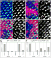
Figure 2. kay/fos is required for R3/R4 cell fate establishment in developing eye discs
(A-D) Confocal pictures of 3rd instar eye discs. Photoreceptors are marked with anti-Elav (blue), and the R3 and R4 cells by psq>GFP (green; monochrome in right panels). Note high levels of psq>GFP in R3 and low levels in R4 (some examples are marked with “3” in green and “4” in yellow). (A) wild-type disc. (B-D) Clones of indicated genotype, mutant tissue is marked by loss of red marker (arm-lacZ). (B) fzR52 mosaic eye disc. Note that in fz mosaic R3/R4 pairs the wild-type cell always expresses GFP at high levels, presumably adopting R3 fate, and the ommatidial cluster rotates according to that decision (see Suppl. Fig. S3 for quantification of rotation behavior). Some examples that show either switched fates or symmetrical clusters are indicated by green [R3 fate] or yellow [R4 fate] asterisks. Within fz mutant clones ommatidial clusters can also express the R3/R4 specific psq>GFP marker in more than 2 cells per cluster (examples indicated by orange arrowheads). (C-D) Expression of the psq>GFP marker in kay mutant ommatidia reflects R3/R4 fate and chirality defects; (C) kay1644 and (D) kayED6315. Specific features are highlighted as in (B). (E) and (F) Quantification comparing fzR52 null and kayED6315 hypomorphic mutant effects in mosaic R3/R4 pairs. The graphs summarize the analysis of mosaic R3/R4 pairs in eye discs and the effect on chirality establishment. If R3 precursors were wild-type and R4 precursors mutant for fz (E) or kay/fos (F) 84% of R3/R4 pairs expressed psq>GFP correctly. If R3 was fz mutant and R4 wild-type, 78% of the pairs showed inverted psq>GFP expression (E), which is in accordance with previously published analysis in adult eyes (Zheng et al., 1995). (F) In kayED6315 mosaics with a mutant R3, 16% of clusters show inverted expression (6.7% for kayP54) and 50% show equal, low expression (27% for kayP54, not shown), indicating that kay/fos is required in R3. Note that such clusters are underrepresented (n=38) as compared to the opposite mosaic combination (n=63), which confirms a genetic requirement of kay/fos in R3 not only in PCP specification.
Figure 3
Figure 3. Fos is sufficient to induce PCP defects in the eye
(A-B) Adult eye sections, with respective schematic representation in lower panels (arrows as in Fig. 1, symmetric ommatidia are indicated by green arrows). (C) Developing eye disc marked as in Fig. 2. (A) Wild-type phenotype as seen in tub-fos/+ rescue of kay1644/kay1. (B) sev-Gal4/+, fosEP[EY12710]/+ eyes show on average 40% symmetrical and 3% inverted chirality (n=394) at 29°C. (C) In tub-fos/Y eye discs stained for Elav (blue) and psq>GFP (green), chirality defects are apparent as indicated (green stars: high level GFP, R3 fate and yellow stars:low level GFP, R4), indicating inverted chirality. (D) Summary of Fos overexpression in single R-cell precursors. Panel lists which R-cells expresses Fos exogenously and the effect this has on R4 fate (m∂-lacZ marker). Only the R4 precursor of the R3/R4 pair is sensitive to elevated Fos levels loosing m∂-lacZ expression and R4 fate.
Figure 4
Figure 4
yan and pointed affect polarity establishment in the eye and cooperate with AP-1 in this process. (A-C) Adult eye sections with respective schematic representations in lower panels (marked as in Fig. 3). (D, F) Confocal pictures of eye discs of genotypes indicated stained with anti-Elav (red: all photoreceptors), psq>GFP (green R3/R4 in D) and m∂-lacZ (green R4 in F; monochrome in lower panels). (B-F) For quantification of defects and further genotypes analyzed see Methods. (A) Wild-type phenotype of mostly kay2 mutant tissue indicated by loss of pigment. (B) kay2 clones in yanE2d/+ mutant background display chirality, rotation and photoreceptor number defects. Note that kay2 clones do not show a PCP phenotype by themselves (panel A). (C) junRC46 clones in a pnt19099 heterozygous background display increased number of PCP phenotypes as compared to jun clones alone. kay2/fos clones were generated in yan−/+ and jun clones in pn−/+ because of their chromosomal location. kay2 clones in yanE2d/+ showed 31.5% defective ommatidia and 4.1% with aberrant chirality. 539 ommatidia in 8 eyes were evaluated. jun clones in pnt heterozygous background displayed increased number of PCP phenotypes as compared to jun clones alone. 9.4% chirality defects were observed in 328 ommatidia in mosaic areas from 6 eyes, junRC46, junIA109 and pntd88 or pnt19099 alleles were used. kayP54 clones in yanE2d/+ showed too many photoreceptor number defects and were thus not scorable; kay2 clones in yan1/+ showed a mild effect. (D,D’) pnt1277/1230 mutant disc; such discs displayed 10% inverted and 9% symmetric clusters as indicated by psq>GFP expression levels (asterisks are as in Fig. 3A-D). The psq>GFP marker highlights clusters with inverted or even expression levels as indicated by asterisks. Clusters with more than 2 cells expressing the marker were not scored for chirality. The orange doted line indicates the equator. pnt1277/Δ88 discs showed 6.7% inverted and 6% symmetrical clusters with psq>GFP (not shown). m∂-lacZ, serving as a R4 specific marker confirmed PCP defects in pnt1230/Δ88 and pntKG04968/Δ88 discs, with 26.4% and 9.5% chirality defects, respectively (not shown). (E) Graph summarizing chirality defects observed in yan1/E2d mutant discs stained for the photoreceptor R4 fate with m∂-Gal4,UAS-GFP. Clusters were counted according to lost marker expression reflected by yellow bar, equal R3 and R4 expression by orange and R3 expression by red bar. Discs were co-stained for all photoreceptors (Elav) and R8 (Senseless). Control yan/+ discs had 6.9% defects. yan1/P discs showed 9.4% chirality defects as monitored by psq>GFP, controls had 3% defects (not shown). (F) Example of a mosaic yanXE18 disc, wild-type tissue is marked in blue. Two clusters, which have lost yan function in R3 (indicated by arrowheads) express the R4 specific marker m∂-lacZ in R3, compare Table 1A. If several photoreceptors in a cluster loose yan function, multiple cells express m∂-lacZ (see upper right area). For graphs and percentages the sum of 3 discs and 110–428 ommatidia were evaluated for each genotype.
Figure 5
Figure 5
kay/fos alleles dominantly suppress sev-Dsh. (A-C) Eye sections (upper panels) and schematic representations (lower panels; arrows as in Fig. 4). (A) 2xsev-dsh/+ caused on average 42% symmetric ommatidia and inverted chirality. (B) 2xsev-dsh/+, kay1/+ eyes: occurrence of symmetric ommatidia is reduced to 16%, no inverted chirality is observed and the equator is restored (note that the latter effects of suppression are not represented in graph, panel D). (C) yanE2d/+; 2xsev-dsh/+ eyes show 28% symmetric ommatidia. The equator is absent. (D) Quantification of interactions with sev-dsh as scored by counting symmetric ommatidia only. Reducing jun function (control, Boutros et al. 1998) or kay/fos by one copy suppresses all chirality defects in sev-dsh and restores the equator. yan reduces symmetrical ommatidia formation, but clusters with inverted chirality are still present and no equator can be determined. pnt caused loss of photoreceptors in sev-Dsh, which precluded the analysis. When cell death was inhibited by GMR-p35 it was apparent that pnt had no effect (~400 ommatidia from 3 eyes were counted each).
Figure 6
Figure 6. Egfr is required for R4 fate specification
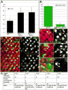
(A-B) The fz and Egfr associated PCP phenotypes are suppressed by dosage reduction in the other gene. (A) Reducing a genomic copy of fz (fzP21 and fzR52) suppresses the defects of Egfr mutant eyes as indicated by increased percentage of correctly oriented wild-type ommatidia. (B) Reducing a genomic copy of Egfr suppresses the occurrence of symmetric ommatidia in fz mutant eyes. 3–4 eyes were counted and ~400 ommatidia evaluated for each genotype. Egfr fz mutant analysis was also evaluated for topCO/1 and topCO/+ with fzP21/+ and fzR52/+. In both cases fz dominantly suppressed the rotation defects and loss of R-cells. In topCO/+, rotation defects were suppressed by fz heterozygosity from 26% to 12% or 18%, respectively. (C-F) Confocal pictures of 3rd instar eye discs. Photoreceptors are marked with anti-Elav in red. (C) sep-GAL4, UAS-Egfr[DN]/+; m∂-lacZ/+ disc. Examples of 5 cell ommatidial preclusters which lost m-lacZ expression are marked with arrowheads (16.8% of total ommatidia. Note that only clusters with the full complement of R-cells were counted). R8 is marked with anti-Sens (blue) and R4 fate with m∂-lacZ (green). (D-F) Mosaic tissue containing cells expressing dominant negative Egfr marked by GFP (green) and the R4 marker m∂-lacZ (blue). White arrowheads mark R4 precursors, yellow arrowheads R3 precursors. Note that R4 cells expressing Egfr[DN] loose m∂-lacZ in R4 and then R3 expresses m∂-lacZ (arrowheads in D and E). Egfr[DN] expression in R3 (or other R-cells) does not affect R4 fate (arrowheads in F, quantif. in G). Egfr[DN] expression in both R3 and R4 cells leads to loss of m∂-lacZ (orange arrow in E). (G) Summary of mosaic analyses with Egfr[DN] in ommatidial preclusters. Individual R-cells positive for Egfr[DN] in otherwise normal 5 cell preclusters were counted and the m∂-lacZ R4 fate marker was registered. Only the R4 precursor was sensitive to Egfr[DN].
Figure 7
Figure 7. PntP2 is sufficient to induce R4 fate. yan and jun antagonize pntP2
(A) sev-GAL4, UAS-pntP2/+ eye section with schematic representation on the right (arrows drawn as in Fig. 1). Symmetric ommatidia are represented by green arrows. sev-GAL4, UAS-pntP2/+ eyes showed 70% symmetric ommatidia. (B) Many ommatidial preclusters in sev-GAL4, UAS-pntP2/+ show both cells of the R3/R4 pair expressing m∂-lacZ, the R4 fate marker. Elav indicates all photoreceptors in red. (C) Graph summarizing average defects of over expression combinations. Ommatidial clusters were scored for wild-type (black), symmetric (green) and wrong (inverted) chirality (grey). Genotypes are indicated below graph. Yan antagonizes the PntP2 effect, and Jun does mildly. 400–500 ommatidia of 4 eyes were evaluated each. Single over expression of wt Jun, Fos and Yan did not cause PCP defects at 18°C and 25°C. (C) Model of combinatorial input into R3/R4 specification (see text for details). Note that lower levels of Egfr and Pnt are required for cell survival/induction of all R-cells (indicated by smaller grey Egfr writing).

Similar articles

Cited by

References

    1. Adler PN. Planar signaling and morphogenesis in Drosophila. Dev Cell. 2002;2:525–535. - PubMed
    1. Bassuk AG, Leiden JM. A direct physical association between ETS and AP-1 transcription factors in normal human T cells. Immunity. 1995;3:223–37. - PubMed
    1. Boutros M, et al. Dishevelled activates JNK and discriminates between JNK pathways in planar polarity and wingless signaling. Cell. 1998;94:109–118. - PubMed
    1. Brunner D, et al. The ETS domain protein Pointed-P2 is a target of MAP kinase in the Sevenless signal transduction pathway. Nature. 1994;370:386–389. - PubMed
    1. Casci T, Freeman M. Control of EGF receptor signalling: lessons from fruitflies. Cancer Metastasis Rev. 1999;18:181–201. - PubMed

Publication types

MeSH terms

Substances