. 2008 Mar 18;152(2):338-45.
doi: 10.1016/j.neuroscience.2007.11.043. Epub 2007 Dec 8.

Reimplantation of avulsed lumbosacral ventral roots in the rat ameliorates injury-induced degeneration of primary afferent axon collaterals in the spinal dorsal columns

Affiliations

Reimplantation of avulsed lumbosacral ventral roots in the rat ameliorates injury-induced degeneration of primary afferent axon collaterals in the spinal dorsal columns

A J Bigbee et al. Neuroscience. .

Abstract

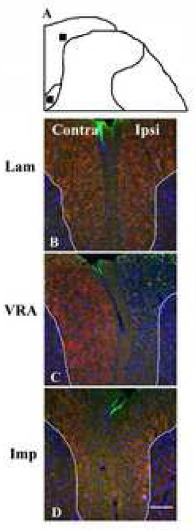
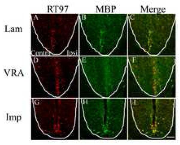
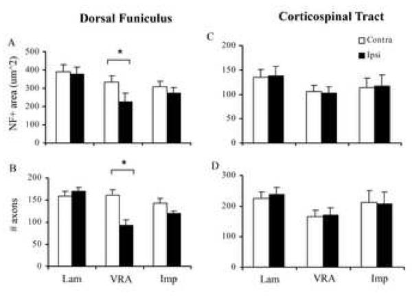
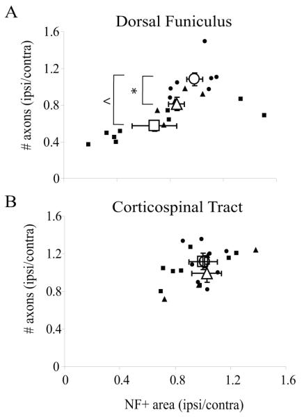
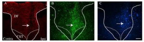
Injuries to the cauda equina/conus medullaris portion of the spinal cord can result in motor, sensory, and autonomic dysfunction, and neuropathic pain. In rats, unilateral avulsion of the motor efferents from the lumbosacral spinal cord results in at-level allodynia, along with a corresponding glial and inflammatory response in the dorsal horn of the spinal cord segments immediately rostral to the lesion. Here, we investigated the fate of intramedullary primary sensory projections following a motor efferent lesion. The lumbosacral (L6 and S1) ventral roots were unilaterally avulsed from the rat spinal cord (VRA; n=9). A second experimental group had the avulsed roots acutely reimplanted into the lateral funiculus (Imp; n=5), as this neural repair strategy is neuroprotective, and promotes the functional reinnervation of peripheral targets. A laminectomy-only group served as controls (Lam; n=7). At 8 weeks post-lesion, immunohistochemical examination showed a 42% reduction (P<0.001) in the number of RT97-positive axons in the ascending tracts of the dorsal funiculus of the L4-5 spinal segment in VRA rats. Evidence for degenerating myelin was also present. Reimplantation of the avulsed roots ameliorated axon and myelin degeneration. Axons in the descending dorsal corticospinal tract were unaffected in all groups, suggesting a specificity of this lesion for spinal primary sensory afferents. These results show for the first time that a lesion restricted to motor roots can induce the degeneration of intramedullary sensory afferents. Importantly, reimplantation of the lesioned motor roots ameliorated sensory axon degeneration. These data further support the therapeutic potential for reimplantation of avulsed ventral roots following trauma to the cauda equina/conus medullaris.


Figures

Fig. 1
Fig. 1
VRA-induced sensory afferent degeneration in the DF is ameliorated by reimplantation. Schematic shows the regions of the dorsal funiculus (DF) and corticospinal tract (CST) that were quantitatively evaluated for neurofilament-positive axons (A). Transverse sections of the L5 dorsal horn from a Lam control shows axons (200 kD neurofilament or NF; red) and myelin (MBP; green) in the contralateral and ipsilateral DF (B). At 8 wks post-ventral root avulsion (VRA), the presence of axons is sparse ipsilateral to the lesion, concomitant with a visible increase in the number of DAPI-positive nuclei (C). Rats which underwent ventral root avulsion plus reimplantation of the avulsed roots (Imp) exhibited a qualitatively more symmetrical distribution of axons between the contralateral an ipsilateral DF, similar to that exhibited in Lam control rats (D). Calibration bar = 100 μm.
Fig. 2
Fig. 2
Morphological characteristics of the ipsilateral DF at 8 weeks post-lesion. The Lam DF (A-D), showed neurofilament positive axons (red; A) that were commonly surrounded by myelin sheaths (green; B). Some Dapi-positive nuclei were also observed (C). At 8 weeks post-VRA, NF labeling was sparse (E), and remaining axons were often observed without a visible myelin sheath (arrow in E-H; however, see Results section for caveat). There was also a distinct reduction in the presence of ring-like myelin-positive structures (compare F to B), while some remaining myelin-positive structures were not associated with any visible neurofilament (arrowheads in E-H). VRA rats also exhibited a qualitative increase in the number of Dapi-positive nuclei (G). Imp rats exhibited neurofilament and myelin-positive structures in the ipsilateral DF that are qualitatively similar to Lam controls. Arrows highlight the location of NF-positive structures, while arrowheads highlight myelin-positive structures in the absence of NF, which exhibit characteristics of degradation. Calibration bar = 30 μm.
Fig. 3
Fig. 3
NF-positive axons in the corticospinal tract. The contralateral (Contra) and ipsilateral (Ipsi) dorsal corticospinal tracts (CST) are shown for Lam (A-C), VRA (D-F), and Imp rats (G-I). NF and MBP are shown in red and green, respectively. Qualitatively, Contra and Ipsi NF and MBP immunopositive labeling is symmetrical. Calibration bar = 50 μm.
Fig. 4
Fig. 4
Using the contralateral dorsal horn as an unlesioned, internal control, the ipsilateral DF of VRA rats exhibited a significant reduction in the total NF+ area (A) and the number of neurofilament-positive axons (B) within a 2500 μm2 region. On the contrary, ipsilateral Lam and Imp NF values did not differ from their respective contralateral sides. In the same sized region of the corticospinal tract, the ipsilateral NF+ area and the number of NF+ structures were unaffected in all groups relative to their contralateral, internal control levels (C and D). Data shown are the mean values ± S.E.M. *, significantly different at p<0.05
Fig. 5
Fig. 5
The relationship between the number of remaining NF+ profiles and the NF+ area are shown for the dorsal funiculus (A) and the corticospinal tract (B). Values presented are the ratio of the ipsilateral divided by the contralateral values in a 2500 um2 region. Individual subject values (closed symbols) are shown, as are the group mean values (open symbols) ± S.E.M. •, Lam; ■, VRA; ▲, Imp. *, significantly different from Lam at p<0.05; ^, significantly different from Lam at p<0.001.
Fig. 6
Fig. 6
VRA affects ascending sensory afferent pathways multiple segments rostral to the lesioned spinal segments. Adjacent spinal segments from the T13-L1 segments of the spinal cord at 8 wks post-L6-S1 VRA show a somatotopic loss of NF+ axons in the ipsilateral DF (red; A,), concomitant with an increased activation of astrocytes (GFAP, green; B) and microglia (AIF-1, pseudo-colored blue; C), suggesting that VRA-induced sensory afferent degeneration extends rostrally from the segmental level of the lesion. The arrows point to the affected portion of the DF. Note the absence of a glial response in the dorsal CST regions. Contra and Ipsi refer to the contralateral and ipsilateral sides of the spinal cord relative to the VRA, respectively. Calibration bar = 50 μm.

References

    1. Aldskogius H, Cerne H, Holmberg A. The effect of sciatic nerve transection on myelinated fibers in the L5 dorsal root and lumbar dorsal column. A Marchi study in the rat. Anat Embryol. 1985;171:181–186. - PubMed
    1. Baker KA, Hagg T. An adult rat spinal cord contusion model of sensory axon degeneration: the estrus cycle or a preconditioning lesion do not affect outcome. J Neurotrauma. 2005;22:415–428. - PubMed
    1. Bigbee AJ, Hoang TX, Havton LA. At-level neuropathic pain is induced by lumbosacral ventral root avulsion injury and ameliorated by root reimplantation into the spinal cord. Exp Neurol. 2007;204:273–282. - PMC - PubMed
    1. Blight AR, Decrescito V. Morphometric analysis of experimental spinal cord injury in the cat: the relation of injury intensity to survival of myelinated axons. Neuroscience. 1986;19:321–341. - PubMed
    1. Carlstedt T, Anand P, Hallin R, Misra PV, Noren G, Seferlis T. Spinal nerve root repair and reimplantation of avulsed ventral roots into the spinal cord after brachial plexus injury. J Neurosurg. 2000;93:237–247. - PubMed

Publication types

MeSH terms