

Progesterone and estrogen regulate oxidative metabolism in brain mitochondria
- PMID: 18292191
- PMCID: PMC2408802
- DOI: 10.1210/en.2007-1227
Progesterone and estrogen regulate oxidative metabolism in brain mitochondria
Abstract
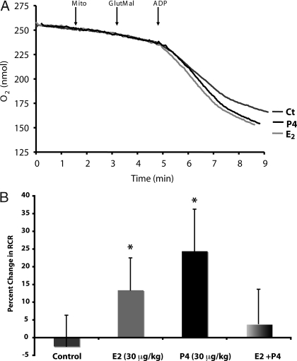
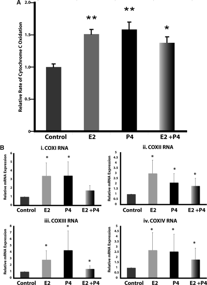
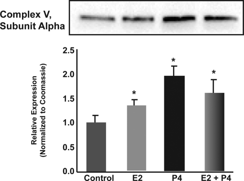
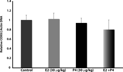
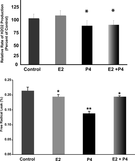
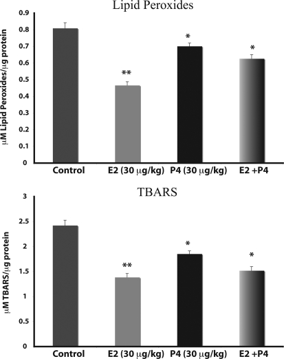
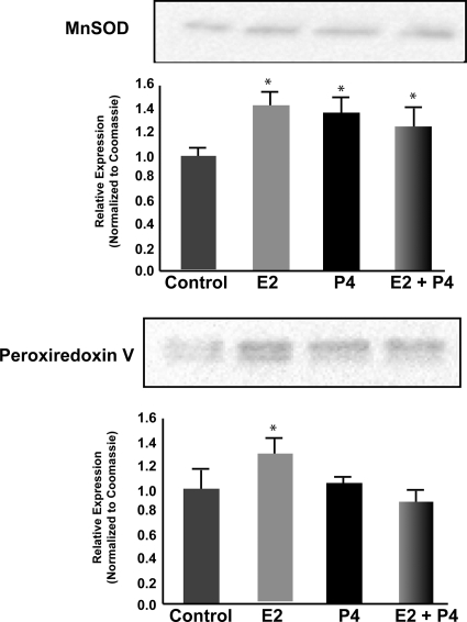
The ovarian hormones progesterone and estrogen have well-established neurotrophic and neuroprotective effects supporting both reproductive function and cognitive health. More recently, it has been recognized that these steroids also regulate metabolic functions sustaining the energetic demands of this neuronal activation. Underlying this metabolic control is an interpretation of signals from diverse environmental sources integrated by receptor-mediated responses converging upon mitochondrial function. In this study, to determine the effects of progesterone (P4) and 17beta-estradiol (E2) on metabolic control via mitochondrial function, ovariectomized rats were treated with P4, E2, or E2 plus P4, and whole-brain mitochondria were isolated for functional assessment. Brain mitochondria from hormone-treated rats displayed enhanced functional efficiency and increased metabolic rates. The hormone-treated mitochondria exhibited increased respiratory function coupled to increased expression and activity of the electron transport chain complex IV (cytochrome c oxidase). This increased respiratory activity was coupled with a decreased rate of reactive oxygen leak and reduced lipid peroxidation representing a systematic enhancement of brain mitochondrial efficiency. As such, ovarian hormone replacement induces mitochondrial alterations in the central nervous system supporting efficient and balanced bioenergetics reducing oxidative stress and attenuating endogenous oxidative damage.
Figures
Similar articles
-
Medroxyprogesterone acetate antagonizes estrogen up-regulation of brain mitochondrial function.Endocrinology. 2011 Feb;152(2):556-67. doi: 10.1210/en.2010-1061. Epub 2010 Dec 15. Endocrinology. 2011. PMID: 21159850 Free PMC article.
-
Dehydroepiandrosterone and alpha-estradiol limit the functional alterations of rat brain mitochondria submitted to different experimental stresses.Neuroscience. 2002;115(2):415-24. doi: 10.1016/s0306-4522(02)00416-5. Neuroscience. 2002. PMID: 12421607
-
Estradiol affects liver mitochondrial function in ovariectomized and tamoxifen-treated ovariectomized female rats.Toxicol Appl Pharmacol. 2007 May 15;221(1):102-10. doi: 10.1016/j.taap.2007.02.006. Epub 2007 Feb 23. Toxicol Appl Pharmacol. 2007. PMID: 17397887
-
Sex differences in brain mitochondrial metabolism: influence of endogenous steroids and stroke.J Neuroendocrinol. 2018 Feb;30(2). doi: 10.1111/jne.12497. J Neuroendocrinol. 2018. PMID: 28650095 Review.
-
Oestrogenic Regulation of Mitochondrial Dynamics.Int J Mol Sci. 2022 Jan 20;23(3):1118. doi: 10.3390/ijms23031118. Int J Mol Sci. 2022. PMID: 35163044 Free PMC article. Review.
Cited by
-
Early, but not late onset estrogen replacement therapy prevents oxidative stress and metabolic alterations caused by ovariectomy.Antioxid Redox Signal. 2014 Jan 10;20(2):236-46. doi: 10.1089/ars.2012.5112. Epub 2013 Jul 20. Antioxid Redox Signal. 2014. PMID: 23725100 Free PMC article.
-
EVALUATION OF OXIDATIVE STRESS MARKERS IN GIRLS WITH PREMATURE THELARCHE AND PRECOCIOUS PUBERTY.Acta Endocrinol (Buchar). 2024 Jan-Mar;20(1):5-11. doi: 10.4183/aeb.2024.5. Epub 2024 Oct 3. Acta Endocrinol (Buchar). 2024. PMID: 39372292 Free PMC article.
-
Sex differences and estrogen effects in cardiac mitochondria in human aortic stenosis and in the mouse heart.Front Endocrinol (Lausanne). 2023 Oct 17;14:1181044. doi: 10.3389/fendo.2023.1181044. eCollection 2023. Front Endocrinol (Lausanne). 2023. PMID: 37916152 Free PMC article.
-
Steroid Hormones and Their Action in Women's Brains: The Importance of Hormonal Balance.Front Public Health. 2018 May 23;6:141. doi: 10.3389/fpubh.2018.00141. eCollection 2018. Front Public Health. 2018. PMID: 29876339 Free PMC article. Review.
-
Analysis of arsenic-modulated expression of hypothalamic estrogen receptor, thyroid receptor, and peroxisome proliferator-activated receptor gamma mRNA and simultaneous mitochondrial morphology and respiration rates in the mouse.PLoS One. 2024 May 16;19(5):e0303528. doi: 10.1371/journal.pone.0303528. eCollection 2024. PLoS One. 2024. PMID: 38753618 Free PMC article.
References
-
- Nilsen J, Diaz Brinton R 2002 Impact of progestins on estrogen-induced neuroprotection: synergy by progesterone and 19-norprogesterone and antagonism by medroxyprogesterone acetate. Endocrinology 143:205–212 - PubMed
-
- Nicholls DG, Budd SL 2000 Mitochondria and neuronal survival. Physiol Rev 80:315–360 - PubMed
-
- Cadenas E 2004 Mitochondrial free radical production and cell signaling. Mol Aspects Med 25:17–26 - PubMed
Publication types
MeSH terms
Substances
Grants and funding
LinkOut - more resources
Full Text Sources
Other Literature Sources
Research Materials
