. 2008 Jun;149(6):2840-52.
doi: 10.1210/en.2007-1192. Epub 2008 Feb 21.

Tumor necrosis factor (TNF)-alpha persistently activates nuclear factor-kappaB signaling through the type 2 TNF receptor in chromaffin cells: implications for long-term regulation of neuropeptide gene expression in inflammation

Affiliations

Tumor necrosis factor (TNF)-alpha persistently activates nuclear factor-kappaB signaling through the type 2 TNF receptor in chromaffin cells: implications for long-term regulation of neuropeptide gene expression in inflammation

Djida Ait-Ali et al. Endocrinology. 2008 Jun.

Abstract

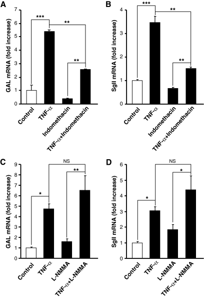
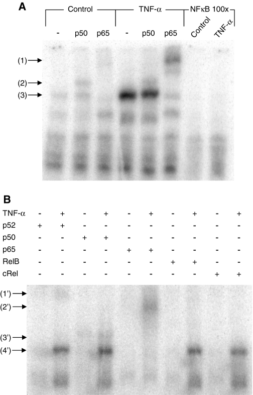
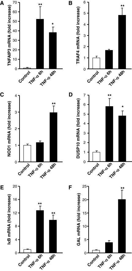
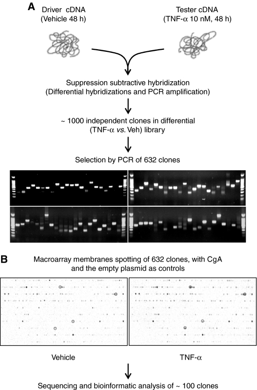
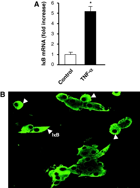
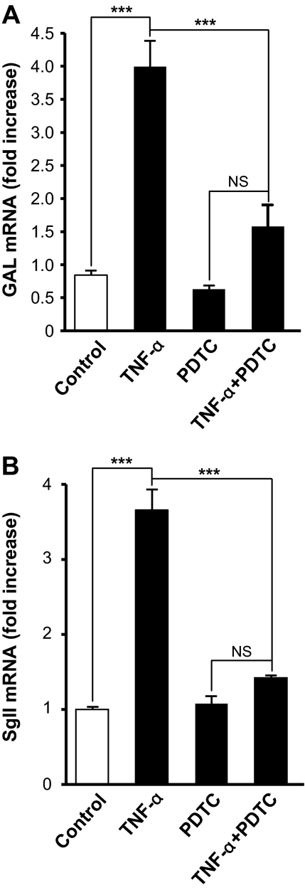
Chromaffin cells of the adrenal medulla elaborate and secrete catecholamines and neuropeptides for hormonal and paracrine signaling in stress and during inflammation. We have recently documented the action of the cytokine TNF-alpha on neuropeptide secretion and biosynthesis in isolated bovine chromaffin cells. Here, we demonstrate that the type 2 TNF-alpha receptor (TNF-R2) mediates TNF-alpha signaling in chromaffin cells via activation of nuclear factor (NF)-kappaB. Microarray and suppression subtractive hybridization have been used to identify TNF-alpha target genes in addition to those encoding the neuropeptides galanin, vasoactive intestinal polypeptide, and secretogranin II in chromaffin cells. TNF-alpha, acting through the TNF-R2, causes an early up-regulation of NF-kappaB, long-lasting induction of the NF-kappaB target gene inhibitor kappaB (IkappaB), and persistent stimulation of other NF-kappaB-associated genes including mitogen-inducible gene-6 (MIG-6), which acts as an IkappaB signaling antagonist, and butyrate-induced transcript 1. Consistent with long-term activation of the NF-kappaB signaling pathway, delayed induction of neuropeptide gene transcription by TNF-alpha in chromaffin cells is blocked by an antagonist of NF-kappaB signaling. TNF-alpha-dependent signaling in neuroendocrine cells thus leads to a unique, persistent mode of NF-kappaB activation that features long-lasting transcription of both IkappaB and MIG-6, which may play a role in the long-lasting effects of TNF-alpha in regulating neuropeptide output from the adrenal, a potentially important feedback station for modulating long-term cytokine effects in inflammation.


Figures

Figure 1
Figure 1
Adrenochromaffin cells express the TNF-R2. A, Autoradiogram of a bovine adrenal gland transverse section hybridized with 35S-labeled bovine antisense TNF-R2 riboprobe showing expression of TNF-R2 in both cortex and medulla (left panel). Hybridization with the sense probe was used as a negative control (right panel). B, Colocalization studies using a bovine antisense CgA riboprobe labeled with digoxigenin (dark staining in the medulla) and 35S-labeled TNF-R2 riboprobe (arrowheads in cortex and medulla) showed that TNF-R2 is expressed in chromaffin cells. C, Hybridization with TNF-R2 and CgA sense probes showing only background labeling.
Figure 2
Figure 2
Effect of inhibiting prostaglandin or nitric oxide production on TNF-α induction of neuropeptide gene expression. Chromaffin cells were incubated for 48 h in control conditions or with 10 nm TNF-α, in the absence or presence of 1 μm indomethacin or 2 mm l-NMMA (C and D) as described in Materials and Methods. GAL (A) and SgII (B) mRNA levels, determined by Q-RT-PCR, are expressed as fold increase over corresponding control values, and represent means ± sem of four determinations for each condition from one experiment representative of three different experiments. *, P < 0.05; **, P < 0.01; ***, P < 0.001 vs. the corresponding control (one-way ANOVA test, Bonferroni posttest). NS, Not significant.
Figure 3
Figure 3
TNF-α activates NF-κB transcription factor in adrenochromaffin cells. A, Chromaffin cells were incubated with TNF-α (10 nm) for 2 h, and nuclear extracts were prepared and assayed for their binding to the 32P-labeled NF-κB site. Supershift assays of the NF-κB-bound complex were obtained in the presence of vehicle-treated or TNF-α-treated nuclear extracts, using antibodies directed against two NF-κB subunits, p50 and p65. The symbol (−) denotes control supershift reactions with no antibody. The binding specificity of the NF-κB site to vehicle-treated or TNF-α-treated nuclear extracts was verified by adding a 100-fold excess of unlabeled NF-κB oligonucleotide. The supershifted complexes formed by p65 and p50 are indicated by the arrows 1 and 2, whereas arrow 3 indicates the shift of NF-κB. B, Supershift assays of the NF-κB-bound complex obtained in the presence of vehicle-treated or TNF-α-treated nuclear extracts, using antibodies directed against five subunits p52, p50, p65, RelB, and cRel. The supershifted p52, p50, and p65 complexes are indicated by the arrows 1′, 2′, and 3′, respectively. The arrow 4′ indicates the shift of NF-κB.
Figure 4
Figure 4
Time course of TNFAIP3, TRAF4, DUSP10, NOD1, and IκB gene stimulation by TNF-α determined by Q-RT-PCR. Cultured BCCs were incubated for 6 and 48 h in control conditions or with 10 nm TNF-α, and TNFAIP3 (A), TRAF4 (B), NOD1 (C), DUSP10 (D), IκB (E), and GAL (F) mRNA levels were measured by Q-RT-PCR as described in Materials and Methods. Values are the mean ± sem of four determinations for each condition. *, P < 0.05; **, P < 0.01 vs. the corresponding control (one-way ANOVA test, Bonferroni posttest).
Figure 5
Figure 5
Generation and validation by hybridization of a SSH library from TNF-α-treated BCCs. A, Schematic depiction of generation of SSH clone set. Chromaffin cells were incubated with TNF-α (10 nm) for 48 h, RNA was extracted, reverse transcribed, ligated to amplification primer-adapters (tester cDNA), denatured, and hybridized to excess control BCC cDNA (driver cDNA). Doubly primer-adapted (subtractive hybridization nonsuppressed) cDNA was amplified and cloned. The TNF-α vs. control differential library contained more than 1000 clones from which 632 inserts, ranging in size from about 500 to about 1500 bp were amplified. B, The 632 amplified inserts were spotted on nylon membranes, and the macroarrays obtained were hybridized with labeled cDNA from chromaffin cells treated with TNF-α (right) or vehicle (left) for 48 h. Four differentially expressed clones are circled, for purposes of illustration, from a total of 100 individual clones found to be differentially expressed based on densitometric analysis of hybridization results generated from four independent experiments similar to the one shown here. These clones were sequenced and identified through bioinformatics analysis and found to represent 23 distinct differentially expressed transcripts.
Figure 6
Figure 6
Regulation of IκB by TNF-α in BCCs. A, Chromaffin cells were incubated for 48 h in control conditions or with 10 nm TNF-α, and IκB mRNA levels were measured by Q-RT-PCR as described in Materials and Methods. Values are the mean ± sem of four separate determinations for each condition from one experiment representative of three different experiments and are expressed as a fold increase over control untreated cells. *, P < 0.05 vs. control (Mann-Whitney U test). B, Chromaffin cells treated with 10 nm TNF-α for 48 h were fixed and processed for immunofluorescence. IκB was visualized by using an appropriate antibody and Alexa-488-conjugated goat antirabbit IgGs. IκB cytoplasmic staining is indicated by arrowheads in a representative field.
Figure 7
Figure 7
Implication of NF-κB in the effect of TNF-α on neuropeptide gene expression. Chromaffin cells were incubated for 48 h in control conditions or with 10 nm TNF-α, in the presence or absence of 100 mm PDTC as described in Materials and Methods. GAL (A) and SgII (B) mRNA levels, determined by Q-RT-PCR, are expressed as fold increase over corresponding control values and represents means ± sem of four determinations for each condition from one experiment representative of three different experiments. NS, Not significant (P > 0.05); ***, P < 0.001 vs. TNF-α alone (one-way ANOVA test, Bonferroni posttest).

References

    1. Wrona D 2006 Neural-immune interactions: an integrative view of the bidirectional relationship between the brain and immune systems. J Neuroimmunol 172:38–58 - PubMed
    1. Andreis PG, Tortorella C, Ziolkowska A, Spinazzi R, Malendowicz LK, Neri G, Nussdorfer GG 2007 Evidence for a paracrine role of endogenous adrenomedullary galanin in the regulation of glucocorticoid secretion in the rat adrenal gland. Int J Mol Med 19:511–515 - PubMed
    1. Ait-Ali D, Turquier V, Grumolato L, Yon L, Jourdain M, Alexandre D, Eiden LE, Vaudry H, Anouar Y 2004 The proinflammatory cytokines tumor necrosis factor-α and interleukin-1 stimulate neuropeptide gene transcription and secretion in adrenochromaffin cells via activation of extracellularly regulated kinase 1/2 and p38 protein kinases, and activator protein-1 transcription factors. Mol Endocrinol 18:1721–1739 - PubMed
    1. Bornstein SR, Haidan A, Ehrhart-Bornstein M 1996 Cellular communication in the neuro-adrenocortical axis: role of vasoactive intestinal polypeptide (VIP). Endocr Res 22:819–829 - PubMed
    1. Kruppa G, Thoma B, Machleidt T, Wiegmann K, Kronke M 1992 Inhibition of tumor necrosis factor (TNF)-mediated NF-κB activation by selective blockade of the human 55-kDa TNF receptor. J Immunol 148:3152–3157 - PubMed

Publication types

MeSH terms