. 2008 May;26(5):1253-64.
doi: 10.1634/stemcells.2007-0691. Epub 2008 Feb 21.

Reciprocal intraepithelial interactions between TP63 and hedgehog signaling regulate quiescence and activation of progenitor elaboration by mammary stem cells

Affiliations

Reciprocal intraepithelial interactions between TP63 and hedgehog signaling regulate quiescence and activation of progenitor elaboration by mammary stem cells

Na Li et al. Stem Cells. 2008 May.

Abstract

TP63 is required for preservation of epithelial regenerative stasis and regulates the activity of diverse genetic pathways; however, specific effector pathways are poorly understood. Data presented here indicate that reciprocal regulatory interactions between hedgehog signaling and TP63 mediate stage-specific effects on proliferation and clonigenicity of separable enriched mammary stem and progenitor fractions. Analysis of DeltaN-p63 and TA-p63 indicates segregated expression in mammary stem and progenitor fractions, respectively, demonstrating that differential TP63 promoter selection occurs during elaboration of mammary progenitors by mammary stem cells. This segregation underlies mammary progenitor-specific expression of Indian Hedgehog, identifying it as a binary transcriptional target of TP63. Hedgehog activation in vivo enhances elaboration of mammary progenitors and decreases label retention within mammary stem cell-enriched fractions, suggesting that hedgehog exerts a mitogenic effect on mammary stem cells. Hedgehog signaling promotes differential TP63 promoter usage via disruption of Gli3 or Gli3(R) accumulation, and shRNA-mediated disruption of Gli3 expression was sufficient to alter TP63 promoter usage and enhance clonigenicity of mammary stem cells. Finally, hedgehog signaling is enhanced during pregnancy, where it contributes to expansion of the mammary progenitor compartment. These studies support a model in which hedgehog activates elaboration and differentiation of mammary progenitors via differential TP63 promoter selection and forfeiture of self-renewing capacity.


Conflict of interest statement

Disclosure of Potential Conflicts of Interest

The authors indicate no potential conflicts of interest.

Figures
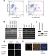
Figure 1
Figure 1
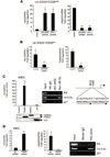
Segregated expression of ΔN-p63, TA-p63, and Ihh in the mammary regenerative hierarchy. (A): Fluorescence-activated cell sorting of mouse MECs on the basis of CD24/CD29 (left) and CD24/CD49f (right). Nomenclature used in the right panel is as previously described [2]. (B): Reverse transcription-polymerase chain reaction analysis indicates segregation of ΔN-p63 from TA-p63 and Ihh in mammary epithelial fractions derived from CD24/CD29-based sorting (left) and CD24/CD49f–based sorting (right). (C): Western blot analysis of Lin/CD24+/CD29high and Lin/CD24+/ CD29low with antibodies specific against ΔN-p63, TA-p63, and β-actin confirms the segregated expression of TP63 isoforms. (D): Immunocytofluorescent analysis of ΔN-p63 and Ihh in Lin/CD24+/CD29high and Lin/CD24+/CD29low confirms the segregated expression of ΔN-p63 and Ihh. (E): Two-color immunofluorescence demonstrates segregated expression of ΔN-p63 and Ihh in human primary mammary ducts (left) and smaller ducts and lobules (right). The image at the right was captured by confocal microscopy and includes nuclear staining with DAPI. Abbreviations: DAPI, 4,6-di-amidino-2-phenylindole; Dhh, desert hedgehog; GAPDH, glyceraldehyde-3-phosphate dehydrogenase; Ihh, Indian hedgehog; Ma-CFC, mammary colony-forming cells; MRU, mammary regenerative units; MYO, myoepithelia; No-RT, no reverse transcription; Shh, sonic hedgehog.
Figure 2
Figure 2
Ihh is a direct bidirectional target of ΔN-p63 and TA-p63. (A): shRNA-mediated suppression of ΔN-p63 (right) in Lin/CD24+/CD29high cells leads to increased Ihh mRNA levels (left). p63-directed shRNAs were targeted to the DBD (p63DBD) or the α-specific carboxy terminus (p63-α). (B): shRNA-mediated suppression of TA-p63 (right) in Lin/CD24+/CD29low cells leads to decreased Ihh mRNA levels (left). (C): Ectopic myc-tagged TA-p63-γ activates expression of Ihh in IMECs. Left panel shows the fold activation of Ihh relative to GAPDH, and Western blot (below) confirms the expression of ectopic TA-p63-γ, ΔN-p63-α, and p53. Center panel shows chromatin immunoprecipitation (ChIP) indicating binding of myc-tagged TA-p63-γ to a putative p63-binding element located at −1,392 to −1,412 relative to the initiator ATG of Ihh. Right panel shows the sequence of the putative p63-binding element above the consensus sequence. Bases shown in red represent divergence from the canonical p53/p63-binding element. (D): siRNA-mediated ablation of ΔN-p63-α (middle) in IMECs leads to enhanced expression of Ihh (left). ChIP indicates direct binding of endogenous ΔN-p63-α to a region of the Ihh promoter located at −1,392 to −1,412 relative to the initiator ATG of the human Ihh gene (right). Error bars represent the SEM derived from triplicate experimental data. Throughout the figure, p values <.001 are indicated by ***p values <.01 are indicated by **, and p values <.05 are indicated by *. Abbreviations: DBD, DNA binding domain; GAPDH, glyceraldehyde-3-phosphate dehydrogenase; Ihh, Indian hedgehog; IMEC, immortalized mammary epithelial cell; NS, nonspecific; scr, scrambled.
Figure 3
Figure 3
Activation of hedgehog signaling leads to enhanced elaboration of MPs by MSCs. (A): CD24/CD29-based sorting of mouse MECs from wild-type and Ptch1−/+mice indicates an expansion of the Lin/CD24+CD29low population in Ptch1−/+mice. (B): Reverse transcription-polymerase chain reaction (PCR) confirms the segregated expression of ΔN-p63 from TA-p63 and Ihh in Lin/CD24+CD29high and Lin/CD24+/CD29low cells from Ptch1−/mice. (C): Quantification of BrdU uptake following a 3-hour labeling indicates enhanced proliferation in Lin/CD24+/CD29low and Lin/CD24+/CD29high in Ptch−/+mice relative to wild-type littermates. Data represent quantification of 10 microscopic fields per mouse and three mice. Error bars represent the SEM. (D): Enhanced BrdU uptake and Ki-67 expression in Ptch−/+ mice. The first two columns are representative images of Lin/CD24+/ CD29high and Lin/CD24+/CD29low fractions that were fixed to glass and stained with anti-BrdU (green) and DAPI (blue). The third column is in situ detection of BrdU in wt and Ptch1−/+ virgin mice. The last two columns are representative images of Lin/CD24+/CD29high and Lin/CD24+/CD29low fractions that were fixed to glass and stained with anti-Ki-67. (E): Quantification of BrdU retention following a 10-week chase indicates that Lin/CD24+/CD29high cells from wild-type but not Ptch1−/+ are able to retain BrdU. Data represent quantification of 10 microscopic fields per mouse and four mice. Error bars represent the SEM. Images at right are representative of enrichment of BrdU retaining cells (white arrowheads) in the Lin/CD24+/CD29high fraction of wild-type but not Ptch1−/+ mice. (F): Semiquantitative PCR analysis of GATA3 indicates that expression of GATA3 is enhanced in the stem cell-enriched fractions from Ptch1−/+. Throughout the figure, p values <.001 are indicated by ***, p values <.01 are indicated by **, and p values <.05 are indicated by *. Abbreviations: BrdU, bromodeoxyuridine; DAPI, 4,6-diamidino-2-phenylindole; GAPDH, glyceraldehyde-3-phosphate dehydrogenase; Ihh, Indian hedgehog; MG, mammary gland; MP, mammary progenitor; MSC, mammary stem cell; WT, wild-type; wt, wild-type.
Figure 4
Figure 4
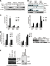
Hedgehog activation causes differential TP63 promoter usage via disruption of Gli3R accumulation. (A): Western blot analysis of Gli3 and Gli3R expression in response to Shh-cm in IMECs indicates that hedgehog activation disrupts Gli3R accumulation in a manner that is sensitive to 5E1. (B): Ectopic expression of Ihh in IMECs diminishes expression of ΔN-p63-α and Gli3R. (C): Ectopic expression of Ihh causes differential TP63 promoter selection in IMECs. RNA from IMEC-EV, IMEC-Ihh1, and IMEC-Ihh2 was harvested at 24, 48, and 72 h after plating, and expression of ΔN-p63 and TA-p63 was evaluated by quantitative polymerase chain reaction (Q-PCR). Data represent the mean of triplicate points, and error bars represent the SEM. (D): siRNA-mediated ablation of Gli3 leads to differential TP63 promoter usage in IMECs. RNA was harvested at 24, 48, and 72 h following transfection with a scr or Gli3-directed siRNA, and expression of ΔN-p63 and TA-p63 was measured by Q-PCR. Data represent the mean of triplicate points and error bars represent the SEM. Western blot at right demonstrates the efficacy of the Gli3-directed siRNA. (E): Ablation of Gli3 from Lin/CD24+/CD29high leads to differential TP63 promoter usage and expression of Ihh. Cells were isolated and plated on Bio-Coat prior to infection with lentiviruses expressing a nonspecific shRNA or a Gli3-directed shRNA. RNA was harvested 48 h after infection, and expression of ΔN-p63, TA-p63, and Ihh was measured by reverse transcription-polymerase chain reaction. Western blot at right demonstrates the efficacy of the Gli3-directed shRNA in IMECs. Abbreviations: GAPDH, glyceraldehyde-3-phosphate dehydrogenase; h, hours; Ihh, Indian hedgehog; IMEC, immortalized mammary epithelial cell; scr, scrambled; Shh-cm, sonic hedgehog-conditioned medium.
Figure 5
Figure 5
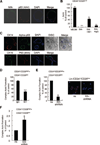
Differential effects of hedgehog on clonigenicity of enriched mammary stem and progenitor fractions. (A): Lin/CD24+/CD29low cells form bilayered acinar structures with distinct patterns of expression in specific cell layers. Following 10 days of growth, acini were stimulated with prolactin, fixed, and stained with anti-milk, anti-p63α, and DAPI. (B): Hedgehog ablates acini formation by Lin/CD24+/CD29low cells. Cells were isolated and cultured under acini-forming conditions in the presence of Shh-cm −/+ 5E1 or a nonspecific mouse IgG. After 10 days, acini were counted. Data represent the mean of four experiments, and error bars represent the SEM. (C): Cells from the Lin/CD24+/CD29high fraction form complex multilobular structures. Top row shows expression of basal/myoepithelial markers CK14 and p63 merged with phase images of complex acini. Bottom row shows a single lobe from one of the complex acini expressing the luminal marker CK19 and the basal/myoepithelial marker p63 in two distinct cell layers. (D): Lin/CD24+/CD29low and Lin/CD24+/CD29high cells were cocultured under acini-forming conditions in the presence or absence of 5E1, and complex acini were counted. Data represent the mean of four experiments, and error bars represent the SEM. (E): Ablation of Ihh from Lin/CD24+/CD29low cells restricts their ability to stimulate complex acini formation by Lin/CD24+/CD29high cells. Isolated Lin/CD24+/CD29low cells were infected with lentiviruses encoding nonspecific shRNA or Ihh-directed shRNA. Following infection, cells were cocultured under acini-forming conditions with Lin/CD24+/CD29high cells, and complex acini were counted. Data represent the mean of three distinct experiments, and error bars represent the SEM. Immunofluorescence of Lin/CD24+/CD29low cells (right) demonstrates the efficacy of the Ihh-directed shRNA. (F): Disruption of Gli3R accumulation in Lin/CD24+/CD29high cells promotes complex acini formation. Lin/CD24+/CD29high cells were infected with lentiviruses encoding a nonspecific shRNA or a Gli3-directed shRNA. Following infection, cells were cultured under acini-forming conditions, and complex acini were counted. Data represent the mean of three distinct experiments, and error bars represent SEM. Throughout the figure, p values <.001 are indicated by ***, p values <.01 are indicated by **, and p values <.05 are indicated by *. Abbreviations: DAPI, 4,6-diamidino-2-phenylindole; Ihh, Indian hedgehog; Shh, sonic hedgehog.
Figure 6
Figure 6
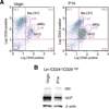
Hedgehog signaling is active during pregnancy-associated expansion of the mammary gland. (A): Sorting of MECs from virgin and P14 mice on the basis of CD24/CD49f indicated that the MRU-containing fraction remained constant while the MYO fraction was expanded. Results also indicate expansion of the Ma-CFC fraction. (B): Western blot analysis of Gli3 and Gli3R indicated reduced levels of Gli3R in the Lin/CD24+/CD29High fraction of P14 mice relative to virgin mice. Abbreviations: Ma-CFC, mammary colony-forming cells; MRU, mammary regenerative units; MYO, myoepithelial; P14, progeny day 14.
Figure 7
Figure 7
Proposed model for the role of reciprocal interactions between TP63 and hedgehog signaling in the mammary regenerative hierarchy. Hedgehog stimulation of mammary stem cells is mitogenic and promotes elaboration of mammary progenitors and differential TP63 promoter use. Differential TP63 promoter usage in mammary stem and progenitor cells underlies the progenitor-specific expression of Ihh, which feeds back upon mammary stem cells to promote further elaboration of mammary progenitors. Hedgehog activation of Lin/CD24+/CD29Low cells restricts their proliferative capacity and clonigenicity, suggesting a role in directing terminal differentiation of mammary progenitors. Abbreviation: HH, hedgehog; Ihh, Indian hedgehog.

Similar articles

Cited by

References

    1. Lechler T, Fuchs E. Asymmetric cell divisions promote stratification and differentiation of mammalian skin. Nature. 2005;437:275–280. - PMC - PubMed
    1. Tataria M, Perryman SV, Sylvester KG. Stem cells: Tissue regeneration and cancer. Semin Pediatr Surg. 2006;15:284–292. - PubMed
    1. Beachy PA, Karhadkar SS, Berman DM. Tissue repair and stem cell renewal in carcinogenesis. Nature. 2004;432:324–331. - PubMed
    1. Krtolica A. Stem cell: Balancing aging and cancer. Int J Biochem Cell Biol. 2005;37:935–941. - PubMed
    1. Pardal R, Clarke MF, Morrison SJ. Applying the principles of stem-cell biology to cancer. Nat Rev Cancer. 2003;3:895–902. - PubMed

Publication types

MeSH terms