. 2008 Mar 4;105(9):3551-6.
doi: 10.1073/pnas.0712140105. Epub 2008 Feb 21.

Intestinal alkaline phosphatase is a gut mucosal defense factor maintained by enteral nutrition

Affiliations

Intestinal alkaline phosphatase is a gut mucosal defense factor maintained by enteral nutrition

Ross F Goldberg et al. Proc Natl Acad Sci U S A. .

Abstract

Under conditions of starvation and disease, the gut barrier becomes impaired, and trophic feeding to prevent gut mucosal atrophy has become a standard treatment of critically ill patients. However, the mechanisms responsible for the beneficial effects of enteral nutrition have remained a mystery. Using in vitro and in vivo models, we demonstrate that the brush-border enzyme, intestinal alkaline phosphatase (IAP), has the ability to detoxify lipopolysaccharide and prevent bacterial invasion across the gut mucosal barrier. IAP expression and function are lost with starvation and maintained by enteral feeding. It is likely that the IAP silencing that occurs during starvation is a key component of the gut mucosal barrier dysfunction seen in critically ill patients.


Conflict of interest statement

The authors declare no conflict of interest.

Figures

Fig. 1.
Fig. 1.
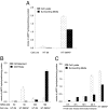
Localization of the IAP enzyme. (A) Gel image of RT-PCR showing IAP mRNA only expressed in the IAP stable transfectant cell line compared with empty vector as a control. (B) Cellular fractions were purified to separate the cytosolic and membranous components. IAP activity was determined in the fraction from parent HT-29 and HT-29/IAP stable cell lines. (C) IAP and MAPK activity in membranous and cytosolic fractions of HT-29/IAP stable cells. Data are presented as mean ± SD.
Fig. 2.
Fig. 2.
The IAP enzyme is secreted by the cell. (A) IAP enzyme activities are depicted in cell lysate and surrounding medium, comparing the parent and transfected HT-29 cells overexpressing IAP. (B) The conditioned medium was filtered through different pore sizes, the results showing that the IAP enzyme is between 50 and 100 kDa (the enzyme is known to be ≈60 kDa in size). (C) Endogenous vs. ectopically produced IAP. Cell lysates and conditioned medium were assayed for IAP enzyme activity. Upon treatment with butyrate, endogenous IAP enzyme activity increases in the lysates and the surrounding medium. The activities are similar to that seen in the case of the ectopic IAP produced in the stable cell line (HT29/IAP). Data are presented as mean ± SD.
Fig. 3.
Fig. 3.
IAP inhibits the NF-κB pathway. (A) IAP blocks LPS-activated NF-κB nuclear translocation. HT-29 WT, vector, and IAP-overexpressing cells were exposed ±LPS, then fixed and stained for immunofluorescence studies. Staining with antibodies for RelA/p65 (part of the NF-κB complex translocated into the nucleus) and DAPI (cell nucleus) is shown. Only the IAP-overexpressing cells were able to block the effects of LPS, preventing NF-κB nuclear translocation. (B) IAP protects the cell from LPS exposure. WT and IAP-expressing IEC-6 cells were exposed to LPS at varying concentrations. NF-κB-Luc activity was determined as the readout for the cellular effects of LPS. Data refer to mean ± SD. (C) IAP specifically blocks LPS activation of the NF-κB pathway in IEC-6 cells. Western blotting was performed with a specific antibody to IκBα phosphorylation, a critical step in the NF-κB pathway. IkBα did not become phosphorylated in the case of the IAP-overexpressing cells exposed to LPS. The β-actin staining was used to confirm the relative amounts of protein in each sample. (D) IAP blocks phosphorylation of components of the NF-κB pathway in LPS-treated T84 cells. Western blotting was performed with specific antibodies to the phosphorylated forms of the IκBα and RelA/p65 proteins. The β-actin staining was used to confirm the relative amounts of protein in each sample.
Fig. 4.
Fig. 4.
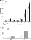
LPS-dephosphorylating activity. (A) Biological activity is present in the transfected but not parent HT-29 cells, the magnitude greatest in the cell lysate > membrane > medium (all significant, P < 0.01). There was no statistically significant difference in LPS-dephosphorylating activity in the cytosol between the transfected and parent cells. (B) The LPS-dephosphorylating activity is compared in the endogenous (butyrate-treated) and ectopically produced (transfected cells) conditions. The increases in the lysates became significant (P < 0.01) at 12 and 24 h of butyrate exposure and in the medium at 24 h. Data are presented as mean ± SD.
Fig. 5.
Fig. 5.
IAP enzyme dephosphorylates LPS in vivo. (A) AP assay for WT and IAP KO mice groups that were fed (n = 5), fasted (starved for 2 days, n = 5), and refed (starved for 2 days, refed for 2 days, n = 4). Starvation causes a significant decrease in IAP activity in the WT animals, down to levels similar to those in the KO mice. Refeeding stimulates IAP expression in the WT animals. Starvation and refeeding appear to have minimal effect on IAP expression in the IAP KO mice. *, P < 0.05, comparing fasted with the fed and refed WT animals. As expected, KO AP levels are significantly lower than those in the WT animals. (B) A similar pattern was seen in the LPS-dephosphorylating activity of the fed, fasted, and refed WT and KO groups. Starvation dramatically reduced the LPS-dephosphorylating ability of the WT animal, whereas refeeding returned it to normal levels. *, P < 0.05, comparing fasted with the fed and refed WT animals. KO levels are significantly lower than those in the WT animals. (C) A comparison of luminal contents (n = 5) and stool (n = 4) of the WT and IAP KO animals reveals that the WT animals having a significantly higher IAP enzyme activity in both their luminal contents and stool compared with the KO animals. *, P < 0.05, comparing WT and KO contents. (D) LPS-dephosphorylating activity is also much higher in the lumen and stool of the WT compared with the KO animals. *, P < 0.05, comparing WT and KO contents. Data in this figure are presented as mean ± SEM.
Fig. 6.
Fig. 6.
IAP protects the mice from gut bacterial translocation. (A) Direct gut I/R. WT and IAP KO mice were exposed to 45 min of SMA clamping followed by varying times of reperfusion. Sham laparotomy and no intervention were used as controls. Mesenteric tissues were harvested, and bacterial counts in the nodes were determined. Data are based on experiments repeated on multiple occasions, n = 4 for no surgery, sham laparotomy, 0 and 4-h groups; n = 7 for 24-, 48-, and 120-h groups. *, P < 0.05, comparing the values with previous time points. **, P < 0.05, comparing KO with WT mice. (B) Remote trauma. After hind-limb I/R, mesenteric tissues were harvested, and bacterial counts in the nodes were determined. Sham mice were used for control purposes in all experiments. *, P < 0.05, comparing KO with WT mice. Data in this figure are presented as mean ± SEM.

References

    1. Hollander D. Intestinal permeability, leaky gut, and intestinal disorders. Curr Gastroenterol Rep. 1999;1:410–416. - PubMed
    1. Nagler-Anderson C. Man the barrier! Strategic defences in the intestinal mucosa. Nat Rev Immunol. 2001;1:59–67. - PubMed
    1. Muller CA, Autenrieth IB, Peschel A. Innate defenses of the intestinal epithelial barrier. Cell Mol Life Sci. 2005;62:1297–1307. - PMC - PubMed
    1. Fink MP. Intestinal epithelial hyperpermeability: Update on the pathogenesis of gut mucosal barrier dysfunction in critical illness. Curr Opin Crit Care. 2003;9:143–151. - PubMed
    1. Fink MP, Delude RL. Epithelial barrier dysfunction: A unifying theme to explain the pathogenesis of multiple organ dysfunction at the cellular level. Crit Care Clin. 2005;21:177–196. - PubMed

Publication types