. 2008 May;22(5):1078-92.
doi: 10.1210/me.2007-0547. Epub 2008 Feb 21.

A natural polymorphism in peroxisome proliferator-activated receptor-alpha hinge region attenuates transcription due to defective release of nuclear receptor corepressor from chromatin

Affiliations

A natural polymorphism in peroxisome proliferator-activated receptor-alpha hinge region attenuates transcription due to defective release of nuclear receptor corepressor from chromatin

Mei Hui Liu et al. Mol Endocrinol. 2008 May.

Abstract

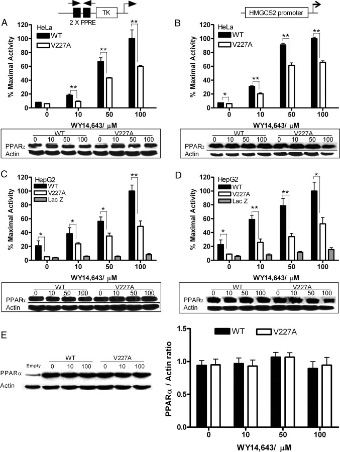
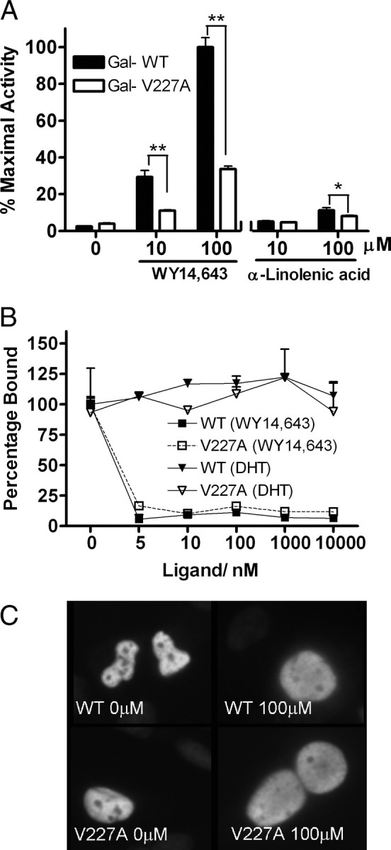
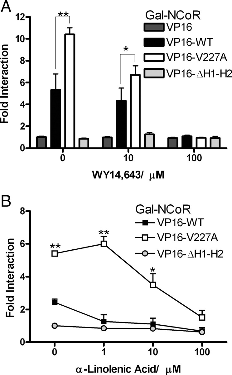
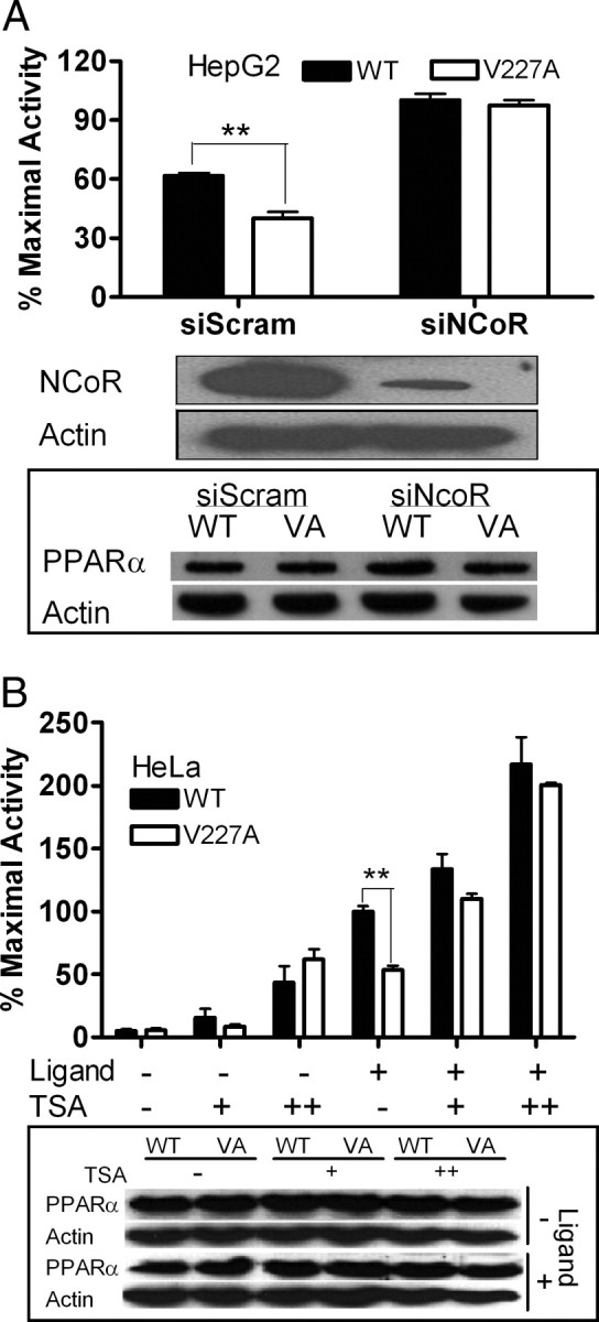
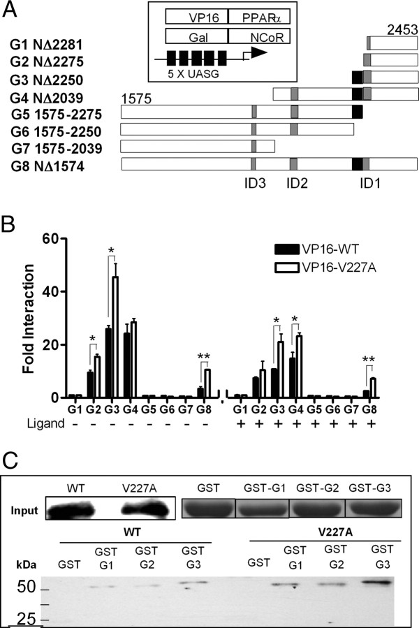
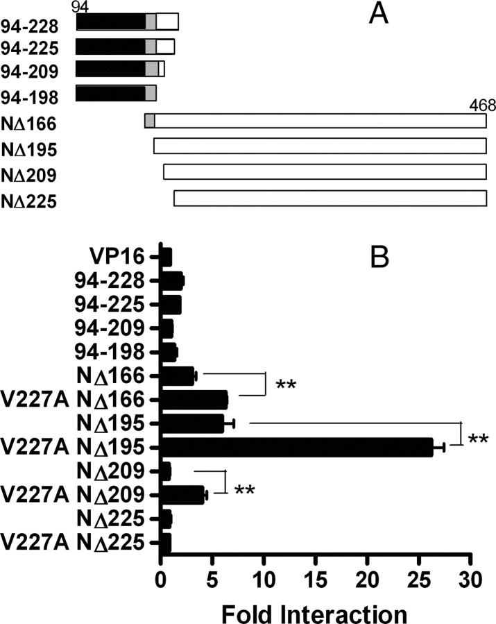
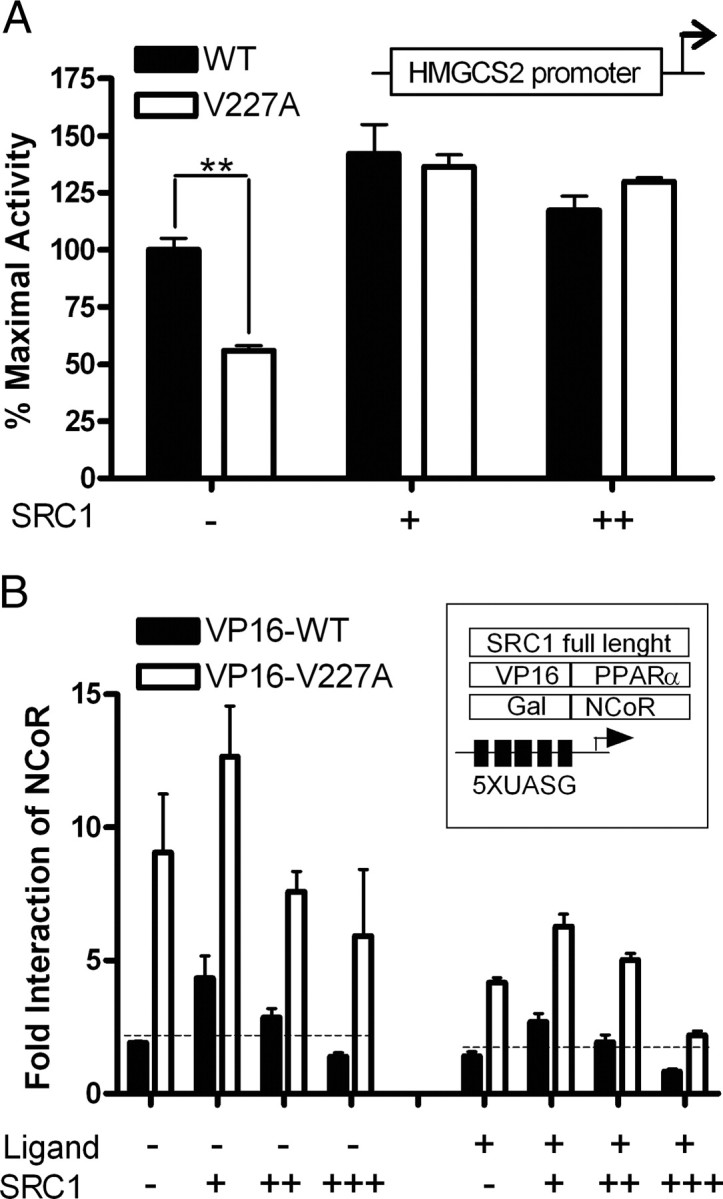
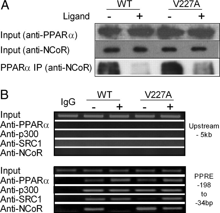
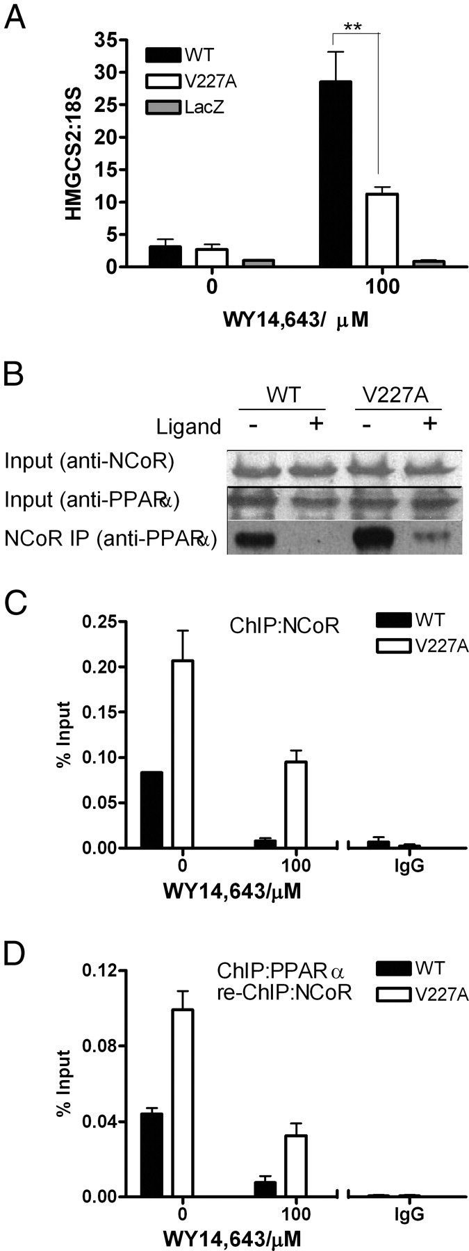
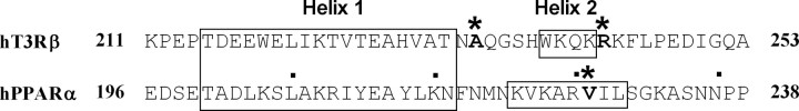
Peroxisome proliferator-activated receptor-alpha (PPARalpha) is a central regulator of lipid metabolism. Fibrate drugs act on PPARalpha to modulate dyslipidemias. A natural variant (V227A) affecting the PPARalpha hinge region was associated with perturbations in blood lipid levels in Asian populations. In this study, we investigated the functional significance of the V227A substitution. The variant significantly attenuated PPARalpha-mediated transactivation of the cytochrome P450 4A6 and mitochondrial 3-hydroxy-3-methylglutaryl-CoA synthase (HMGCS2) genes in the presence of fibrate ligands. Screening of a panel of PPARalpha coregulators revealed that V227A enhanced recruitment of the nuclear corepressor NCoR. Transactivation activity of V227A could be restored by silencing NCoR or by inhibition of its histone deacetylase activity. Deletion studies indicated that PPARalpha interacted with NCoR receptor-interacting domain 1 (ID1) but not ID2 or ID3. These interactions were dependent on the intact consensus nonapeptide nuclear receptor interaction motif in NCoR ID1 and were enhanced by the adjacent 24 N-terminal residues. Novel corepressor interaction determinants involving PPARalpha helices 1 and 2 were identified. In hepatic cells, the V227A substitution stabilized PPARalpha/NCoR interactions and caused defective release of NCoR in the presence of agonists on the HMGCS2 promoter. These results provide the first indication that defective function of a natural PPARalpha variant was due, at least partially, to increased corepressor binding. Our data suggest that the PPARalpha/NCoR interaction is physiologically relevant and can produce a discernable phenotype when the magnitude of the interaction is altered by a naturally occurring variation.


Figures

Fig. 1.
Fig. 1.
Transactivation Activity of PPARα V227A Variant on a Consensus CYP4A6-PPRE and the Mitochondria HMGCS2 Promoter HeLa cells were cotransfected with reporter vector CYP4A6-PPRE-Luc (100 ng) (A) or a reporter construct driven by residues −1081 to +22 of the HMGCS2 promoter (100 ng) (B) and full-length WT or V227A PPARα (50 ng) before treatment with indicated doses of WY14,643. HepG2 cells were infected with adenovirus expressing WT PPARα, V227A, or LacZ before transfection with CYP4A6-PPRE-Luc (100 ng) (C) or HMGCS2-Luc (100 ng) (D) and treatment with WY14,643 as above. PPARα and actin protein levels from total protein lysates (20–25 μg) of representative replicates were detected with specific antibodies. Values are mean ± sd of three replicates and expressed as percentage of maximal WT activity. *, P < 0.05; **, P < 0.01. E, Western blot for PPARα protein content. HepG2 cells were transfected with empty vector and 50 ng of WT or V227A PPARα, treated with increasing doses of WY14,643, and harvested 48 h after treatment. PPARα protein was detected with specific antibody and quantified using ScionImage analyzer. PPARα expression was normalized with actin and expressed as the ratio of PPARα and actin. Values are mean ± sd of eight replicates done on different occasions. TK, Thymidine kinase basal promoter.
Fig. 2.
Fig. 2.
Effect of V227A Variant on LBD Function, Ligand Binding, and Nuclear Localization A, HeLa cells were cotransfected with chimeric Gal-PPARαWT-LBD or Gal-PPARαV227A-LBD (50 ng) together with 100 ng (UASG)5-Luc reporter before treatment with ligands in the mammalian one-hybrid assay. Values are mean ± sd of three replicates and expressed as percentage of maximal WT activity. *, P < 0.05; **, P < 0.01. B, Ligand-binding affinity. WT or V227A PPARα in HeLa cells was exposed to 3 nm [3H]WY14,643 alone or with increasing concentrations of unlabeled WY14,643. The control was DHT. The amount of [3H]WY14,643 specifically bound after 24 h incubation was measured and expressed as a percentage relative to controls not exposed to cold hormone. C, Subcellular localization of PPARα proteins. Full-length WT or V227A PPARα (50 ng) was transfected into HepG2 cells before treatment with WY14,643. The resulting cells were stained with PPARα polyclonal antibody and goat antirabbit antibody conjugated with FITC. Nucleus was stained with 4′,6-diamidino-2-phenylindole.
Fig. 3.
Fig. 3.
Interactions of NCoR in the Mammalian Two-Hybrid Assay HeLa (A) and HepG2 (B) cells were cotransfected with chimeric VP16-WT or VP16-V227A (100 ng) and Gal-NCoR (100 ng) together with (UASG)5-Luc reporter (500 ng). VP16-ΔH1-H2 (100 ng) was used as a negative control. Cells were exposed to indicated doses of WY14,643 (A) or α-linolenic acid (B) for 48 h. Interaction was expressed as fold (mean ± sd of three replicates) of VP16 alone. *, P < 0.05; **, P < 0.01.
Fig. 4.
Fig. 4.
Effect of NCoR Silencing and HDAC Inhibition on Transcriptional Activity of V227A A, NCoR silencing. HepG2 cells were transiently infected by pLenti6-GW/U6-scrambleshRNA (siScram) or pLenti6-GW/U6-NCoRshRNA (siNCoR) and cotransfected with either full-length WT or V227A (VA) PPARα (50 ng) and reporter vector CYP4A6-PPRE-Luc (100 ng) before exposure to WY14,643 (100 μm). Cell lysates were immunoblotted with antibodies to NCoR, PPARα, and actin to evaluate effects of NCoR knockdown. B, Effects of HDAC inhibitor. HeLa cells overexpressing WT or V227A PPARα (50 ng) together with CYP4A6-PPRE-Luc (100ng) were exposed to an increasing dose of TSA (100 and 200 nm) in the absence or presence of WY14,643 (100 μm). Cell lysates were immunoblotted with antibodies to PPARα and actin to evaluate effects of TSA on protein content. Values are mean ± sd of three replicates and expressed as percentage of maximal WT activity. *, P < 0.05; **, P < 0.01.
Fig. 5.
Fig. 5.
Interactions of PPARα with Receptor ID of NCoR A, Schematic diagram of Gal-NCoR truncation fragments and the mammalian two-hybrid system. Gray boxes represent unique CoRNR box sequences within each receptor ID, residues 2277–2285, 2073–2081, and 1949–1953 for ID1, ID2, and ID3, respectively. Black boxes represents the first 24 amino acids immediately N-terminal of the ID1 CoRNR box. B, HeLa cells were cotransfected with 100 ng PPARα LBD chimeras, VP16-WT or VP-V227A (100 ng), and indicated C-terminal Gal-NCoR truncated fragments (100 ng) with the (UASG)5-Luc reporter gene (500 ng) in the absence or presence of WY14,643 (10 μm). Fold interaction (mean ± sd of three replicates) was expressed as fold of G1 at 0 μm. *, P < 0.05; **, P < 0.01. C, GST pulldown assay. GST-NCoR truncated fragments were incubated with equal amounts of in vitro translated full-length PPARα WT or V227A and GST pulldown performed using Sepharose 4B beads. Bound proteins were identified using anti-PPARα rabbit polyclonal antibody. Input shows the immunoblot for in vitro translated PPARα and the Coomassie blue staining of GST proteins expressed.
Fig. 6.
Fig. 6.
Interactions of NCoR with Deletion Fragments of PPARα Hinge A, Schematic diagram of VP16-PPARα truncation fragments. Black boxes represent DBD (residues 94–166); gray boxes represent residues 167–195 before helix 1. White boxes represent residues 196–468 of helix 1 to helix 12. B, WT or V227A PPARα VP16 truncated fragments (100 ng) were coexpressed with G3 (NΔ2250) (Fig. 6) (100 ng) and the (UASG)5-Luc reporter gene (500 ng) in the mammalian two-hybrid assay. Fold interaction (mean ± sd of three replicates) was expressed as fold of VP16 empty vector. *, P < 0.05; **, P < 0.01.
Fig. 7.
Fig. 7.
Ternary Interactions between SRC-1, NCoR, and PPAR A, SRC-1 overexpression. HeLa cells were cotransfected with either full-length WT or V227A PPARα (50 ng), a reporter construct driven by residues −1081 to +22 of the HMGCS2 promoter (100 ng) and increasing amounts of full-length SRC-1 (50 and 100 ng) with 100 μm WY 14,643. Values (mean ± se of three replicates) are expressed as percentage of maximal WT activity. B, Effects of SRC-1 on NCoR/PPARα interactions. HeLa cells were cotransfected with chimeric VP16-WT, VP16-V227A (100 ng), and Gal-NCoR (100 ng) together with the (UASG)5-Luc reporter (500 ng). Cells were exposed to WY14,643 (10 μm) at increasing doses (50, 100, and 200 ng) of full-length SRC-1. Fold interaction (mean ± sd of three replicates) was expressed as fold of VP16 empty vector. *, P < 0.05; **, P < 0.01.
Fig. 8.
Fig. 8.
Effect of V227A on the Recruitment of Coregulators to the Mitochondria HMG-CoA Promoter A, Coimmunoprecipitation. HepG2 cells transfected with full-length PPARα-WT or V227A (24 μg) were incubated with anti-PPARα rabbit monoclonal antibody in the absence or presence of 100 μm WY14,643. Precipitates were probed with anti-NCoR rabbit polyclonal antibody. Inputs were 5 and 10% cell lysate Western blotted for PPARα and NCoR expression, respectively. B, ChIP. HepG2 cells were transfected with 24 μg full-length PPARα-WT or V227A, with or without 100 μm WY14,643. ChIP analysis was carried out using antibodies against PPARα, p300, SRC-1, or NCoR. PCR for the HMGCS2 PPRE were performed up to 33 cycles. Amplification (36 cycles) of a fragment 5 kb upstream of the PPRE served as control (upper panels). The input was a representative of soluble chromatin (1%) that was reverse cross-linked and amplified under the same PCR conditions.
Fig. 9.
Fig. 9.
Correlation of PPARα-Regulated Gene Expression with Recruitment of NCoR to PPRE in the HMGCS2 Promoter HepG2 cells were infected with adenovirus expressing WT PPARα, V227A, or empty LacZ vector and treated with, or without, WY14,643 for 24 h. A, HMGCS2 mRNA levels were measured by quantitative RT-PCR and normalized to 18S rRNA. Values are mean ± sd of three replicates and expressed in relative quantities to LacZ in the absence of ligand. B, Immunoprecipitation. Cells from representative replicates in A were incubated with anti-NCoR rabbit polyclonal antibody overnight, and precipitates were probed with anti-PPARα mouse monoclonal antibody. The input was a representative of soluble chromatin (1%) that was reverse cross-linked and amplified under the same PCR conditions. C, Recruitment of NCoR to HMGCS2 promoter. ChIP analyses were performed on parallel replicates depicted in A, using antibodies against NCoR or rabbit IgG. Real-time PCR was used to quantify amount of PPRE of the HMGCS2 promoter bound to NCoR. D, ChIP-re-ChIP experiments. ChIP experiments were carried out as in C, and PPARα ChIP complexes were eluted and subjected again to the ChIP procedure using NCoR antibody. Values are mean ± sd of three replicates.
Fig. 10.
Fig. 10.
Sequence Comparison of TRβ and PPARα Sequence alignment is shown of helices 1 and 2 of human TRβ (hTR3β) and PPARα (hPPARα). The V227A PPARα variants TRβ (A234, R243) mutants associated with resistance to thyroid hormones are marked with asterisks.

Similar articles

Cited by

References

    1. Michalik L, Auwerx J, Berger JP, Chatterjee VK, Glass CK, Gonzalez FJ, Grimaldi PA, Kadowaki T, Lazar MA, O’Rahilly S, Palmer CN, Plutzky J, Reddy JK, Spiegelman BM, Staels B, Wahli W 2006. International Union of Pharmacology. LXI. Peroxisome proliferator-activated receptors. Pharmacol Rev 58:726–741 - PubMed
    1. Lefebvre P, Chinetti G, Fruchart JC, Staels B 2006. Sorting out the roles of PPARα in energy metabolism and vascular homeostasis. J Clin Invest 116:571–580 - PMC - PubMed
    1. Desvergne B, Wahli W 1999. Peroxisome proliferator-activated receptors: nuclear control of metabolism. Endocr Rev 20:649–688 - PubMed
    1. Germain P, Staels B, Dacquet C, Spedding M, Laudet V 2006. Overview of nomenclature of nuclear receptors. Pharmacol Rev 58:685–704 - PubMed
    1. Krey G, Keller H, Mahfoudi A, Medin J, Ozato K, Dreyer C, Wahli W 1993. Xenopus peroxisome proliferator activated receptors: genomic organization, response element recognition, heterodimer formation with retinoid X receptor and activation by fatty acids. J Steroid Biochem Mol Biol 47:65–73 - PubMed

Publication types

MeSH terms