. 2008 Mar 1;180(5):3535-42.
doi: 10.4049/jimmunol.180.5.3535.

Dipeptidyl peptidase I-dependent neutrophil recruitment modulates the inflammatory response to Sendai virus infection

Affiliations

Dipeptidyl peptidase I-dependent neutrophil recruitment modulates the inflammatory response to Sendai virus infection

Antonina M Akk et al. J Immunol. .

Abstract

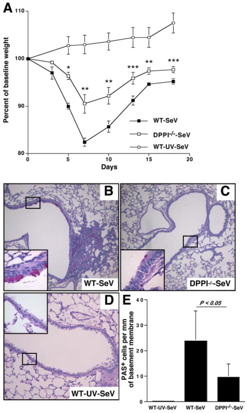
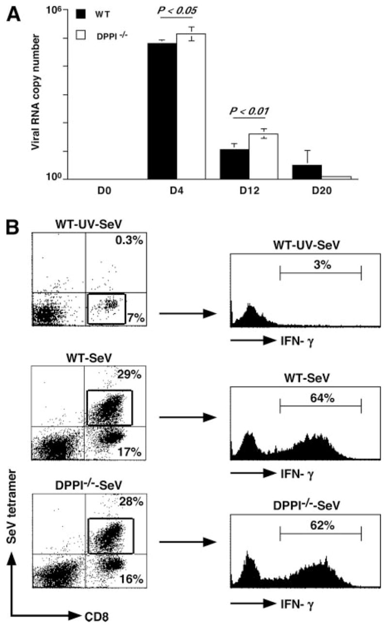
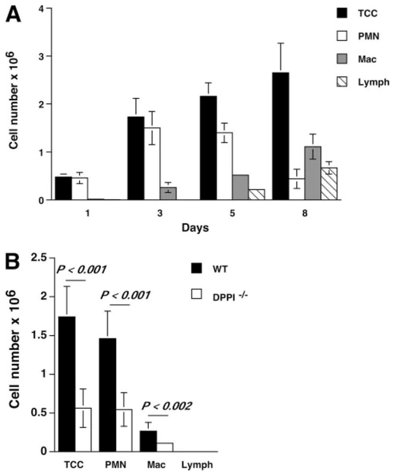
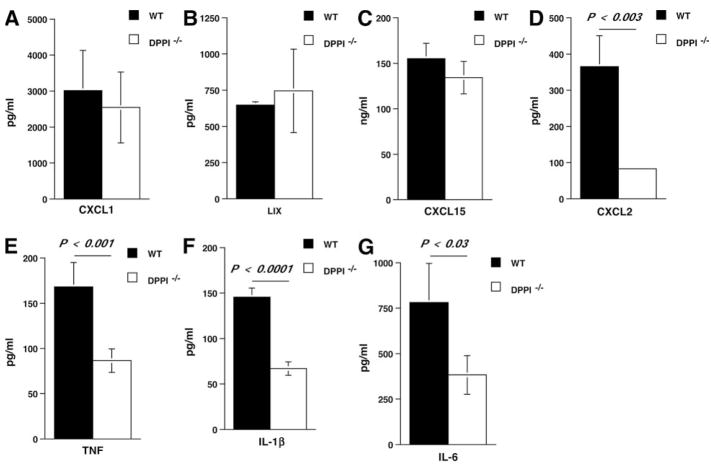
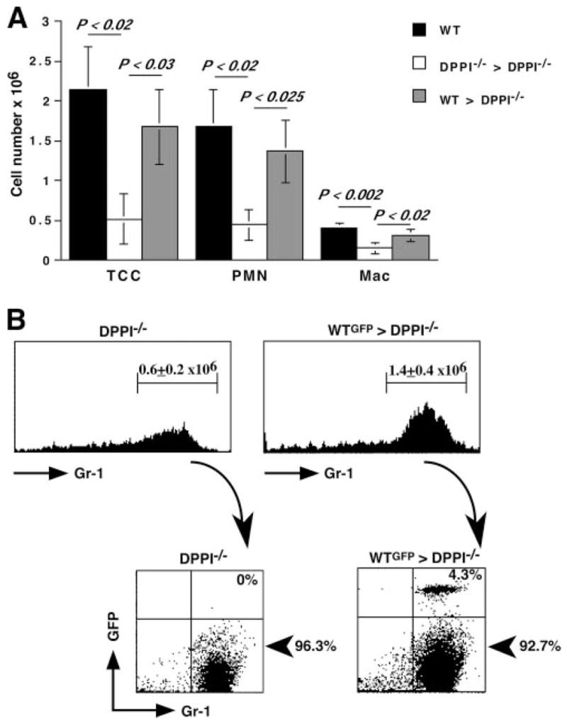
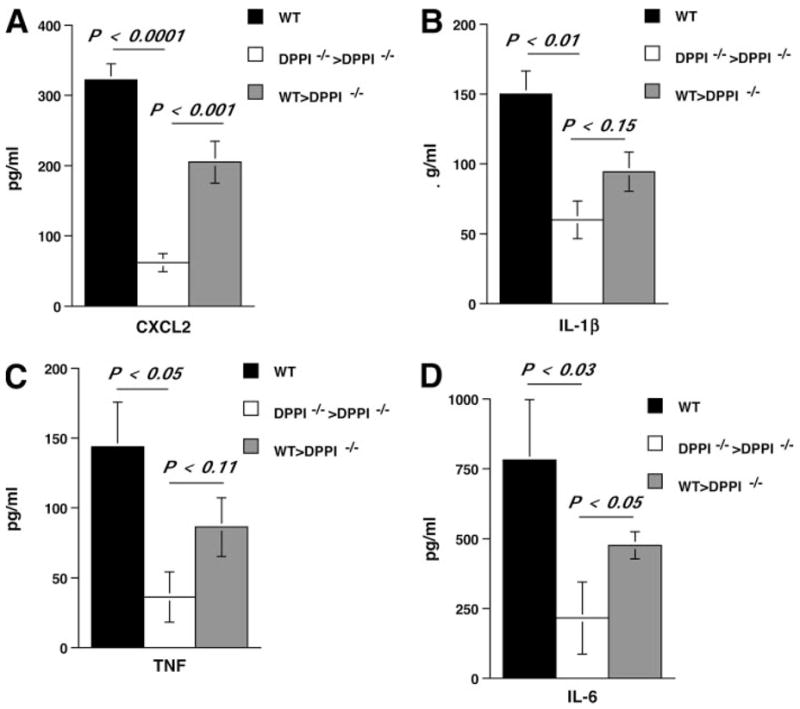
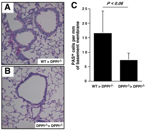
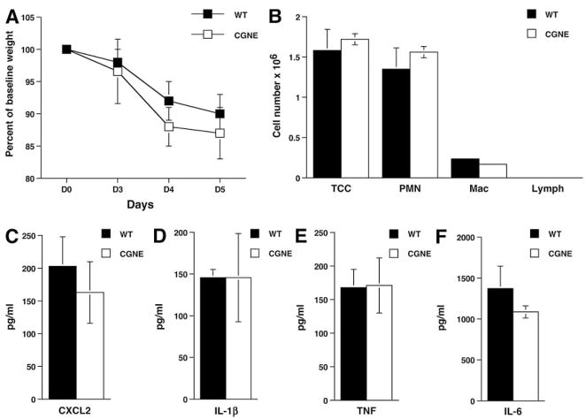
The role of innate immunity in the pathogenesis of asthma is unclear. Although increased presence of neutrophils is associated with persistent asthma and asthma exacerbations, how neutrophils participate in the pathogenesis of asthma remains controversial. In this study, we show that the absence of dipeptidyl peptidase I (DPPI), a lysosomal cysteine protease found in neutrophils, dampens the acute inflammatory response and the subsequent mucous cell metaplasia that accompanies the asthma phenotype induced by Sendai virus infection. This attenuated phenotype is accompanied by a significant decrease in the accumulation of neutrophils and the local production of CXCL2, TNF, IL-1beta, and IL-6 in the lung of infected DPPI-/- mice. Adoptive transfer of DPPI-sufficient neutrophils into DPPI-/- mice restored the levels of CXCL2 and enhanced cytokine production on day 4 postinfection and subsequent mucous cell metaplasia on day 21 postinfection. These results indicate that DPPI and neutrophils play a critical role in Sendai virus-induced asthma phenotype as a result of a DPPI-dependent neutrophil recruitment and cytokine response.


Figures

FIGURE 1
FIGURE 1
Absence of DPPI protects against SeV-induced severe weight loss and mucous cell hyperplasia. A, WT and DPPI−/− mice were infected with SeV, and their weight was followed over time. Data are presented as weight change from pretreatment baseline weight. Values represent mean ± SEM of five mice per condition from three independent experiments. Mice infected with UV-SeV did not lose weight. **, p < 0.001; **, p < 0.01; ***, p < 0.02. Persistence of peribronchial inflammation and mucous cell metaplasia on day 21 PI. Lung sections were stained with PAS, and representative photomicrographs are shown for SeV-infected WT mice (B), SeV-infected DPPI−/− mice (C), and UV-SeV-infected WT mice (D). Insets, Represent higher magnification of boxed areas. E, Quantification of results shown in B and C using values for PAS+ cells/mm basement membrane. Values represent mean ± SEM (n = 8–9 airways from 3 to 4 mice per condition).
FIGURE 2
FIGURE 2
Absence of DPPI delays viral clearance, but does not alter cytotoxic T cell response. A, Lungs were harvested on the indicated days after SeV infection, and viral RNA copy number was determined by real-time RT-PCR for SeV nucleocapsid protein and corrected for GAPDH control. Values represent mean ± SEM of four to five mice per genotype per time point (B). Cells isolated from post-BAL lung homogenates on day 8 PI were surface stained with SeV tetramers and anti-CD8 Abs, and their IFN-γ level was determined by intracellular staining. Note that there was a significant increase in the total number of CD8+ T cells recovered from SeV-infected lungs on day 8 PI compared with control mice (WT-SeV-UV mice = 0.15 ± 0.04 × 10⁶ cells), but no difference was detected between WT (1.58 ± 0.21 × 10⁶ cells) and DPPI−/− mice (1.71 ± 0.51 × 10⁶ cells). Values for CD8+ T cells represent mean ± SEM derived from n = 3–4 mice per condition.
FIGURE 3
FIGURE 3
Leukocyte recruitment to the alveolar space is defective in DPPI−/− mice after SeV infection. A, Groups of WT mice were infected with SeV, and their BAL fluid was subjected to total and differential cell counts. B, BAL fluid differential cell counts on SeV PI day 3. The absolute number of each cell type was calculated by multiplying the total number of cells from each lung by the percentage of positive staining cells. Values represent mean ± SEM of at least 10 mice per genotype. TCC, total cell count; PMN, neutrophil; Mac, macrophage; Lymph, lymphocyte.
FIGURE 4
FIGURE 4
Dampened chemokine/cytokine release in DPPI−/− mice following SeV infection. At day 3 PI, BAL fluid was obtained from each group of mice and analyzed for chemokines CXCL1 (A), LIX (B), CXCL15 (C), and CXCL2 (D), and cytokine IL-1β (F), TNF (E), and IL-6 (G) levels. Values represent mean ± SEM of four to five mice per genotype for LIX and CXCL15, and 11–12 mice per genotype for others.
FIGURE 5
FIGURE 5
Leukocyte recruitment into the alveolar space is dependent on WT neutrophils. A, Mice were infected with SeV on day 0, followed by i.v. transfer of 107 purified WT or DPPI−/− neutrophils on days 1, 2, and 3 PI. On day 4 PI, mice were sacrificed and their lungs were lavaged. Adoptive transfer of WT neutrophils into DPPI−/− mice after SeV infection led to an absolute increase in the number of cells recruited to the alveolar space. In contrast, transfer of DPPI−/− neutrophils failed to increase the number of cells localizing to the alveolar space. Values represent mean ± SEM (n = 4–5 mice per condition). B, To confirm that adoptively transferred neutrophils localized to the lungs, GFP+ WT neutrophils were purified and adoptively transferred into DPPI−/− mice on days 1, 2, and 3 PI. Twenty-four hours after the last transfer, mice were sacrificed and the cells recovered from their BAL fluid were analyzed by flow cytometry. Transfer of GFP+ WT neutrophils into DPPI−/− mice led to an increase in the total number of neutrophils that localized to the alveolar space, as revealed by flow cytometric analysis of Gr1-positive cells. Of these, only a small percentage of cells was from the donor pool, as revealed by the presence of GFP+ GR1+ cells. Representative flow cytometry plots are shown. The absolute number of neutrophils was calculated by multiplying the total number of cells obtained from the BAL fluid by the percentage of GR1+ cells. Values represent mean ± SEM of four mice per condition. TCC, total cell count; PMN, neutrophil.
FIGURE 6
FIGURE 6
Adoptive transfer of WT neutrophils restores CXCL2 levels. Mice were infected on day 0 and received 107 purified WT or DPPI−/− neutrophils i.v. on days 1, 2, and 3 PI. On day 4 PI, the mice were sacrificed and their lungs were lavaged. Adoptive transfer of WT neutrophils, but not DPPI−/− neutrophils, led to a significant increase in the levels of CXCL2 (A) and enhanced the levels of IL-1β (B), TNF (C), and IL-6 (D) in the alveolar space. Values represent mean ± SEM of three to four mice per condition.
FIGURE 7
FIGURE 7
WT neutrophils contribute to mucous cell metaplasia post-SeV infection. DPPI−/− mice were infected with SeV on day 0, followed by i.v. transfer of 107 WT or DPPI−/− neutrophils on days 1, 2, and 3 PI. On day 21 PI, mice were sacrificed and their lungs were harvested for analysis. Lung sections from DPPI−/− mice that received WT neutrophils showed an increase in the number of PAS+ cells (A) compared with mice that received DPPI−/− neutrophils (B), although this increase did not reach statistical significance (C). Values represent mean ± SEM of three mice per condition.
FIGURE 8
FIGURE 8
Absence of CG and NE does not protect mice against SeV-induced weight loss and proinflammatory chemokine/cytokine release. Mice deficient in both CG and NE (CGNE) had the same weight loss curve as WT mice infected with SeV (A) and released the same levels of chemokines and cytokines into the alveolar space on day 3 PI (BF). Values represent mean ± SEM of four mice per genotype. TCC, total cell count; PMN, neutrophil; Mac, macrophage; Lymph, lymphocyte.

References

    1. Robinson DS, Hamid Q, Ying S, Tsicopoulos A, Barkans J, Bentley AM, Corrigan C, Durham SR, Kay AB. Predominant TH2-like bronchoalveolar T-lymphocyte population in atopic asthma. N Engl J Med. 1992;326:298–304. - PubMed
    1. Humbert M, Durham SR, Ying S, Kimmitt P, Barkans J, Assoufi B, Pfister R, Menz G, Robinson DS, Kay AB, Corrigan CJ. IL-4 and IL-5 mRNA and protein in bronchial biopsies from patients with atopic and nonatopic asthma: evidence against “intrinsic” asthma being a distinct immunopathologic entity. Am J Respir Crit Care Med. 1996;154:1497–1504. - PubMed
    1. Temann UA, Prasad B, Gallup MW, Basbaum C, Ho SB, Flavell RA, Rankin JA. A novel role for murine IL-4 in vivo: induction of MUC5AC gene expression and mucin hypersecretion. Am J Respir Cell Mol Biol. 1997;16:471–478. - PubMed
    1. Webb DC, McKenzie AN, Koskinen AM, Yang M, Mattes J, Foster PS. Integrated signals between IL-13, IL-4, and IL-5 regulate airways hyperreactivity. J Immunol. 2000;165:108–113. - PubMed
    1. Temann UA, Geba GP, Rankin JA, Flavell RA. Expression of interleukin 9 in the lungs of transgenic mice causes airway inflammation, mast cell hyperplasia, and bronchial hyperresponsiveness. J Exp Med. 1998;188:1307–1320. - PMC - PubMed

Publication types

MeSH terms