Review
. 2008 Mar;9(3):206-18.
doi: 10.1038/nrm2346.

The Rpd3/Hda1 family of lysine deacetylases: from bacteria and yeast to mice and men

Affiliations
Review

The Rpd3/Hda1 family of lysine deacetylases: from bacteria and yeast to mice and men

Xiang-Jiao Yang et al. Nat Rev Mol Cell Biol. 2008 Mar.

Abstract

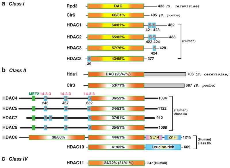
Protein lysine deacetylases have a pivotal role in numerous biological processes and can be divided into the Rpd3/Hda1 and sirtuin families, each having members in diverse organisms including prokaryotes. In vertebrates, the Rpd3/Hda1 family contains 11 members, traditionally referred to as histone deacetylases (HDAC) 1-11, which are further grouped into classes I, II and IV. Whereas most class I HDACs are subunits of multiprotein nuclear complexes that are crucial for transcriptional repression and epigenetic landscaping, class II members regulate cytoplasmic processes or function as signal transducers that shuttle between the cytoplasm and the nucleus. Little is known about class IV HDAC11, although its evolutionary conservation implies a fundamental role in various organisms.


Figures

Figure 1
Figure 1. Domain organization of classical HDACs from yeast and humans
The deacetylases are grouped into different classes according to sequence similarity to yeast prototypes. In mammals, class I members (Rpd3-like) include HDAC1, 2, 3 and 8; class II members are HDAC4, 5, 6, 7, 9 and 10 (similar to Hda1); and class IV is made up of HDAC11 (Fig. 1). Class II is further divided into two subclasses, IIa (HDAC4, 5, 7 and 9) and IIb (HDAC6 and 10). The total number of amino acid residues in each deacetylase is shown to the right of the figure. Many of the deacetylases have isoforms that result from alternative splicing. For simplicity, the number refers to the longest isoform. The deacetylase (DAC) domain is depicted by a rectangle containing a yellow (class I, (a)), clear (class II, (b)), or light green (class IV, (c)) centre flanked by orange shading, and the percentage amino acid sequence identity/similarity to that of Rpd3 (for class I) or Hda1 (class II/IV) is shown. The sequence identity/similarity of Hda1 and HDAC11 to Rpd3 is given in brackets. The C-terminal domains (shaded rectangles) of Hda1 and Clr3 are homologous (identity/similarity, 26/57%). Dark bars represent similar N-terminal domains and C-terminal tails of class IIa histone deacetylases (HDACs). Myocyte enhancer factor 2 (MEF2)-binding motifs are depicted as small green boxes, whereas 14-3-3 binding motifs are indicated by small boxes labelled with the letter “S” (for serine). SE14, Ser-Glu-containing tetradecapeptide repeats; ZnF, ubiquitin-binding zinc finger.
Figure 2
Figure 2. HDAC4 and homologues as signal transducers
a Multiple kinases (calcium/calmodulin-dependent protein kinase (CaMK), protein kinase D (PKD), microtubule affinity-regulating kinase (MARK), salt-inducible kinase (SIK), checkpoint kinase 1 (CHK1) and other kinases) mediate specific phosphorylation of human HDAC4 on three 14-3-3-binding sites. Myosin phosphatase-targeting subunit-1 (MYPT1)/protein phosphatase (PP)1β and PP2A can act on these sites. The association of 14-3-3 proteins with HDAC4 retains it in the cytoplasm and prevents its interaction with transcription factors such as myocyte enhancer factor 2 (MEF2), thereby releasing them for transcriptional activation. This model can also be extended to other class IIa members, although some of the kinases and phosphatases have only been shown for HDAC4, 5, 7 and/or 9 (see text for references). At the bottom of the panel, the sequence surronding the major 14-3-3 binding site (S246) of HDAC4 is compared with the corresponding regions of its paralogues (HDAC5, 7 and 9) as well as orthologues from D. melanogaster and C. elegans. The key residues crucial for phosphorylation and 14-3-3 binding are highlighted. This 14-3-3 site constitutes a sequence conservation island. The nuclear localization signal, which can be impeded by 14-3-3 binding, is also shown. b Signal-responsive transcriptional repression by HDAC4 (left) is analogous to transcriptional activation by transforming growth factor-β (TGFβ)-stimulated SMADs (middle) and cytokine-activated signal transducer and activator of transcription (STAT) proteins (right). 14-3-3 proteins bind to HDAC4 and retain it in the cytoplasm, thereby inhibiting its corepressor activity; upon dephosphorylation, HDAC4 translocates to the nucleus for transcriptional repression. The letter P circled in red denotes phosphorylation. For simplicity, only two 14-3-3 molecules are shown. This schematic also applies to HDAC5, 7 and 9.
Figure 3
Figure 3. HDAC6 is a major deacetylase in the cytoplasm
Through its tandem deacetylase (DAC) domains, HDAC6 deacetylates α-tubulin, cortactin and HSP90 to regulate cell motility, cilium assembly, maturation of the glucocorticoid receptor (GR), and activation of some protein kinases. HDAC6 may also deacetylate the type I interferon receptor (IFNαR). Additional cytoplasmic substrates are likely to be identified. Through its ubiquitin-binding zinc finger (ZnF), HDAC6 binds to ubiquitin and regulates aggresome formation, autophagy, activation of heat-shock factor 1 (HSF1), and epidermal growth factor receptor (EGFR)-induced macropinocytosis. It remains to be established whether HDAC6 has a direct role in the nucleus.

References

    1. Gershey EL, Vidali G, Allfrey VG. Chemical studies of histone acetylation. The occurrence of epsilon-N-acetyllysine in the f2a1 histone. J Biol Chem. 1968;243:5018–5022. - PubMed
    1. Inoue A, Fujimoto D. Enzymatic deacetylation of histone. Biochem Biophys Res Commun. 1969;36:146–150. - PubMed
    1. Candido EP, Reeves R, Davie JR. Sodium butyrate inhibits histone deacetylation in cultured cells. Cell. 1978;14:105–113. - PubMed
    1. Sealy L, Chalkley R. The effect of sodium butyrate on histone modification. Cell. 1978;14:115–121. - PubMed
    1. Vidali G, Boffa LC, Bradbury EM, Allfrey VG. Butyrate suppression of histone deacetylation leads to accumulation of multiacetylated forms of histones H3 and H4 and increased DNase I sensitivity of the associated DNA sequences. Proc Natl Acad Sci USA. 1978;75:2239–2243. - PMC - PubMed

Publication types

Substances

LinkOut - more resources