. 2008 Mar;118(3):879-93.
doi: 10.1172/JCI32865.

Curcumin prevents and reverses murine cardiac hypertrophy

Affiliations

Curcumin prevents and reverses murine cardiac hypertrophy

Hong-Liang Li et al. J Clin Invest. 2008 Mar.

Retraction in

  • Curcumin prevents and reverses murine cardiac hypertrophy.
    Li HL, Liu C, de Couto G, Ouzounian M, Sun M, Wang AB, Huang Y, He CW, Shi Y, Chen X, Nghiem MP, Liu Y, Chen M, Dawood F, Fukuoka M, Maekawa Y, Zhang L, Leask A, Ghosh AK, Kirshenbaum LA, Liu PP. Li HL, et al. J Clin Invest. 2009 Jul;119(7):2113. doi: 10.1172/jci32865r1. J Clin Invest. 2009. PMID: 19603550 Free PMC article. No abstract available.

Abstract

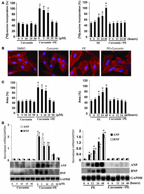
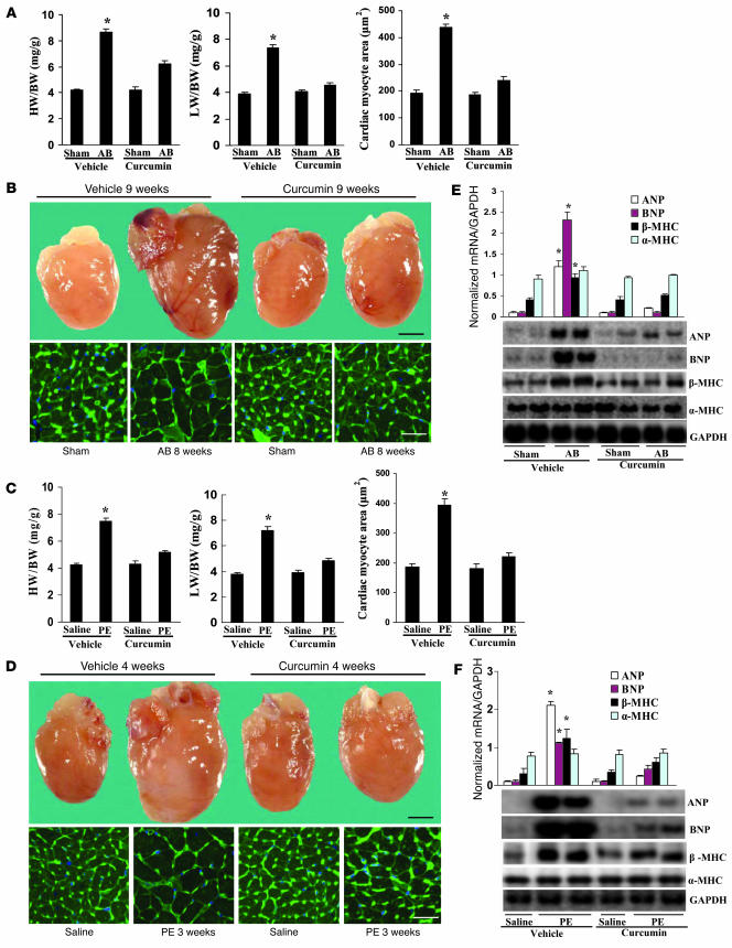
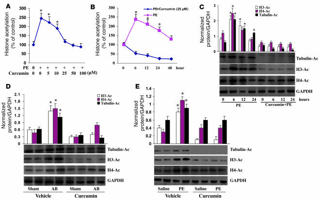
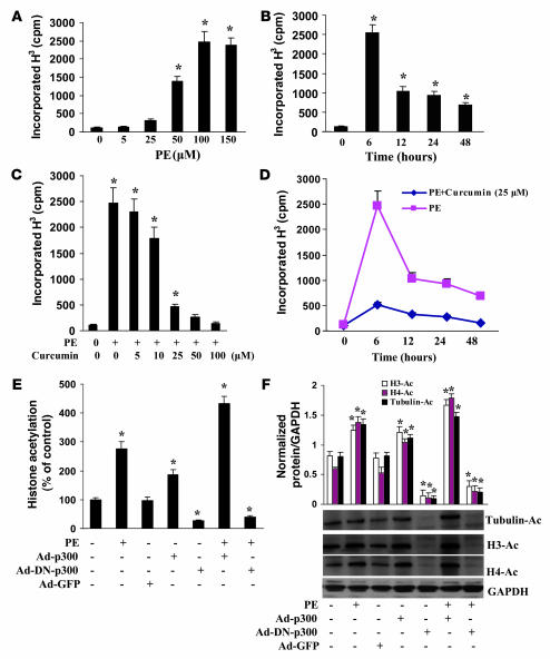
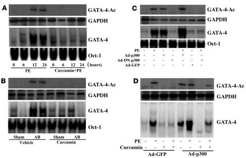
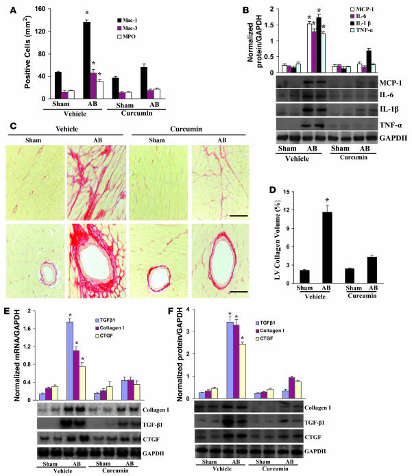
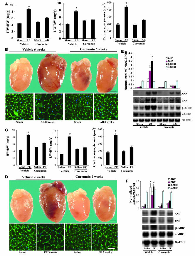
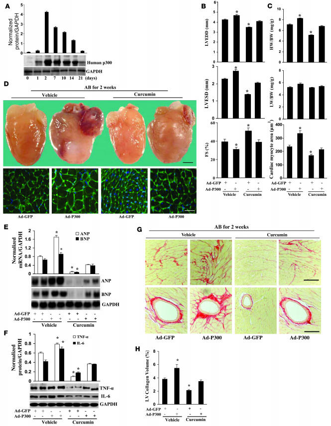
Chromatin remodeling, particularly histone acetylation, plays a critical role in the progression of pathological cardiac hypertrophy and heart failure. We hypothesized that curcumin, a natural polyphenolic compound abundant in the spice turmeric and a known suppressor of histone acetylation, would suppress cardiac hypertrophy through the disruption of p300 histone acetyltransferase-dependent (p300-HAT-dependent) transcriptional activation. We tested this hypothesis using primary cultured rat cardiac myocytes and fibroblasts as well as two well-established mouse models of cardiac hypertrophy. Curcumin blocked phenylephrin-induced (PE-induced) cardiac hypertrophy in vitro in a dose-dependent manner. Furthermore, curcumin both prevented and reversed mouse cardiac hypertrophy induced by aortic banding (AB) and PE infusion, as assessed by heart weight/BW and lung weight/BW ratios, echocardiographic parameters, and gene expression of hypertrophic markers. Further investigation demonstrated that curcumin abrogated histone acetylation, GATA4 acetylation, and DNA-binding activity through blocking p300-HAT activity. Curcumin also blocked AB-induced inflammation and fibrosis through disrupting p300-HAT-dependent signaling pathways. Our results indicate that curcumin has the potential to protect against cardiac hypertrophy, inflammation, and fibrosis through suppression of p300-HAT activity and downstream GATA4, NF-kappaB, and TGF-beta-Smad signaling pathways.


Figures

Figure 1
Figure 1. Pretreatment with curcumin inhibits cardiac hypertrophy in vitro.
(A) Curcumin inhibited PE-induced [3H]leucine incorporation. (B) Representative fields of cardiac myocytes stained with α-actinin. Original magnification, ×200. (C) Quantification of cell cross-sectional area from experiments shown in B by measuring 50 random cells. (D) Curcumin blunted PE-induced ANP and BNP mRNA expression levels by Northern blot. [3H]leucine incorporation and Northern blot were measured as described in Methods. The results were reproducible in 3 separate experiments. *P < 0.05 versus control.
Figure 2
Figure 2. Pretreatment with curcumin blunts cardiac hypertrophy in vivo.
(A and C) Statistical results of HW/BW ratio, LW/BW ratio, and myocyte cross-sectional areas (n = 200 cells per section) at 8 weeks after AB surgery (A; n = 6) or 3 weeks after PE infusion (C; n = 7). (B and D) Gross heart and WGA-FITC staining of sham and AB mice at 8 weeks after surgery (B) or 3 weeks of saline- and PE-infused mice (D) treated with or without curcumin. Scale bars: 20 mm (gross heart); 50 μm (WGA stain). (E and F) Analysis of hypertrophic markers (n = 4). Total RNA was isolated from hearts of mice of the indicated groups, and expression of transcripts for ANP, BNP, β-MHC, and α-MHC induced by AB (E) or PE infusion (F) was determined by Northern blot analysis. *P < 0.05 versus respective vehicle control.
Figure 3
Figure 3. Pretreatment with curcumin inhibits histone acetylation in vitro and in vivo.
(A and B) The dose and time courses of curcumin on the global acetylation of histones induced by PE. The results of 4 parallel experiments are shown. (C) Curcumin inhibited the acetylation of histone H3, histone H4, and tubulin. Experiments were performed in triplicate. The levels of histone H3 acetylation, histone H4 acetylation, and tubulin were quantified and normalized relative to GAPDH. *P < 0.05 versus control. (D and E) Curcumin blocked AB- and PE infusion–mediated acetylation of histone H3, histone H4, and tubulin in mice (n = 5). Densitometric quantification of acetyl-H3, acetyl-H4, and tubulin was normalized to GAPDH. *P < 0.05 versus respective vehicle control.
Figure 4
Figure 4. Pretreatment with curcumin blocks p300-HAT activity.
(A and B) The dose and time course of PE on HAT activity of p300. Cells were treated with different doses of PE (A) or with 100 μM PE for the indicated times (B) and then harvested and subjected to analysis of HAT activity as described in Methods. (C and D) Curcumin inhibited PE-induced p300-HAT activity. Cells were either pretreated for 60 minutes with different doses of curcumin and then incubated with 100 μM PE for 6 hours (C) or pretreated for 60 minutes with 25 μM curcumin and then incubated with 100 μM PE for different times up to 48 hours (D). (E and F) Effect of p300 on histone acetylation. Cells were infected with Ad-p300, Ad-DN-p300, or Ad-GFP for 24 hours and then treated with 100 μM PE for 6 hours. The global acetylation of histones (E) and the acetylation of histone H3, histone H4, and tubulin (F) were determined. Each assay was performed at least 3 times. *P < 0.05 versus control.
Figure 5
Figure 5. Pretreatment with curcumin blocks GATA4 activation.
(A and B) Curcumin blocked the acetylation and DNA-binding activity of GATA4 induced by PE infusion (A) or AB (B). n = 4. Oct-1 DNA-binding activity was used as a control. (C) Effect of p300 on the acetylation and DNA-binding activity of GATA4 induced by PE. Cells were infected with Ad-p300, Ad-DN-p300, or Ad-GFP for 24 hours and then treated with 100 μM PE for 24 hours. Extracts were assayed for GATA4 acetylation and DNA-binding activity. (D) p300 partially reversed the inhibitory effect of curcumin on the acetylation and DNA-binding activity of GATA4 induced by PE. Cells were infected with Ad-p300 or Ad-GFP for 24 hours, treated with 25 μM curcumin for 60 minutes, and then incubated with 100 μM PE for 24 hours. The results were reproducible in 3 separate experiments.
Figure 6
Figure 6. Pretreatment with curcumin inhibits inflammation and fibrosis induced by AB.
(A) Quantitative analysis revealed the population of MPO-, Mac-1–, and Mac-3–positive cells in the hearts of the indicated mice (n = 6). (B) Western blot analysis of TNF-α, IL-1β, IL-6, and MCP-1 protein expression in the myocardia obtained from the indicated mice (n = 6). Each assay was performed in triplicate. (C) PSR staining on histological sections of the LV was performed on each group 8 weeks after AB. Scale bars: 10 μm. (D) Fibrotic areas from histological sections were quantified using an image-analyzing system (n = 6). (E and F) Northern blot and Western blot analyses of TGF-β1, collagen I, and CTGF were performed to determine mRNA (E) and protein (F) expression levels in each group 8 weeks after AB (n = 3). GAPDH was used as the sample loading control. *P < 0.05 versus vehicle-treated sham control.
Figure 7
Figure 7. Curcumin ameliorates established cardiac hypertrophy.
(A and C) Statistical results of HW/BW ratio, LW/BW ratio, and myocyte cross-sectional areas (200 cells per section) at 8 weeks after AB surgery (A; n = 5) or 3 weeks after PE infusion (C; n = 6). (A) Mice began treatment with curcumin at 2 weeks after AB or sham surgery and then sacrificed 8 weeks later. (C) Mice began treatment with curcumin at 1 week after PE or saline infusion and were sacrificed at 3 weeks later. (B and D) Gross heart and WGA-FITC staining of mice 8 weeks after sham or AB surgery (B) or 3 weeks after saline or PE infusion (D) treated with or without curcumin. Scale bars: 20 mm (gross heart); 50 μm (WGA stain). (E and F) Analysis of hypertrophic markers (n = 4). Total RNA was isolated from hearts of the indicated mice, and expression of transcripts for ANP, BNP, β-MHC, and α-MHC induced by AB (E) or PE infusion (F) were determined by Northern blot analysis. *P < 0.05 versus respective vehicle control.
Figure 8
Figure 8. p300 partly reverses the inhibitory effects of curcumin on cardiac hypertrophy, inflammation, and fibrosis.
(A) Human p300 protein expression (n = 4). Western blot showing adenoviral-mediated expression of human p300 protein for up to 21 days compared with Ad-GFP. Adenovirus (2 × 10⁹ pfu) was injected into LV. Representative blots are shown. (B) Echocardiography results from 4 group mice at 2 weeks after AB surgery. (C) Statistical results of HW/BW ratio, LW/BW ratio, and myocyte cross-sectional areas (n = 5; 200 cells per section). (D) Gross heart and WGA staining of AB mice at 2 weeks after surgery infected with Ad-p300 or Ad-GFP. Scale bar: 20 mm (gross heart); 50 μm (WGA stain). (E) Expression of ANP and BNP in hearts isolated from each group (n = 4). Representative blots are shown. (F) p300 partly reversed the inhibitory effects of curcumin on the protein expression of TNF-α and IL-6 (n = 4). Representative blots are shown. (G) PSR staining of the LV from paraffin-embedded histological sections from each group. Scale bars: 10 μm. (H) Quantification of fibrotic area measured by an image-analyzing system (n = 5). *P < 0.05 versus Ad-GFP–infected vehicle control.

Comment in

  • Currying favor for the heart.
    Epstein JA. Epstein JA. J Clin Invest. 2008 Mar;118(3):850-2. doi: 10.1172/JCI34650. J Clin Invest. 2008. PMID: 18292806 Free PMC article.

References

    1. Berk B.C., Fujiwara K., Lehoux S. ECM remodeling in hypertensive heart disease. J. Clin. Invest. 2007;117:568–575. - PMC - PubMed
    1. Heineke J., Molkentin J.D. Regulation of cardiac hypertrophy by intracellular signaling pathways. Nat. Rev. Mol. Cell Biol. 2006;7:589–600. - PubMed
    1. Chen F., et al. Hop is an unusual homeobox gene that modulates cardiac development. Cell. 2002;110:713–723. - PubMed
    1. Backs J., Olson E.N. Control of cardiac growth by histone acetylation/deacetylation. Circ. Res. 2006;98:15–24. - PubMed
    1. Clayton A.L., Hazzalin C.A., Mahadevan L.C. Enhanced histone acetylation and transcription: a dynamic perspective. Mol Cell. 2006;23:289–296. - PubMed

Publication types

MeSH terms