. 2008 Mar;118(3):956-64.
doi: 10.1172/JCI34314.

ChREBP, but not LXRs, is required for the induction of glucose-regulated genes in mouse liver

Affiliations

ChREBP, but not LXRs, is required for the induction of glucose-regulated genes in mouse liver

Pierre-Damien Denechaud et al. J Clin Invest. 2008 Mar.

Abstract

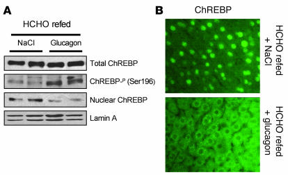
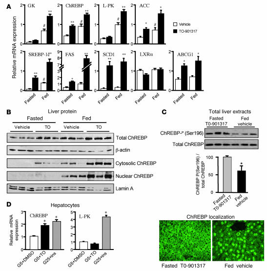
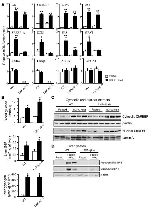
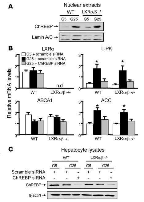
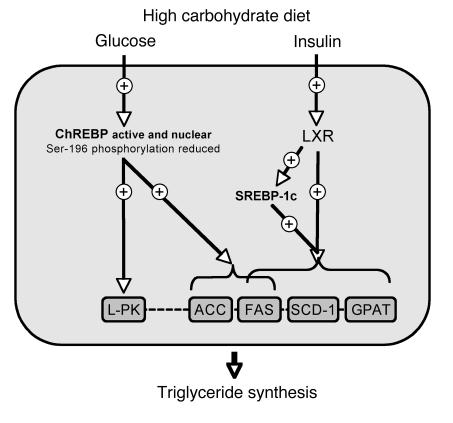
The transcription factor carbohydrate-responsive element-binding protein (ChREBP) has emerged as a central regulator of lipid synthesis in liver because it is required for glucose-induced expression of the glycolytic enzyme liver-pyruvate kinase (L-PK) and acts in synergy with SREBP to induce lipogenic genes such as acetyl-CoA carboxylase (ACC) and fatty acid synthase (FAS). Liver X receptors (LXRs) are also important regulators of the lipogenic pathway, and the recent finding that ChREBP is a direct target of LXRs and that glucose itself can bind and activate LXRs prompted us to study the role of LXRs in the induction of glucose-regulated genes in liver. Using an LXR agonist in wild-type mice, we found that LXR stimulation did not promote ChREBP phosphorylation or nuclear localization in the absence of an increased intrahepatic glucose flux. Furthermore, the induction of ChREBP, L-PK, and ACC by glucose or high-carbohydrate diet was similar in LXRalpha/beta knockout compared with wild-type mice, suggesting that the activation of these genes by glucose occurs by an LXR-independent mechanism. We used fluorescence resonance energy transfer analysis to demonstrate that glucose failed to promote the interaction of LXRalpha/beta with specific cofactors. Finally, siRNA silencing of ChREBP in LXRalpha/beta knockout hepatocytes abrogated glucose-induced expression of L-PK and ACC, further demonstrating the central role of ChREBP in glucose signaling. Taken together, our results demonstrate that glucose is required for ChREBP functional activity and that LXRs are not necessary for the induction of glucose-regulated genes in liver.


Figures

Figure 1
Figure 1. Glucagon injection in vivo promotes ChREBP nuclear delocalization and its phosphorylation on Ser196.
C57BL/6J mice were fasted overnight and refed on a HCHO diet for 18h. (A) Total, Ser196 phosphorylated, and nuclear ChREBP protein in liver extracts from HCHO-refed mice treated with either NaCl or glucagon (0.5 U/kg) for 30 min. Lamin A/C antibody was used as a loading control. (B) ChREBP immunofluorescence analysis in liver sections from HCHO refed mice treated with either NaCl or glucagon (0.5 U/kg) for 30 min. Original magnification, ×400. n = 6 per group. No signal was obtained when liver sections were incubated with the secondary antibody only (data not shown).
Figure 2
Figure 2. Differential regulation of ChREBP by LXRs and glucose.
(A) Quantitative real-time RT-PCR (qRT-PCR) analysis of GK, ChREBP, L-PK, ACC, SREBP-1c, FAS, SCD1, LXRα, and ABCG1 in livers of C57BL/6J mice treated for 3 days with either vehicle or 50 mg/kg body weight of the synthetic LXR agonist T0-901317. After treatment, mice were fasted overnight or maintained in the fed state. Results are mean ± SEM (n = 6 per group). #P < 0.005 vs. fasted; *P < 0.05, **P < 0.001 vs. vehicle. (B) Total, cytosolic, and nuclear ChREBP protein in liver extracts from vehicle- and T0-901317–treated fasted and fed mice. Lamin A/C and β-actin antibodies were used as loading controls. A representative Western blot is shown (n = 6 per group). (C) Ser196 phosphorylation level of the endogenous ChREBP protein. A representative Western blot is shown (n = 6 per group). Quantification of the ratio of Ser196 ChREBP phosphorylation to total ChREBP protein content is shown. *P < 0.05 vs. fasted T0-901317–treated. ChREBP immunofluorescence analysis in liver sections from T0-901317–treated fasted and vehicle-treated fed mice. Original magnification, ×400. n = 6 per group. (D) qRT-PCR analysis of ChREBP and L-PK in mouse hepatocytes incubated in the presence of low glucose (5 mM; G5) plus DMSO (white bars) or 10 μM T0-901317 (black bars) or in the presence of high glucose concentrations (25 mM) plus 100 nM insulin (gray bars) for 24 h. Error bars represent SD (n = 4 independent cultures). *P < 0.005 vs. 5 mM glucose plus DMSO.
Figure 3
Figure 3. Adequate response to glucose occurs in the absence of LXR.
(A) qRT-PCR analysis of GK, ChREBP, L-PK, ACC, SREBP-1c, FAS, SCD1, GPAT, LXRα, LXRβ, ABCG1, and ABCA1 in livers from wild-type and LXRα/β knockout mice either fasted overnight or challenged with a HCHO diet for 18 h. Error bars represent SD. n = 5–8 per group. (B) Blood glucose concentrations and liver G6P and glycogen content in wild-type and LXRα/β knockout mice either fasted or HCHO refed. *P < 0.05, **P < 0.001 versus fasted. n.d., not detectable. (C) Cytosolic and nuclear ChREBP protein in liver extracts from fasted and HCHO-refed wild-type and LXRα/β knockout mice. Lamin A/C and β-actin antibodies were used as loading controls. A representative Western blot is shown (n = 5–8 per group). Lanes were run on the same gel but were noncontiguous. (D) Precursor and mature SREBP-1 protein in liver lysates from fasted and HCHO-refed wild-type and LXRα/β knockout mice. β-Actin was used as a loading control. A representative Western blot is shown (n = 5–8 per group). Lanes were run on the same gel but were noncontiguous.
Figure 4
Figure 4. ChREBP, but not LXR, is required for glucose-regulated gene expression.
(A) ChREBP protein in nuclear extracts from wild-type and LXRα/β knockout hepatocytes. A representative Western blot is shown (n = 4 per group). (B) qRT-PCR analysis of L-PK, ACC, LXRα, and ABCA1 in isolated hepatocytes from wild-type and LXRα/β knockout mice. Hepatocytes from both genotypes were incubated under low glucose (5 mM) plus 100 nM insulin in the presence of a scramble siRNA or under high glucose concentrations (25 mM) plus 100 nM insulin in the presence of either scramble or ChREBP siRNA (8) for 24 h. Error bars represent SD (n = 4 independent cultures). *P < 0.005 vs. 5 mM glucose plus scramble siRNA and 25 mM glucose plus ChREBP siRNA groups. n.d., not detectable. (C) Total ChREBP protein in lysates from wild-type and LXRα/β knockout murine hepatocytes transfected with either scramble or ChREBP siRNA. A representative Western blot is shown. n = 4 independent cultures.
Figure 5
Figure 5. Respective roles of LXR, ChREBP, and SREBP-1c in the transcriptional control of TG synthesis in liver.
The glucose-mediated activation (Ser196 dephosphorylation and nuclear translocation) of ChREBP is required for the transcriptional induction of L-PK and ACC. FAS gene expression is synergistically regulated by ChREBP, LXR, and SREBP-1c. SCD1 and GPAT are under the transcriptional control of SREBP-1c and/or LXRs. While LXRs play a central role in insulin signaling through the transcriptional control of SREBP-1c, we could not find any evidence for direct involvement of LXRs in the glucose signaling pathway through ChREBP activation.

Comment in

References

    1. Decaux J.F., Antoine B., Kahn A. Regulation of the expression of the L-type pyruvate kinase gene in adult rat hepatocytes in primary culture. J. Biol. Chem. 1989;264:11584–11590. - PubMed
    1. Dentin R., Girard J., Postic C. Carbohydrate responsive element binding protein (ChREBP) and sterol regulatory element binding protein-1c (SREBP-1c): two key regulators of glucose metabolism and lipid synthesis in liver. Biochimie. 2005;87:81–86. - PubMed
    1. Dentin R., et al. Liver-specific inhibition of ChREBP improves hepatic steatosis and insulin resistance in ob/ob mice. Diabetes. 2006;55:2159–2170. - PubMed
    1. Dentin R., et al. Polyunsaturated fatty acids suppress glycolytic and lipogenic genes through the inhibition of ChREBP nuclear protein translocation. J. Clin. Invest. 2005;115:2843–2854. - PMC - PubMed
    1. Kabashima T., Kawaguchi T., Wadzinski B.E., Uyeda K. Xylulose 5-phosphate mediates glucose-induced lipogenesis by xylulose 5-phosphate-activated protein phosphatase in rat liver. Proc. Natl. Acad. Sci. U. S. A. 2003;100:5107–5112. - PMC - PubMed

Publication types