. 2008 Mar;118(3):1123-31.
doi: 10.1172/JCI34134.

A critical role for extracellular protein disulfide isomerase during thrombus formation in mice

Affiliations

A critical role for extracellular protein disulfide isomerase during thrombus formation in mice

Jaehyung Cho et al. J Clin Invest. 2008 Mar.

Abstract

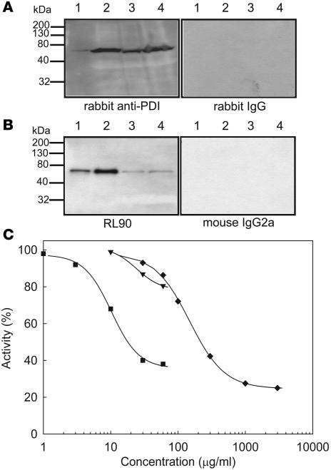
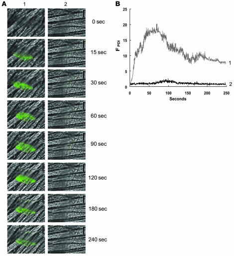
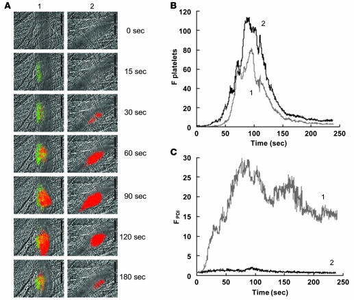
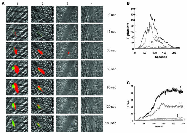
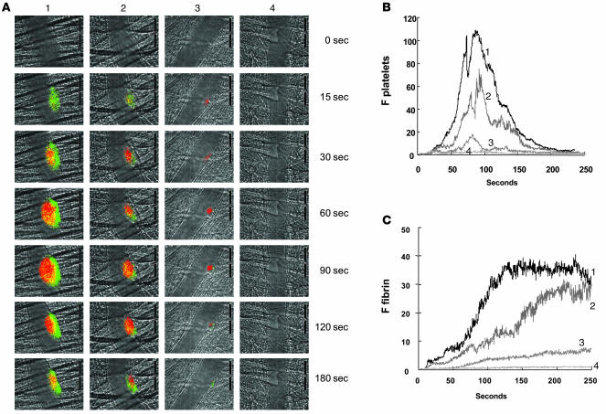
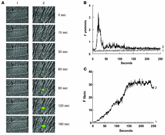
Thiol isomerases, including protein disulfide isomerase (PDI), catalyze disulfide oxidation, reduction, and isomerization, thereby playing an important role in protein synthesis. To determine whether extracellular PDI mediates thrombus formation in an animal model, PDI expression, platelet accumulation, and fibrin generation were monitored in the blood vessels of mice by intravital fluorescence microscopy following laser-induced arteriolar injury. A time-dependent increase in PDI was observed in murine thrombi following injury. Infusion of the PDI inhibitor bacitracin or a blocking monoclonal antibody against PDI inhibited platelet thrombus formation and fibrin generation. Fibrin deposition is normal in mice lacking the G protein-coupled platelet receptor Par4, although there is no stable accumulation of platelets. Infusion of monoclonal antibodies against PDI into the circulation of Par4(-/-) mice prior to vessel wall injury inhibited fibrin generation. These results indicate that PDI is required in vivo in mice for both fibrin generation and platelet thrombus formation.


Figures

Figure 1
Figure 1. Interaction of anti-PDI antibodies with human and mouse PDI.
Lysates of human (1 × 10⁹ platelets/ml) and mouse (1 × 10¹⁰ platelets/ml) platelets were subjected to SDS gel electrophoresis followed by immunoblotting with anti-PDI antibodies. (A) Polyclonal anti-PDI antibodies. Lane 1, recombinant human PDI (20 ng); lane 2, human platelet lysate (20 μl); lane 3, mouse platelet lysate (5 μl); lane 4, mouse platelet lysate (20 μl). Left: Immunoblot developed using affinity-purified rabbit anti-bovine PDI antibody. Right: Immunoblot developed with control nonimmune IgG. (B) Monoclonal antibody RL90 to PDI. Lane 1, recombinant human PDI (1 ng); lane 2, human platelet lysate (1 μl); lane 3, mouse platelet lysate (5 μl); lane 4, mouse platelet lysate (20 μl). Left: Immunoblot developed using the RL90 anti-PDI monoclonal antibody. Right: Immunoblot developed with control IgG2a monoclonal antibody. (C) PDI activity was measured with the insulin transhydrogenase assay in the releasate of thrombin-activated mouse platelets (n = 3) in the presence or absence of 1–60 μg/ml RL90 monoclonal anti-PDI antibody (squares), 10–60 μg/ml rabbit anti-PDI antibody (inverted triangles), or 0.03–3.0 mg/ml bacitracin A (diamonds). Results are shown as a mean of 2 experiments.
Figure 2
Figure 2. Expression of PDI during thrombus formation in vivo.
A nonblocking polyclonal anti-PDI antibody conjugated to Alexa Fluor 488 or an irrelevant IgG conjugated to Alexa Fluor 488 (0.3 μg/g BW) was infused into a mouse 5 min prior to arteriolar injury. (A) Time course of appearance of the anti-PDI antibody (column 1) or the irrelevant IgG (column 2) over 240 s following vessel wall injury. (B) Median integrated PDI fluorescence for 32 thrombi in 3 wild-type mice infused with the anti-PDI antibody (curve 1) and for 28 thrombi in 3 wild-type mice infused with the irrelevant IgG (curve 2) are presented versus time after vessel wall injury. A representative binarized image is shown in A, and the complete data sets of this and multiple identical experiments are presented in B, which plots the integrated fluorescence intensity of all pixels in the image, regardless of their intensity, as a function of time.
Figure 3
Figure 3. Comparison of PDI expression and platelet accumulation.
Rabbit polyclonal anti-PDI antibodies conjugated to Alexa Fluor 488 (0.3 μg/g BW) and Fab fragments of anti-CD41 antibodies conjugated to Alexa Fluor 647 (0.3 μg/g BW) were infused into a mouse 5 min prior to arteriolar injury. (A) Time course of appearance of the fluorescence signals associated with PDI (green) and platelets (red) over 180 s following laser-induced vessel wall injury in wild-type mice within the context of the bright-field microvascular histology (column 1). In control experiments, anti-PDI antibodies were replaced with nonimmune IgG conjugated to Alexa Fluor 488 (column 2). (B and C) Median integrated platelet fluorescence (B) and median integrated PDI fluorescence (C) after infusion of rabbit affinity-purified anti-PDI antibodies (curve 1; 28 thrombi, 3 wild-type mice) or rabbit nonimmune IgG (curve 2; 32 thrombi, 3 wild-type mice) are presented versus time after vessel wall injury. A representative binarized image is shown in A, and the complete data sets of this and multiple identical experiments are presented in B and C, which plot the integrated fluorescence intensity of all pixels in the image, regardless of their intensity, as a function of time.
Figure 4
Figure 4. Inhibition of fibrin formation and platelet accumulation in wild-type mice as a function of increasing concentrations of bacitracin A.
(A) Fibrin was labeled with a fibrin-specific antibody conjugated to Alexa Fluor 488, and platelets were labeled with Fab fragments of anti-CD41 antibodies conjugated to Alexa Fluor 647. The time course of appearance of fluorescence associated with fibrin (green) and platelets (red) is shown over 180 s following laser-induced vessel wall injury in wild-type mice within the context of the bright-field microvascular histology. Bacitracin A was infused into the circulation 5 min prior to injury at 0, 0.3, 1.5, and 5.0 mg per mouse (columns 1–4, respectively). (B and C) Median integrated platelet fluorescence (B) and median integrated fibrin fluorescence (C) for thrombi formed in the presence of increasing doses of bacitracin A — 0 mg (curve 1; 24 thrombi, 3 mice), 0.3 mg (curve 2; 27 thrombi, 3 mice), 1.5 mg (curve 3; 22 thrombi, 3 mice), and 5.0 mg (curve 4; 20 thrombi, 3 mice) — are presented versus time after vessel wall injury. A representative binarized image is shown in A, and the complete data sets of this and multiple identical experiments are presented in B and C, which plot the integrated fluorescence intensity of all pixels in the image, regardless of their intensity, as a function of time.
Figure 5
Figure 5. Inhibition of fibrin formation and platelet accumulation in wild-type mice as a function of increasing concentrations of inhibitory RL90 antibody to PDI.
(A) Fibrin was labeled with a fibrin-specific antibody conjugated to Alexa Fluor 488, and platelets were labeled with Fab fragments of anti-CD41 antibodies conjugated to Alexa Fluor 647. The time course of appearance of fluorescence associated with fibrin (green) and platelets (red) is shown over 180 s following laser-induced vessel wall injury in wild-type mice within the context of the bright-field microvascular histology. Inhibitory monoclonal anti-PDI antibody RL90 was infused into the circulation 5 min prior to injury at 0, 0.3, 1.0, and 3.0 μg/g BW (columns 1–4, respectively). In all experiments when no inhibitory monoclonal anti-PDI antibody was infused, 3.0 μg/g BW of isotype-matched IgG2a was infused instead. (B and C) Median integrated platelet fluorescence (B) and median integrated fibrin fluorescence (C) for thrombi formed in the presence of increasing concentrations of the anti-PDI antibody RL90 — 0 μg (curve 1; 27 thrombi, 3 mice), 0.3 μg (curve 2; 28 thrombi, 3 mice), 1.0 μg (curve 3; 23 thrombi, 3 mice), and 3.0 μg (curve 4; 21 thrombi, 3 mice) — are presented versus time after vessel wall injury. A representative binarized image is shown in A, and the complete data sets of this and multiple identical experiments are presented in B and C, which plot the integrated fluorescence intensity of all pixels in the image, regardless of their intensity, as a function of time.
Figure 6
Figure 6. Inhibition of fibrin formation by an inhibitory antibody to PDI in the Par4–/– mouse.
(A) Fibrin was labeled with a fibrin-specific antibody conjugated to Alexa Fluor 488, and platelets were labeled with Fab fragments of anti-CD41 antibodies conjugated to Alexa Fluor 647. The time course of appearance of fluorescence associated with fibrin (green) and platelets (red) is shown over 250 s following laser-induced vessel wall injury in Par4–/– mice within the context of the bright-field microvascular histology. Inhibitory monoclonal anti-PDI antibody RL90 (2 μg/g BW; column 1) or an irrelevant isotype matched IgG2a antibody (2 μg/g BW; column 2) was infused into the circulation 5 min prior to injury. (B and C) Median integrated platelet fluorescence (B) and median integrated fibrin fluorescence (C) for 24 thrombi in 3 Par4–/– mice infused with the irrelevant IgG2a antibody (curve 1) and for 20 thrombi in 3 Par4–/– mice infused with RL90 antibody (curve 2) are presented versus time after vessel wall injury. A representative binarized image is shown in A, and the complete data sets of this and multiple identical experiments are presented in B and C, which plot the integrated fluorescence intensity of all pixels in the image, regardless of their intensity, as a function of time.
Figure 7
Figure 7. Effect of PDI antibody on tail bleeding time.
(A) Tail bleeding times were determined as described in Methods following infusion of 0.3, 1.0, or 3 μg/g BW RL90 or 3 μg/g BW isotype-matched IgG2a control antibody in 7, 7, 8, and 6 mice, respectively. Data are expressed as the time to the cessation of blood flow for greater than 10 s; horizontal bars represent median tail bleeding time. The experiment was terminated at 15 min after tail cutting. (B) Blood loss during the bleeding time experiments was assayed by measuring the absorbance at 575 nm of hemoglobin (Hb) in phosphate-buffered saline in which tails were immersed. Horizontal bars represent the median of bleeding times based on the absorbance of hemoglobin for each group of animals. **P < 0.01 versus control, Dunn test after ANOVA (nonparametric).

References

    1. Chen V.M., Hogg P.J. Allosteric disulfide bonds in thrombosis and thrombolysis. J. Thromb. Haemost. 2006;4:2533–2541. - PubMed
    1. Chen K., Lin Y., Detwiler T.C. Protein disulfide isomerase activity is released by activated platelets. Blood. 1992;79:2226–2228. - PubMed
    1. Essex D.W., Chen K., Swiatkowska M. Localization of protein disulfide isomerase to the external surface of the platelet plasma membrane. Blood. 1995;86:2168–2173. - PubMed
    1. Thomas G., Skrinska V.A., Lucas F.V. The influence of glutathione and other thiols on human platelet aggregation. Thromb. Res. 1986;44:859–866. - PubMed
    1. Essex D.W., Li M. Redox control of platelet aggregation. Biochemistry. 2003;42:129–136. - PubMed

Publication types

Substances