. 2008 Apr;19(4):385-97.
doi: 10.1007/s00198-007-0543-5. Epub 2008 Feb 22.

FRAX and the assessment of fracture probability in men and women from the UK

Affiliations

FRAX and the assessment of fracture probability in men and women from the UK

J A Kanis et al. Osteoporos Int. 2008 Apr.

Abstract

A fracture risk assessment tool (FRAX) is developed based on the use of clinical risk factors with or without bone mineral density tests applied to the UK.

Introduction: The aim of this study was to apply an assessment tool for the prediction of fracture in men and women with the use of clinical risk factors (CRFs) for fracture with and without the use of femoral neck bone mineral density (BMD). The clinical risk factors, identified from previous meta-analyses, comprised body mass index (BMI, as a continuous variable), a prior history of fracture, a parental history of hip fracture, use of oral glucocorticoids, rheumatoid arthritis and other secondary causes of osteoporosis, current smoking, and alcohol intake 3 or more units daily.

Methods: Four models were constructed to compute fracture probabilities based on the epidemiology of fracture in the UK. The models comprised the ten-year probability of hip fracture, with and without femoral neck BMD, and the ten-year probability of a major osteoporotic fracture, with and without BMD. For each model fracture and death hazards were computed as continuous functions.

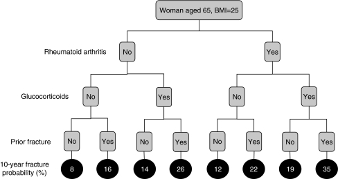
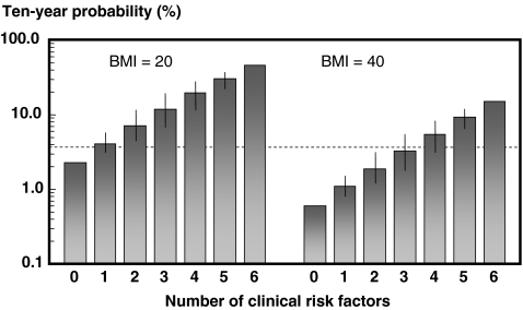
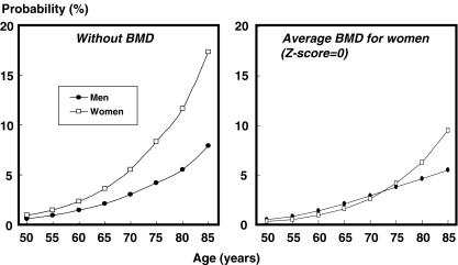
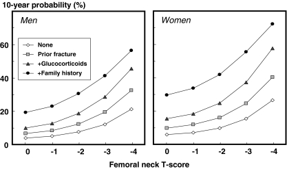
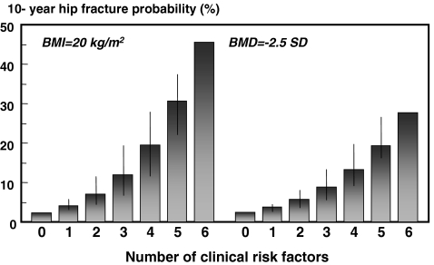
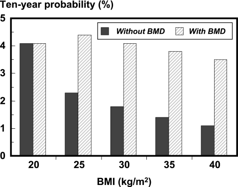
Results: Each clinical risk factor contributed to fracture probability. In the absence of BMD, hip fracture probability in women with a fixed BMI (25 kg/m(2)) ranged from 0.2% at the age of 50 years for women without CRF's to 22% at the age of 80 years with a parental history of hip fracture (approximately 100-fold range). In men, the probabilities were lower, as was the range (0.1 to 11% in the examples above). For a major osteoporotic fracture the probabilities ranged from 3.5% to 31% in women, and from 2.8% to 15% in men in the example above. The presence of one or more risk factors increased probabilities in an incremental manner. The differences in probabilities between men and women were comparable at any given T-score and age, except in the elderly where probabilities were higher in women than in men due to the higher mortality of the latter.

Conclusion: The models provide a framework which enhances the assessment of fracture risk in both men and women by the integration of clinical risk factors alone and/or in combination with BMD.


Figures

Fig. 1
Fig. 1
Input and output for the FRAX™ model
Fig. 2
Fig. 2
Effect of combinations of clinical risk factors on the 10-year probability of a major osteoporotic fracture in women aged 65 years and a BMI of 25 kg/m2. [05Ca201]
Fig. 3
Fig. 3
Ten-year probability of hip fracture (%) in women aged 65 years according to the number of clinical risk factors with a BMI of 20 kg/m2 (left-hand panels) and a BMI of 40 kg/m2 (right-hand panels). The intervals reflect the different weights afforded by different risk factors and provide a range of probabilities. [05Ca070]
Fig. 4
Fig. 4
Ten-year hip fracture probability (%) in men and women with a prior fracture according to age. The left-hand panel gives probabilities in the absence of BMD. In the right-hand panel probabilities are shown at an average BMD for women at each specific age (i.e., a Z-score = 0). A BMI is set at 24 kg/m2. [05Ca065]
Fig. 5
Fig. 5
10-year probability of a major osteoporotic fracture in men and women aged 65 years according to T-score and clinical risk factors. Body mass index is set at 25 kg/m2. [05Ca136]
Fig. 6
Fig. 6
Ten-year probability of hip fracture (%) in women aged 65 years, according to the number of clinical risk factors. The left-hand panel shows the probabilities without BMD at a BMI fixed at 20 kg/m2. The right-hand panel illustrates the effects with BMD fixed at the threshold for osteoporosis. [05Ca073]
Fig. 7
Fig. 7
Effect of variations in BMI on 10-year hip fracture probability (%) in women aged 65 years. Probabilities with BMD are computed at a T-score of −2.8 SD. [05Ca074]

References

    1. {'text': '', 'ref_index': 1, 'ids': [{'type': 'PubMed', 'value': '7941614', 'is_inner': True, 'url': 'https://pubmed.ncbi.nlm.nih.gov/7941614/'}]}
    2. World Health Organization (1994) Assessment of fracture risk and its application to screening for postmenopausal osteoporosis. WHO Technical Report Series 843. WHO, Geneva - PubMed
    1. {'text': '', 'ref_index': 1, 'ids': [{'type': 'PMC', 'value': 'PMC2351094', 'is_inner': False, 'url': 'https://pmc.ncbi.nlm.nih.gov/articles/PMC2351094/'}, {'type': 'PubMed', 'value': '8634613', 'is_inner': True, 'url': 'https://pubmed.ncbi.nlm.nih.gov/8634613/'}]}
    2. Marshall D, Johnell O, Wedel JJ (1996) Meta-analysis of how well measures of bone mineral density predict occurrence of osteoporotic fractures. Br Med J 312:1254–1259 - PMC - PubMed
    1. {'text': '', 'ref_index': 1, 'ids': [{'type': 'DOI', 'value': '10.1359/JBMR.050304', 'is_inner': False, 'url': 'https://doi.org/10.1359/jbmr.050304'}, {'type': 'PubMed', 'value': '15940371', 'is_inner': True, 'url': 'https://pubmed.ncbi.nlm.nih.gov/15940371/'}]}
    2. Johnell O, Kanis JA, Oden A, Johansson H et al (2005) Predictive value of bone mineral density for hip and other fractures. J Bone Miner Res 20:1185–1194 - PubMed
    1. {'text': '', 'ref_index': 1, 'ids': [{'type': 'DOI', 'value': '10.1016/S8756-3282(00)00381-1', 'is_inner': False, 'url': 'https://doi.org/10.1016/s8756-3282(00)00381-1'}, {'type': 'PubMed', 'value': '11062343', 'is_inner': True, 'url': 'https://pubmed.ncbi.nlm.nih.gov/11062343/'}]}
    2. Kanis JA, Johnell O, Oden A, Jönsson B, De Laet C, Dawson A (2000) Risk of hip fracture according to World Health Organization criteria for osteopenia and osteoporosis. Bone 27:585–590 - PubMed
    1. {'text': '', 'ref_index': 1, 'ids': [{'type': 'DOI', 'value': '10.1172/JCI113523', 'is_inner': False, 'url': 'https://doi.org/10.1172/jci113523'}, {'type': 'PMC', 'value': 'PMC442628', 'is_inner': False, 'url': 'https://pmc.ncbi.nlm.nih.gov/articles/PMC442628/'}, {'type': 'PubMed', 'value': '3384952', 'is_inner': True, 'url': 'https://pubmed.ncbi.nlm.nih.gov/3384952/'}]}
    2. Hui SL, Slemenda CW, Johnston CC (1988) Age and bone mass as predictors of fracture in a prospective study. J Clin Invest 81:1804–1809 - PMC - PubMed

Publication types