. 2008 Apr 1;316(1):74-86.
doi: 10.1016/j.ydbio.2008.01.011. Epub 2008 Jan 26.

Pdx-1 and Ptf1a concurrently determine fate specification of pancreatic multipotent progenitor cells

Affiliations

Pdx-1 and Ptf1a concurrently determine fate specification of pancreatic multipotent progenitor cells

Jared S Burlison et al. Dev Biol. .

Abstract

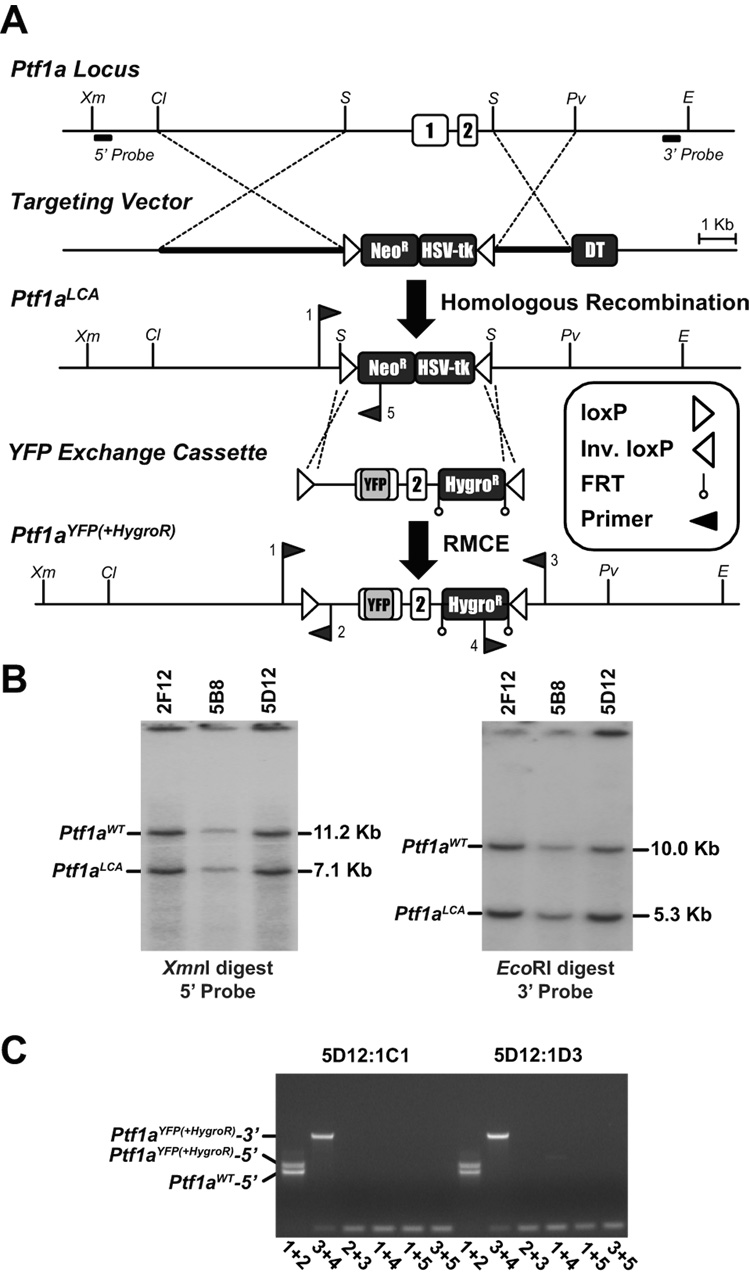
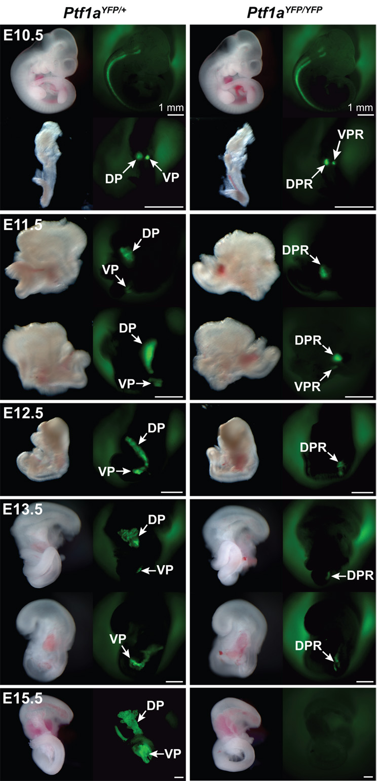
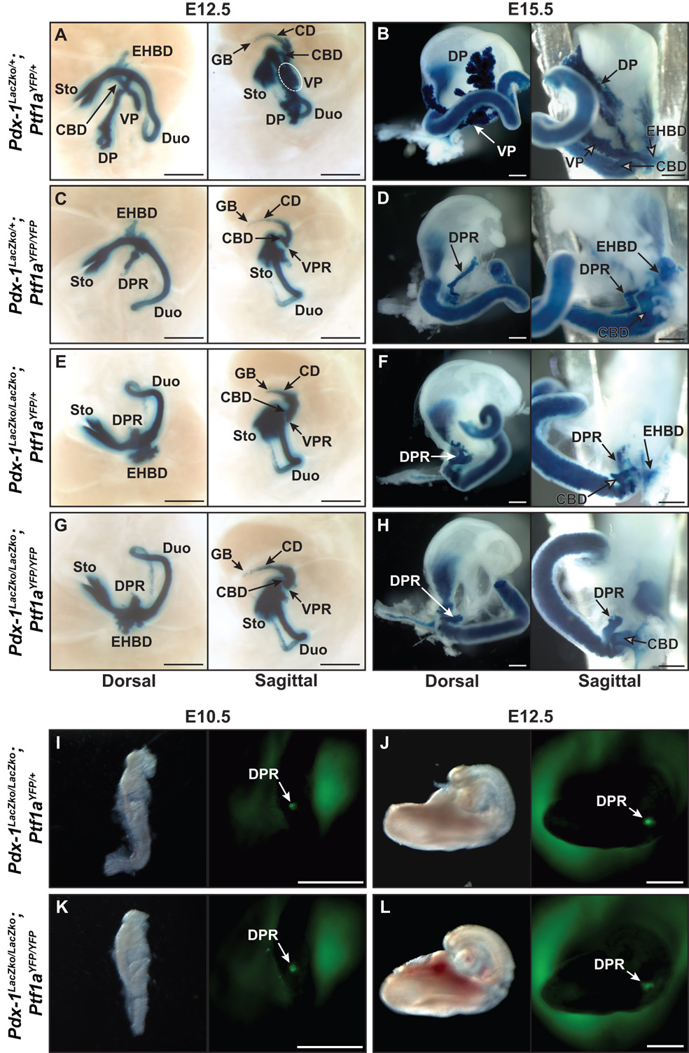
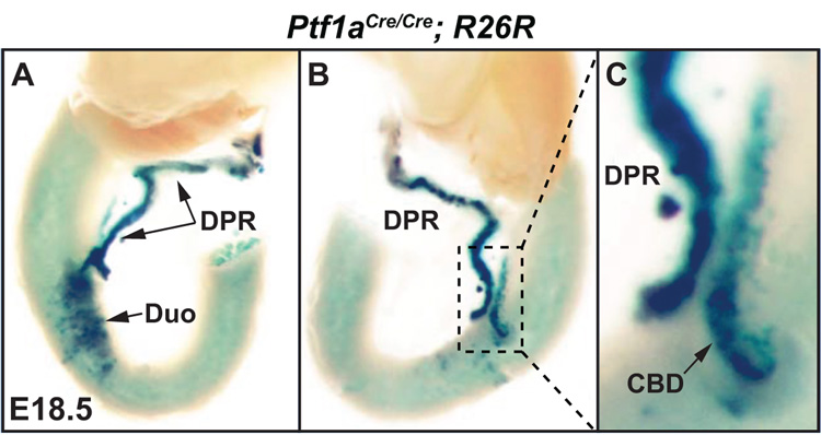
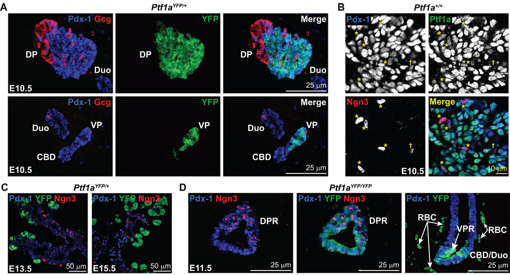
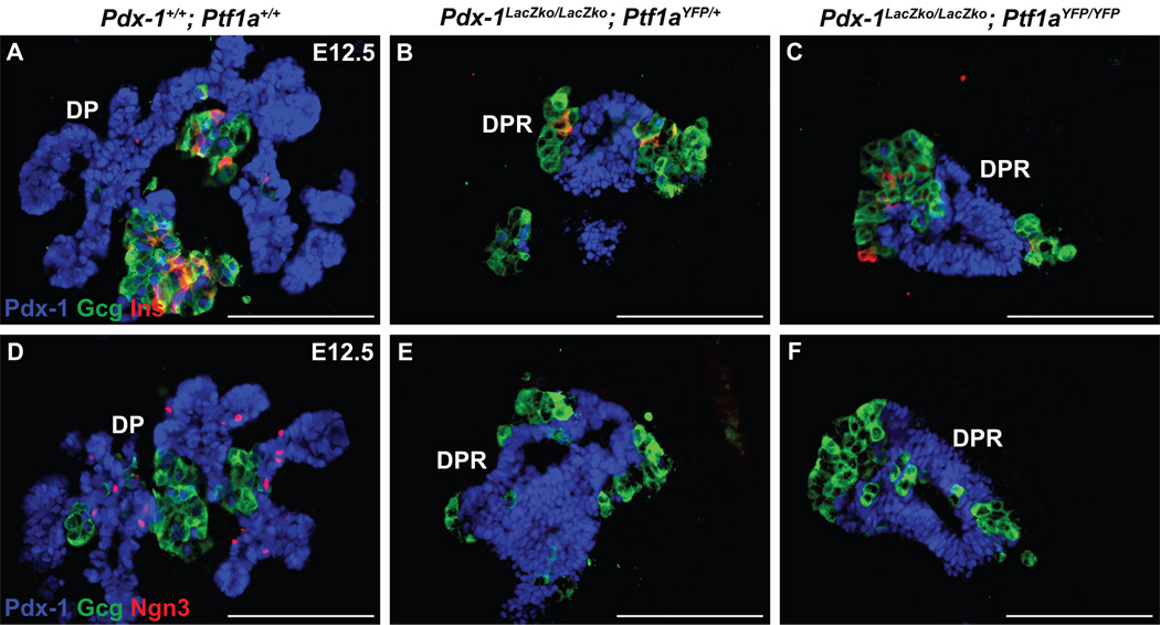
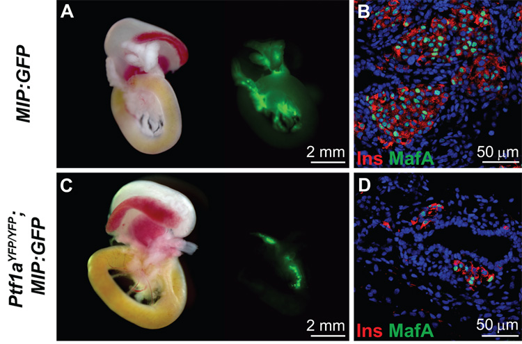
The pancreas is derived from a pool of multipotent progenitor cells (MPCs) that co-express Pdx-1 and Ptf1a. To more precisely define how the individual and combined loss of Pdx-1 and Ptf1a affects pancreatic MPC specification and differentiation we derived and studied mice bearing a novel Ptf1a(YFP) allele. While the expression of Pdx-1 and Ptf1a in pancreatic MPCs coincides between E9.5 and 12.5 the developmental phenotypes of Pdx-1 null and Pdx-1; Ptf1a double null mice are indistinguishable, and an early pancreatic bud is formed in both cases. This finding indicates that Pdx-1 is required in the foregut endoderm prior to Ptf1a for pancreatic MPC specification. We also found that Ptf1a is neither required for specification of Ngn3-positive endocrine progenitors nor differentiation of mature beta-cells. In the absence of Pdx-1 Ngn3-positive cells were not observed after E9.5. Thus, in contrast to the deletion of Ptf1a, the loss of Pdx-1 precludes the sustained Ngn3-based derivation of endocrine progenitors from pancreatic MPCs. Taken together, these studies indicate that Pdx-1 and Ptf1a have distinct but interdependent functions during pancreatic MPC specification.


Figures

Figure 1
Figure 1
Creation of a Ptf1aLCA allele and insertion of YFP by RMCE. A) Schematic representation of the Ptf1a locus, targeting vector, Ptf1aLCA allele, Ptf1a-YFP exchange cassette, and the Ptf1aYFP(+HygroR) allele. A 4.1 kb Ptf1a fragment including both exons was replaced by two pgk-driven selection cassettes, neoR and HSV-tk, flanked by inversely orientated loxP sites during gene targeting. A pgk-driven Diphtheria toxin A cassette is located beyond the end of the short arm of DNA homology. YFP coding sequences replace 5’ UTR and Ptf1a coding sequences in exon 1. Exon 2 is retained in the exchange cassette to supply RNA splicing and poly adenylation signals with a FRT-flanked hygromycin resistance cassette located downstream of Ptf1a sequences for positive selection following RMCE. Insertion of the YFP exchange cassette into the Ptf1aLCA was accomplished by RMCE creating the Ptf1aYFP(+HygroR) allele from which mice were derived. Restriction Endonucleases: ClaI (Cl), EcoRI (E), PvuII (Pv), SalI (S), XmnI (Xm). B) Southern blot analysis of ES cell genomic DNA, using probes indicated in A, confirmed 3 ES cell clones that were correctly targeted for the Ptf1aLCA allele, 2F12, 5B8, and 5D12. Clone 5D12 was chosen for the RMCE experiment. C) PCR screening of Ptf1aYFP(+HygroR) cassette exchanged clones using primer pair combinations that detect cassettes in the forward or reverse orientation on both the 5’ and 3’ ends. Clones 5D12:1C1 and 5D12:1D3 both contain the Ptf1aYFP(+HygroR) cassette in the forward orientation. Primer locations are indicated in A and combinations are indicated in C below their respective lanes.
Figure 2
Figure 2
YFP fluorescence in Ptf1aYFP/+ and Ptf1aYFP/YFP embryos. In Ptf1aYFP/+ embryos, YFP fluorescence can be detected in all sites of bona fide Ptf1a expression by whole mount fluorescence microscopy. Uniform YFP fluorescence is seen in the neural tube as well as in the pancreatic buds at E10.5, which expand and fuse by E12.5. At E13.5, YFP fluorescence is punctate which denotes acinar restriction. At E15.5, exocrine cells have proliferated which results in increased intensity of YFP fluorescence. In Ptf1aYFP/YFP mice, dorsal and ventral Ptf1a expression is initiated at E10.5, but growth of the pancreatic buds is severely retarded by E11.5. Ventral YFP fluorescence is lost by E12.5, but can be observed in the DPR until E13.5. However, by E15.5 YFP fluorescence is extinguished in Ptf1aYFP/YFP mice. Scale bars = 50 µm unless otherwise noted. Dorsal Pancreas (DP), Ventral Pancreas (VP), Dorsal Pancreatic Remnant (DPR), Ventral Pancreatic Remnant (VPR).
Figure 3
Figure 3
Pancreas morphogenesis in Pdx-1 null, Ptf1a null, and Pdx-1, Ptf1a double null embryos. A–H) Histological analysis of β-galactosidase expression from the Pdx-1LacZko allele in Pdx-1 null (Pdx-1LacZko/LacZko; Ptf1aYFP/+), Ptf1a null (Pdx-1LacZko/+; Ptf1aYFP/YFP), and Pdx-1, Ptf1a double null (Pdx-1LacZko/LacZko; Ptf1aYFP/YFP) embryos at E12.5 and E15.5. A) In Pdx-1LacZko/+; Ptf1aYFP/+ E12.5 embryos, the dorsal pancreas is associated with the duodenum, the ventral pancreas is evaginating from the CBD epithelium and the two lobes have fused near the duodenum at their proximal ends. B) At E15.5 in Ptf1aYFP/+; Pdx-1LacZko/+ embryos, normal epithelial branching and lobulation indicates acinar development. X-gal staining also denotes Pdx-1 expression in the CBD, extrahepatic biliary ducts, cystic duct, and gall bladder. C) In Ptf1a null embryos at E12.5, the epithelium of the DPR is growth deficient and does not branch while the VPR is barely discernable. D) In Ptf1a null embryos at E15.5, LacZ expression is observed in the DPR which is minimally branched and extends from the duodenum towards the spleen. The absence of a VPR indicates assimilation of ventral pancreatic MPCs into the CBD and/or duodenum epithelium. E) Pdx-1 null embryos at E12.5 have a considerably smaller DPR than in Ptf1a null embryos at the same stage while they display a similar structure resembling a VPR contiguous with the CBD. F) By E15.5 the DPR of Pdx-1LacZko/LacZko; Ptf1aYFP/+ embryos undergoes limited branching and X-gal staining is more intense in the gall bladder and cystic duct, due to homozygosity of the Pdx-1LacZko alleles, although no gross abnormalities are observed. As in Ptf1a null mice, there appears to be no VPR associated with the CBD in Pdx-1 null mice at E15.5 which suggests ventral pancreatic cells have contributed to the epithelium of the CBD and/or duodenum. G, H) The morphological phenotype of Pdx-1, Ptf1a double null embryos is identical to Pdx-1 null embryos. I–L) Whole mount fluorescence microscopy of YFP (Ptf1aYFP) in Pdx-1 null and Pdx-1, Ptf1a double null embryos at E10.5 and E12.5. I, J) In Pdx-1 null embryos, YFP can be observed in the DPR from E10.5–12.5. YFP is not detectable at E13.5 which indicates a loss of dorsal pancreatic progenitor cells and that Ptf1a expression may be dependent on Pdx-1 after E12.5. K–L) YFP fluorescence in double null embryos indicates that neither Pdx-1 nor Ptf1a is necessary for dorsal expression of Ptf1a or epithelial budding. Scale bars = 50 µm. Duodenum (Duo), Stomach (Sto), Extrahepatic Biliary Ducts (EHBD), Common Bile Duct (CBD), Cystic Duct (CD), Gall Bladder (GB), Dorsal Pancreatic Remnant (DPR), Ventral Pancreatic Remnant (VPR).
Figure 4
Figure 4
Lineage tracing in Ptf1aCre/Cre; R26R embryos at E18.5. A) Dorsal view of the dissected gut from a Ptf1a null embryo at E18.5. X-gal histochemistry was used to detect cells in which the R26R allele had been recombined by the expression of Cre from the Ptf1a locus (Kawaguchi et al., 2002). This analysis indicates as previously reported that Ptf1a-expressing cells contribute to the epithelium of the DPR as well as the duodenum in the absence of a functional Ptf1a allele. B, C) Sagittal views of the dissected gut shown in A. Ptf1a-lineage labeled cells are found in the DPR, duodenum and the CBD. This indicates that the CBD lineage is indeed available to ventral Ptf1a null pancreatic MPCs. Duodenum (Duo), Dorsal Pancreatic Remnant (DPR), Common Bile Duct (CBD).
Figure 5
Figure 5
Developmental compartmentalization of Pdx-1, Ptf1a, and Ngn3. A) Immunofluorescent analysis of Pdx-1 and Ptf1aYFP expression at E10.5 indicates their coincidence throughout the dorsal and ventral pancreatic epithelium with the exception of the first wave endocrine cells. The duodenum and CBD are only Pdx-1-positive and do not express Ptf1a. B) Confocal immunofluorescence analysis for Pdx-1, Ptf1a, and Ngn3 in pancreatic progenitor cells of the dorsal pancreatic bud at E10.5. Ptf1a, Pdx-1, and Ngn3 coincide in scattered cells throughout the pancreatic epithelium at E10.5 (*) although Ptf1a staining in these cells is relatively weak compared to those that do not express Ngn3. Some Ngn3-positive cells are Ptf1a-negative (†), but all Ptf1a-positive and Ngn3-positive cells express Pdx-1. C) Compartmentalization of Ptf1aYFP and Ngn3 expression. By E13.5 and thereafter, Ngn3-positive cells reside in the Pdx-1-positive ductal cords of the pancreas, but do not coincide with YFP which is restricted to pro-acinar structures. Thus, early Ngn3-positive cells express Ptf1a while subsequent generations do not. D) Ptf1a is not necessary for Ngn3 expression. Ngn3-positive cells are found throughout the DPR of Ptf1aYFP/YFP embryos at E11.5 along with Pdx-1 and YFP. Also, YFP-positive cells are observed in a columnar epithelium near the site where the ventral pancreas arises which illustrates a lack of coalescence of Ptf1a-expressing cells into a distinct epithelial bud. This suggests that these cells are already specified to a duodenal and/or CBD fate. Dorsal Pancreas (DP), Ventral Pancreas (VP), Dorsal Pancreatic Remnant (DPR), Ventral Pancreatic Remnant (VPR), Common bile duct (CBD), Duodenum (Duo), Red blood cell (RBC).
Figure 6
Figure 6
Early endocrine cells develop independently of Pdx-1 and Ptf1a. Immunofluorescence analysis for insulin, glucagon and Pdx-1. A) First wave insulin-and glucagon-expressing cells are associated with the pancreatic epithelium of wild type mice at E12.5. These endocrine cells are also present in the DPR of Pdx-1 null (B), and Pdx-1; Ptf1a double null (C) mice which indicates that neither transcription factor is necessary for their formation. D) Ngn3 is observed in the dorsal pancreas of wild type embryos at E12.5 but not in Pdx-1 null (E) or Pdx-1; Ptf1a double null (F) embryos, which suggests that Pdx-1 is required to sustain the specification of pancreatic Ngn3–expressing endocrine progenitors from an MPC pool. Scale bars = 25 µm.
Figure 7
Figure 7
MafA is expressed in insulin-producing cells of Ptf1a null embryos. A) Whole mount fluorescence microscopy of gut sections from E18.5 MIP:GFP embryos indicates β-cell specific GFP fluorescence. B) Confocal immunofluorescence analysis of insulin and MafA in wild type embryos at E18.5. DRAQ5™ was used as a nuclear counterstain. 91.0 ± 2.0% of the insulin-producing cells express MafA in wild type mice indicating that they are mature β-cells. C) Whole mount fluorescence of the MIP:GFP transgene in Ptf1aYFP/YFP embryos at E18.5 allows direct observation of insulin-producing cells since YFP is not expressed past ~E13.5 in Ptf1a null mice. In contrast to previous reports, we do not observe pancreatic endocrine cells in the spleen. Rather they are localized to the DPR. D) Confocal immunofluorescence analysis of insulin and MafA in Ptf1aYFP/YFP embryos at E18.5. DRAQ5™ was used as a nuclear counterstain. 83.0 ± 2.0% of the insulin-producing cells in Ptf1a null embryos express MafA which indicates that the majority also possess properties of mature β-cells. Therefore Ptf1a does not directly contribute to endocrine cell maturation. The ~8.5% decrease of MafA expression in insulin-producing cells is statistically significant (P<0.01), however the apparent reduction in β-cell mass is the more dominant phenotype (compare A and C).

References

    1. Afelik S, et al. Combined ectopic expression of Pdx1 and Ptf1a/p48 results in the stable conversion of posterior endoderm into endocrine and exocrine pancreatic tissue. Genes Dev. 2006;20:1441–1446. - PMC - PubMed
    1. Ahlgren U, et al. The morphogenesis of the pancreatic mesenchyme is uncoupled from that of the pancreatic epithelium in IPF1/PDX1-deficient mice. Development. 1996;122:1409–1416. - PubMed
    1. Apelqvist A, et al. Notch signalling controls pancreatic cell differentiation. Nature. 1999;400:877–881. - PubMed
    1. Chiang MK, Melton DA. Single-cell transcript analysis of pancreas development. Dev Cell. 2003;4:383–393. - PubMed
    1. D'Amour KA, et al. Production of pancreatic hormone-expressing endocrine cells from human embryonic stem cells. Nat Biotechnol. 2006;24:1392–1401. - PubMed

Publication types

MeSH terms

Substances

LinkOut - more resources