. 2008 May;45(9):2629-40.
doi: 10.1016/j.molimm.2007.12.025. Epub 2008 Mar 4.

Decidual endothelial cells express surface-bound C1q as a molecular bridge between endovascular trophoblast and decidual endothelium

Affiliations

Decidual endothelial cells express surface-bound C1q as a molecular bridge between endovascular trophoblast and decidual endothelium

Roberta Bulla et al. Mol Immunol. 2008 May.

Abstract

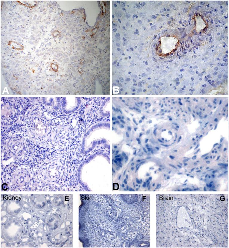
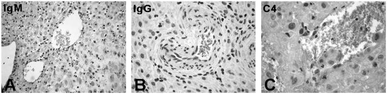
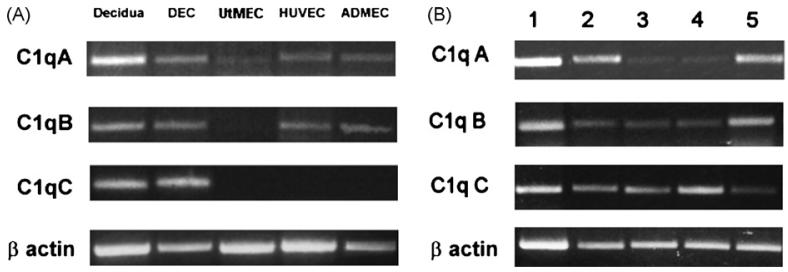
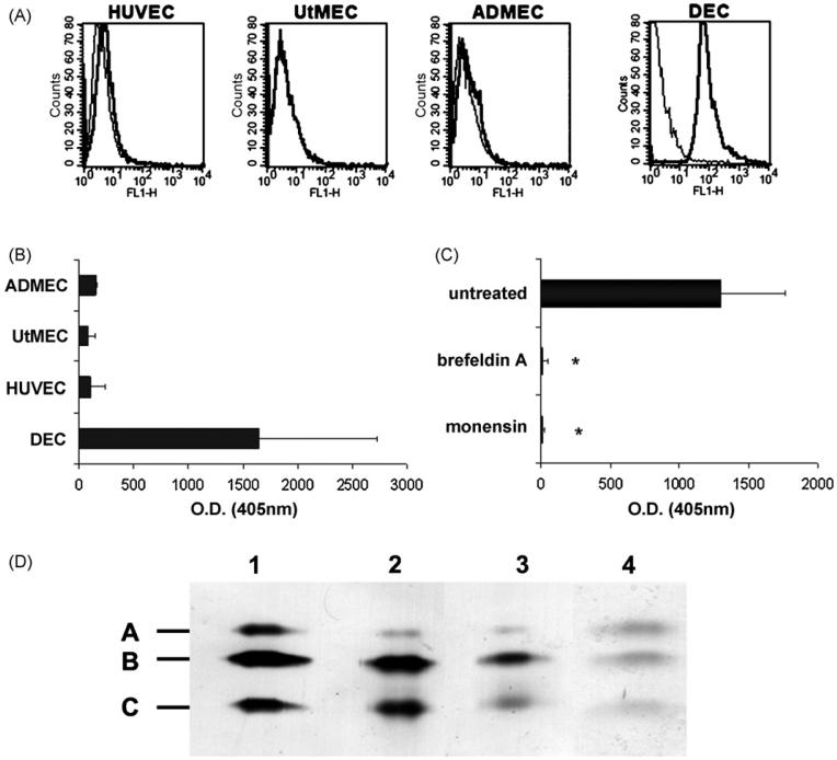
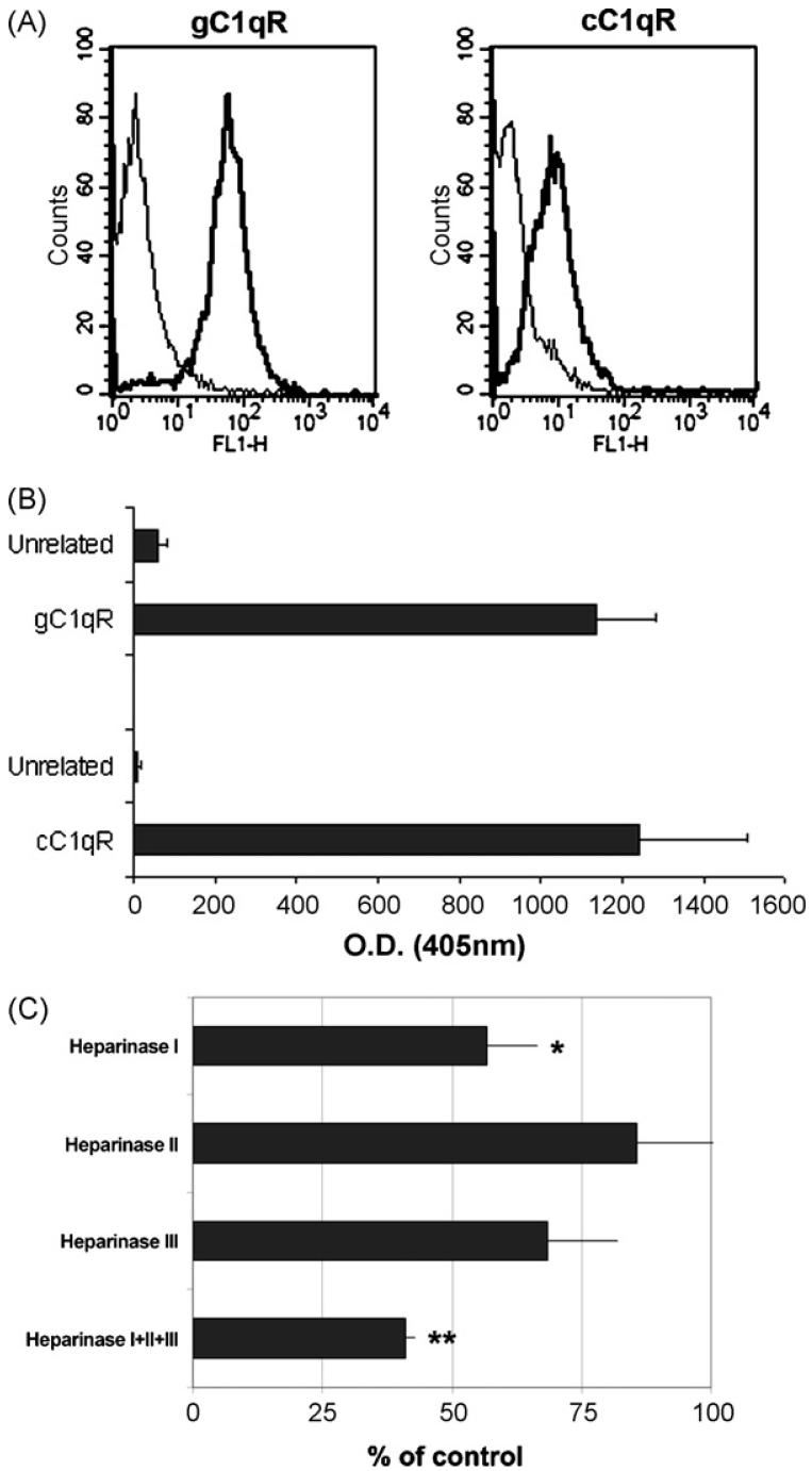
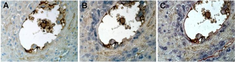
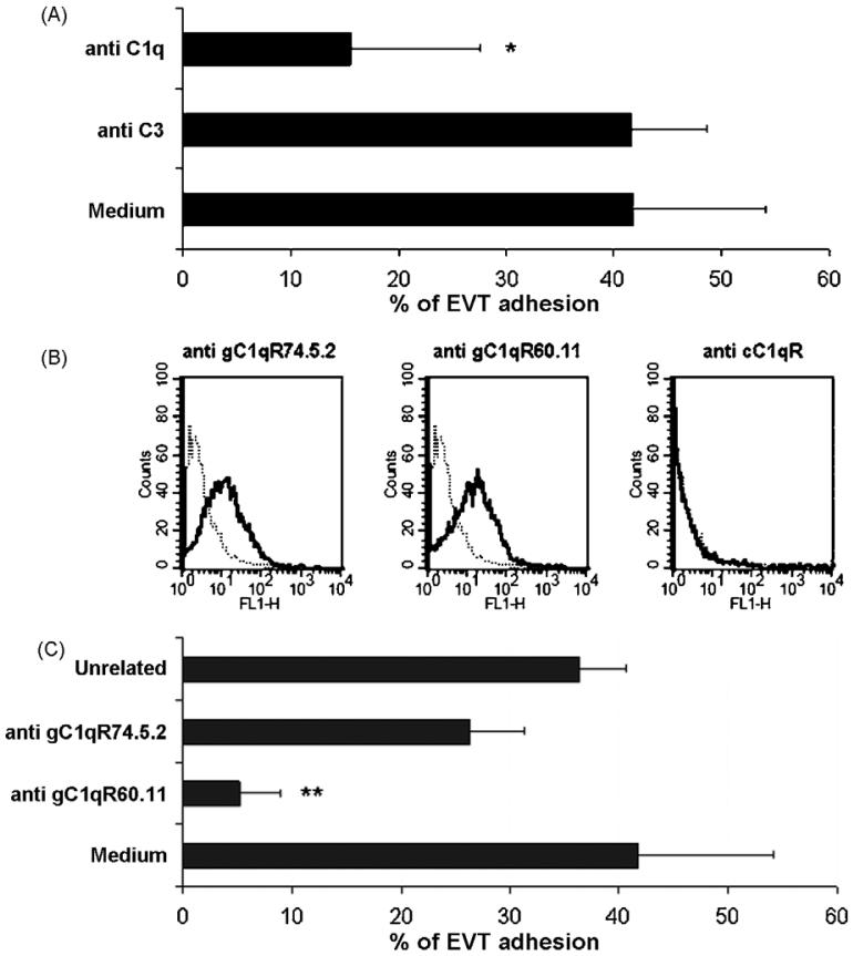
This study was prompted by the observation that decidual endothelial cells (DECs), unlike endothelial cells (ECs) of blood vessels in normal skin, kidney glomeruli and brain, express surface-bound C1q in physiologic pregnancy. This finding was unexpected, because deposits of C1q are usually observed in pathologic conditions and are associated with complement activation. In the case of DECs, we failed to detect immunoglobulins and C4 co-localized with C1q on the cell surface. Surprisingly, DECs expressed mRNA for the three chains of C1q and secreted detectable level of this component in serum-free medium. The ability to synthesize C1q is acquired by DECs during pregnancy and is not shared by ECs obtained from endometrium and from other sources. Cell-associated C1q has a molecular weight similar to that of secreted C1q and is released from DECs following treatment with heparinase or incubation at low pH. This suggests that C1q binds to DECs and it is not constitutively expressed on the cell surface. C1q is localized at contact sites between endovascular trophoblast and DECs and acts as an intercellular molecular bridge because adhesion of endovascular trophoblast to DECs was inhibited by antibodies to C1q and to a receptor recognizing its globular portion expressed on trophoblast.


Figures

Fig. 1
Fig. 1
Detection of C1q on endothelium of vessels in human decidua, endometrium, skin, brain and kidney glomeruli. Sections of paraffin-embedded maternal decidua (A and B), endometrium (C and D) kidney (E), skin (F), and brain (G) were microwaved and stained for human C1q using rabbit IgG anti-human C1q (1/100). Bound antibodies were revealed using the LSAB+ HRP kit and DAB as chromogen. The sections were counterstained with hematoxylin. Original magnifications: (A, C, F and G) 200×; (B, D and E) 400×.
Fig. 2
Fig. 2
Localization of immunoglobulins and C4 on the endothelium of decidual vessels. Sections of maternal decidua were microwaved and incubated with the following antibodies: rabbit IgG anti-human IgM (A) or anti-human IgG (B), both at 1/100, and mAb anti-human C4c (1/100). Bound antibodies were revealed using the LSAB+ HRP kit and DAB as chromogen. The sections were counterstained with hematoxylin. Original magnifications: (A) 200×; (B and C) 400×.
Fig. 3
Fig. 3
RT-PCR analysis of ECs for the expression of mRNA for the three chains of C1q. RNA prepared from first trimester decidua and freshly isolated ECs including DECs, UtMECs, ADMECs, HUVECs) (A) or RNA samples obtained from 5 different populations of DECs (B) were analysed for the expression of C1q A-C. Analysis of mRNA expression for β actin is shown for comparison.
Fig. 4
Fig. 4
Analysis of ECs for surface expression and molecular characterization of C1q. (A) Cytofluorimetric analysis of C1q expression on freshly isolated ECs (DECs, UtMECs, ADMECs, HUVECs) incubated first with mAb for C1q (10 μg/ml) and then with FITC-conjugated F(ab′)2 fragment of goat anti-mouse Ig (1/50). (B) ELISA on ECs using DECs, UtMECs, ADMECs and HUVECs grown to confluence and incubated with mAb anti-human C1q (10 μg/ml) followed by AP-conjugated goat IgG anti-mouse IgG (1/6000). For details see Section 2. (C) ELISA on a confluent monolayer of DECs incubated with 5 μM monensin, 0.5 μM brefeldin A (Sigma-Aldrich) or medium alone for 24 h. The amount of C1q remaining on the cell surface was evaluated as indicated in B. *P < .01 versus untreated cells. (D) Western blot analysis of serum purified C1q from Quidel (track 1), and of C1q affinity-purified from cell supernatant (track 2) or from cell lysate (track 3). The blotted C1q bands were revealed using rabbit IgG anti-human C1q (1/1000) followed by AP-conjugated goat IgG anti rabbit IgG (1/20,000) (Sigma-Aldrich). C1q was also affinity purified from biotin-labeled DEC membranes and revealed with AP-conjugated streptavidine (1/4000) (Sigma-Aldrich) (track 4). The molecular weight of A-C chains of C1q were 29, 27 and 23 kDa respectively.
Fig. 5
Fig. 5
Expression of C1qR on DEC and effect of heparinase on cell-bound C1q. (A) Cytofluorimetric analysis of DECs for the expression of C1q receptors. The cells (5 × 10⁵) were incubated with mAb 74.5.2 to gC1q-R, or rabbit anti-human cC1q-R antibodies. The antibodies were used at the concentration of 10 μg/ml to a total volume of 100 μl in PBS containing 1% BSA and their binding was revealed by 1/50 FITC-conjugated goat F(ab′)2 fragments to mouse IgG or swine IgG anti rabbit Ig. (B) Biotinylated DEC membranes were added to microtiter plate wells coated with pAb anti cC1qR, mAb anti-gC1qR74.5.2 or species-matched control IgG (10 μg/ml). The captured proteins were then detected using AP-conjugated streptavidin and visualized by reaction with p-nitrophenyl phosphate. (C) Heparinase treatment of DECs. Confluent monolayer of DECs was separately incubated with heparinase I, II or III (each at 3 × 10⁻² Units/ml) or treated with the mixture of the three enzymes for 2 h at 37 °C to remove cell-surface glycosaminoglycans. Cells incubated with medium alone served as a control. The residual cell-bound C1q was measured by ELISA. Data of C1q expression on treated cells are given as percent of C1q expressed on untreated cells and presented as mean ± S.D. of triplicate samples from 3 independent experiments. *P < .01, **P < .001 versus control.
Fig. 6
Fig. 6
Localization of C1q at the contact site between endovascular trophoblast and DECs. (A) Single staining (brown) of section of maternal decidua with rabbit IgG anti-human-C1q (1/100) using the LSAB+ HRP kit to reveal bound antibodies and DAB as chromogen (brown); (B) double staining of the same section with rabbit IgG anti-human-C1q as above (brown) and mAb OU-TL 12/30 to human cytokeratin 7 (1/50) developed with APAAP Complex (Dako) and NBT/BCIP as chromogen (blue); (C) triple staining of the same section with rabbit IgG anti-human-C1q (brown), mAb OU-TL 12/30 to human cytokeratin 7 (blue) and mAb QBEnd/10 anti-human-CD34 using the PAP, Mouse Monoclonal Complex (Dako) as a revealing system and AEC as chromogen (red). The sections were counterstained with hematoxylin. Original magnifications: (A-C) 400×.
Fig. 7
Fig. 7
C1q mediates trophoblast-DEC interaction (A) CTBs were labeled with Fast DiI and allowed to adhere to confluent DECs for 30 min at 37 °C in the presence of goat IgG to C1q and C3 (40 μg/ml) or medium alone. The results are given as percent of cell adhesion. (B) Cytofluorimetric analysis of CTBs purified from first trimester placenta for the expression of C1q receptors. The cells (5 × 10⁵) were incubated with mAb 74.5.2 or mAb 60.11, both directed to gC1q-R, or rabbit anti-human cC1q-R antibodies. All the antibodies were used at the concentration of 10 μg/ml to a total volume of 100 μl in PBS containing 1% BSA. Binding of the antibodies was revealed by FITC-conjugated goat F(ab′)2 fragments to mouse IgG or swine IgG anti rabbit Ig. Both antibodies were used at 1/50. (C) Percent adhesion of labeled CTBs to confluent DECs in the presence of mAb 60.11, mAb 74.5.2 or mouse IgG1 isotype control (40 μg/ml). The data in A and C are presented as mean ± S.D. of triplicate samples from at least 4 independent experiments. *P < .01, **P < .001 versus cells incubated with anti-C3 antibody (A) or mouse IgG1 (C).

Similar articles

Cited by

References

    1. Almeda S, Rosenberg RD, Bing DH. The binding properties of human complement component C1q Interaction with mucopolysaccharides. J. Biol. Chem. 1983;258:785–791. - PubMed
    1. Asch AS, Kinoshita T, Jaffe EA, Nussenzweig V. Decay-accelerating factor is present on cultured human umbilical vein endothelial cells. J. Exp. Med. 1986;163:221–226. - PMC - PubMed
    1. Bensa JC, Reboul A, Colomb MG. Biosynthesis in vitro of complement subcomponents C1q C1s and C1 inhibitor by resting and stimulated human monocytes. Biochem. J. 1983;216:385–392. - PMC - PubMed
    1. Bing DH, Spurlock SE, Bern MM. Synthesis of the first component of complement by primary cultures of human tumors of the colon and urogenital tract and comparable normal tissue. Clin. Immunol. Immunopathol. 1975;4:341–351. - PubMed
    1. Bossi F, Fischetti F, Pellis V, Bulla R, Ferrero E, Mollnes TE, Regoli D, Tedesco F. Platelet-activating factor and kinin-dependent vascular leakage as a novel functional activity of the soluble terminal complement complex. J. Immunol. 2004;173:6921–6927. - PubMed

Publication types

MeSH terms