

In vivo static creep loading of the rat forelimb reduces ulnar structural properties at time-zero and induces damage-dependent woven bone formation
- PMID: 18295561
- PMCID: PMC2441934
- DOI: 10.1016/j.bone.2008.01.004
In vivo static creep loading of the rat forelimb reduces ulnar structural properties at time-zero and induces damage-dependent woven bone formation
Abstract
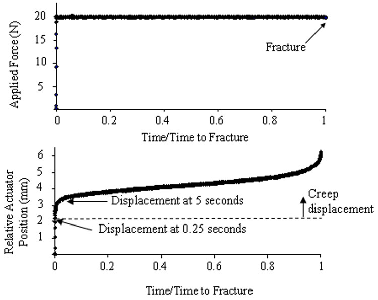
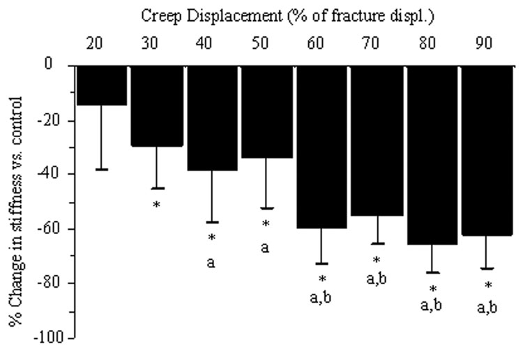
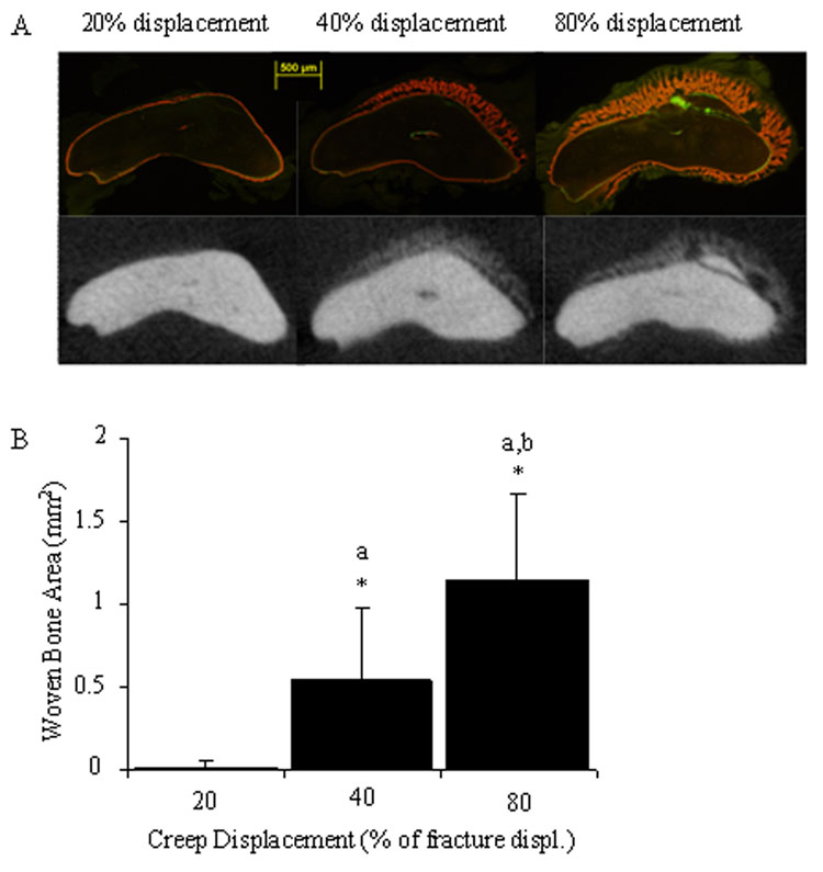
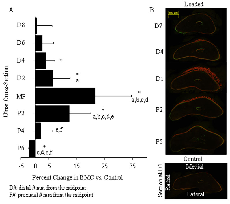
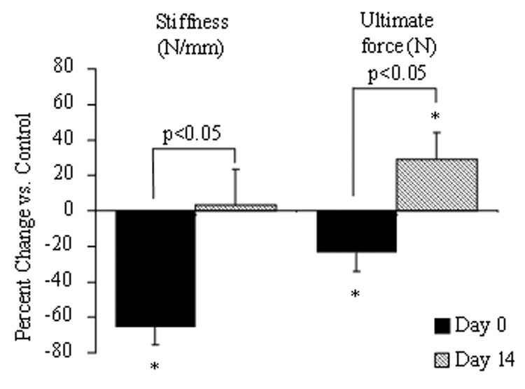
Periosteal woven bone forms in response to stress fractures and pathological overload. The mechanical factors that regulate woven bone formation are poorly understood. Fatigue loading of the rat ulna triggers a woven bone response in proportion to the level of applied fatigue displacement. However, because fatigue produces damage by application of cyclic loading it is unclear if the osteogenic response is due to bone damage (injury response) or dynamic strain (adaptive response). Creep loading, in contrast to fatigue, involves application of a static force. Our objectives were to use static creep loading of the rat forelimb to produce discrete levels of ulnar damage, and subsequently to determine the bone response over time. We hypothesized that 1) increases in applied displacement during loading correspond to ulnae with increased crack number, length and extent, as well as decreased mechanical properties; and 2) in vivo creep loading stimulates a damage-dependent dose-response in periosteal woven bone formation. Creep loading of the rat forelimb to progressive levels of sub-fracture displacement led to progressive bone damage (cracks) and loss of whole-bone mechanical properties (especially stiffness) at time-zero. For example, loading to 60% of fracture displacement caused a 60% loss of ulnar stiffness and a 25% loss of strength. Survival experiments showed that woven bone formed in a dose-dependent manner, with greater amounts of woven bone in ulnae that were loaded to higher displacements. Furthermore, after 14 days the mechanical properties of the loaded limb were equal or superior to control, indicating functional repair of the initial damage. We conclude that bone damage created without dynamic strain triggers a woven bone response, and thus infer that the woven bone response reported after fatigue loading and in stress fractures is in large part a response to bone damage.
Figures
Similar articles
-
Bone formation after damaging in vivo fatigue loading results in recovery of whole-bone monotonic strength and increased fatigue life.J Orthop Res. 2007 Feb;25(2):252-61. doi: 10.1002/jor.20320. J Orthop Res. 2007. PMID: 17106875
-
Stress fracture healing: fatigue loading of the rat ulna induces upregulation in expression of osteogenic and angiogenic genes that mimic the intramembranous portion of fracture repair.Bone. 2009 Feb;44(2):320-30. doi: 10.1016/j.bone.2008.09.010. Epub 2008 Oct 7. Bone. 2009. PMID: 18950737 Free PMC article.
-
Damaging fatigue loading stimulates increases in periosteal vascularity at sites of bone formation in the rat ulna.Calcif Tissue Int. 2007 Jun;80(6):391-9. doi: 10.1007/s00223-007-9031-3. Epub 2007 Jun 6. Calcif Tissue Int. 2007. PMID: 17551770 Free PMC article.
-
In vivo fatigue loading of the rat ulna induces both bone formation and resorption and leads to time-related changes in bone mechanical properties and density.J Orthop Res. 2002 Jul;20(4):764-71. doi: 10.1016/S0736-0266(01)00161-9. J Orthop Res. 2002. PMID: 12168665
-
Multiscale computational and experimental approaches to elucidate bone and ligament mechanobiology using the ulna-radius-interosseous membrane construct as a model system.Technol Health Care. 2012;20(5):363-78. doi: 10.3233/THC-2012-0686. Technol Health Care. 2012. PMID: 23079942 Review.
Cited by
-
Vertebral deformity arising from an accelerated "creep" mechanism.Eur Spine J. 2012 Sep;21(9):1684-91. doi: 10.1007/s00586-012-2279-y. Epub 2012 Mar 25. Eur Spine J. 2012. PMID: 22447410 Free PMC article.
-
Modeling Complex Orthopedic Trauma in Rodents: Bone, Muscle and Nerve Injury and Healing.Front Pharmacol. 2021 Feb 1;11:620485. doi: 10.3389/fphar.2020.620485. eCollection 2020. Front Pharmacol. 2021. PMID: 33597884 Free PMC article. Review.
-
Increased variability of bone tissue mineral density resulting from estrogen deficiency influences creep behavior in a rat vertebral body.Bone. 2012 Nov;51(5):868-75. doi: 10.1016/j.bone.2012.08.124. Epub 2012 Aug 27. Bone. 2012. PMID: 22944606 Free PMC article.
-
Diffuse microdamage in bone activates anabolic response by osteoblasts via involvement of voltage-gated calcium channels.J Bone Miner Metab. 2020 Mar;38(2):151-160. doi: 10.1007/s00774-019-01042-8. Epub 2019 Sep 6. J Bone Miner Metab. 2020. PMID: 31493248
-
Adaptive and Injury Response of Bone to Mechanical Loading.Bonekey Osteovision. 2012 Oct 10;1:192. doi: 10.1038/bonekey.2012.192. Bonekey Osteovision. 2012. PMID: 23505338 Free PMC article.
References
-
- Beck TJ, Ruff CB, Mourtada FA, Shaffer RA, Maxwell-Williams K, Kao GL, Sartoris DJ, Brodine S. Dual-energy X-ray absorptiometry derived structural geometry for stress fracture prediction in male U.S. Marine Corps recruits. J Bone Miner Res. 1996;11(5):645–653. - PubMed
-
- Bentolila V, Boyce TM, Fyhrie DP, Drumb R, Skerry TM, Schaffler MB. Intracortical remodeling in adult rat long bones after fatigue loading. Bone. 1998;23(3):275–281. - PubMed
-
- Bowman SM, Guo XE, Cheng DW, Keaveny TM, Gibson LJ, Hayes WC, McMahon TA. Creep contributes to the fatigue behavior of bovine trabecular bone. J Biomech Eng. 1998;120(5):647–654. - PubMed
-
- Bowman SM, Keaveny TM, Gibson LJ, Hayes WC, McMahon TA. Compressive creep behavior of bovine trabecular bone. J Biomech. 1994;27(3):301–310. - PubMed
-
- Caler WE, Carter DR. Bone creep-fatigue damage accumulation. J. Biomech. 1989;22(67):625–635. - PubMed
Publication types
MeSH terms
Grants and funding
LinkOut - more resources
Full Text Sources
