

doi: 10.1016/j.cell.2008.02.002.
Origins and fates of cardiovascular progenitor cells
Affiliations
- PMID: 18295570
- PMCID: PMC2507768
- DOI: 10.1016/j.cell.2008.02.002
Item in Clipboard
Origins and fates of cardiovascular progenitor cells
Cell.
.
Abstract
Multipotent cardiac progenitor cells are found in the fetal and adult heart of many mammalian species including humans and form as intermediates during the differentiation of embryonic stem cells. Despite similar biological properties, the molecular identities of these different cardiac progenitor cell populations appear to be distinct. Elucidating the origins and lineage relationships of these cell populations will accelerate clinical applications such as drug screening and cell therapy as well as shedding light on the pathogenic mechanisms underlying cardiac diseases.
Figures
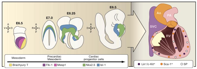
Cells from the developing mesoderm of the mouse embryo are marked by the expression of Brachyury T at E6.5 of embryonic development (yellow). As they transition into precardiac mesoderm, they start to express Mesp1 (pink) and Flk-1 (blue). As these cardiac precursor cells reach the anterior and lateral plate mesoderm, they commit irreversibly to become cardiac progenitor cells by expressing Nkx2.5 (green) or Isl-1 (red) at E7.5. Midline fusion of lateral plate mesoderm and differentiation of these two cell populations results in the formation of Nkx2.5+ embryonic heart tube (green; E8.25 and E8.75) and Isl+ pharyngeal mesoderm (red; E8.25 and E8.75). Some of the pharyngeal mesoderm cells also express Nkx2.5 (not shown). As the heart progresses through embryonic and postnatal development, it acquires a four-chambered identity and is functionally integrated with the systemic vasculature. Within the adult heart reside several different populations of cardiac stem cells including side population (SP) cells and cells that express c-Kit or Sca-1. (Ao, aorta; SVC, superior vena cava; PA, pulmonary artery; RV, right ventricle).
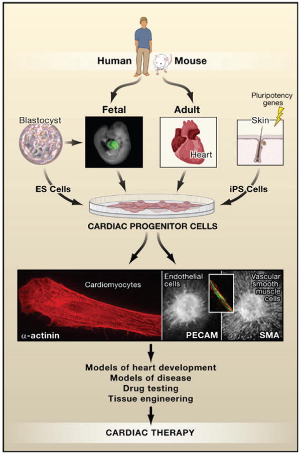
Progenitor cells have been described in fetal and adult heart in multiple species including humans. They also form as an intermediate in the differentiation of embryonic stem (ES) cells giving rise to cardiomyocytes, smooth muscle cells, and endothelial cells. Cardiac progenitors may be uni-, bi-, or tri- potent, depending on their molecular signatures although this relationship is still under investigation. Cardiomyocytes are morphologically readily identifiable by α-actinin staining (red, left panel) of sarcomeric structures, endothelial cells by expression of surface markers such as PE-CAM (green, middle panel and inset), and vascular smooth muscle cells that surround blood vessels by smooth muscle actin (sma; red, right panel). Cells expressing Nkx2.5 in the fetal mouse heart are indicated by green fluorescent protein (GFP) in a transgenic Nkx2.5-GFP mouse. Photos courtesy of C. Mummery (blastocyst; cardiomyocyte); F. Lebrin, D. Ward, and L. Tertoolen (endothelial cell and vascular smooth muscle cell); Sean Wu (fetal heart).
References
-
- Anderson D, Self T, Mellor IR, Goh G, Hill SJ, Denning C. Mol Ther. 2007;15:2027–2036. - PubMed
-
- Assmus B, Honold J, Schachinger V, Britten MB, Fischer-Rasokat U, Lehmann R, Teupe C, Pistorius K, Martin H, Abolmaali ND, et al. N Engl J Med. 2006;355:1222–1232. - PubMed
-
- Beqqali A, Kloots J, Ward-van Oostwaard D, Mummery C, Passier R. Stem Cells. 2006;24:1956–1967. - PubMed
-
- Beltrami AP, Barlucchi L, Torella D, Baker M, Limana F, Chimenti S, Kasahara H, Rota M, Musso E, Urbanek K, et al. Cell. 2003;114:763–776. - PubMed
Publication types
MeSH terms
Grants and funding
LinkOut - more resources
Full Text Sources
Other Literature Sources
Medical
