. 2008 Apr 15;472(2):100-4.
doi: 10.1016/j.abb.2008.02.012. Epub 2008 Feb 14.

Retinal isoforms of inosine 5'-monophosphate dehydrogenase type 1 are poor nucleic acid binding proteins

Affiliations

Retinal isoforms of inosine 5'-monophosphate dehydrogenase type 1 are poor nucleic acid binding proteins

Dong Xu et al. Arch Biochem Biophys. .

Abstract

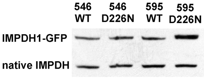
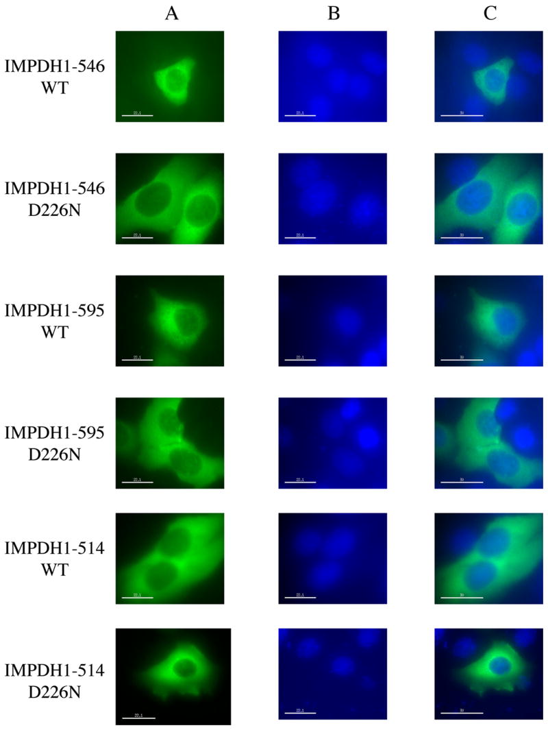
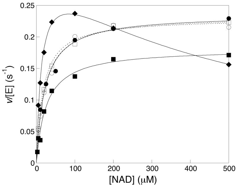
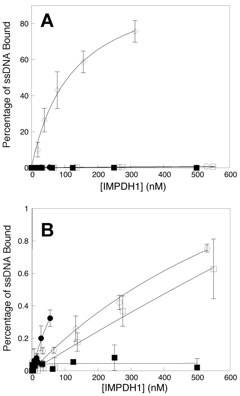
The RP 10 form of autosomal dominant retinitis pigmentosa (adRP) is caused by mutations in the widely expressed protein inosine 5'-monophosphate dehydrogenase type 1 (IMPDH1). These mutations have no effect on the enzymatic activity of IMPDH1, but do perturb the association of IMPDH1 with nucleic acids. Two newly discovered retinal-specific isoforms, IMPDH1(546) and IMPDH1(595), may provide the key to the photoreceptor specificity of disease [S.J. Bowne, Q. Liu, L.S. Sullivan, J. Zhu, C.J. Spellicy, C.B. Rickman, E.A. Pierce, S.P. Daiger, Invest. Ophthalmol. Vis. Sci. 47 (2006) 3754-3765]. Here we express and characterize the normal IMPDH1(546) and IMPDH1(595), together with their adRP-linked variants, D226N. The enzymatic activity of the purified IMPDH1(546), IMPDH1(595) and the D226N variants is indistinguishable from the canonical form. The intracellular distribution of IMPDH1(546) and IMPDH1(595) is also similar to the canonical IMPDH1 and unaffected by the D226N mutation. However, unlike the canonical IMPDH1, the retinal specific isoforms do not bind significant fractions of a random pool of oligonucleotides. This observation indicates that the C-terminal extension unique to the retinal isoforms blocks the nucleic acid binding site of IMPDH1, and thus uniquely regulates protein function within photoreceptors.


Figures

Figure 1
Figure 1
Expression of retinal IMPDH1 isoforms. Soluble extracts of HEK293 cells transiently transfected with plasmids encoding retinal IMPDH1-GFP were analyzed by Western blot. From left to right: IMPDH1(546), IMPDH1(546)-D226N, IMPDH1(595), IMPDH1(595)-D226N.
Figure 2
Figure 2
Localization of the retinal IMPDH1 isoforms. HeLa cells transiently expressing IMPDH1-GFP were viewed under fluorescence microscopy. A) Distribution of various forms of IMPDH1 as revealed by the GFP fluorescence; B) Nucleic staining of the same cells by DAPI; C) Overlays of images in A and B.
Figure 3
Figure 3
Steady state kinetics of retinal IMPDH1 isoforms. The rate of NADH formation in the presence of saturating IMP (final concentration 500 μM) was divided by the monomeric concentration of IMPDH1 to obtain v/[E]. The values were plotted against the concentration of NAD+. The results obtained on the canonical IMPDH(514) were designated with closed diamonds (◆); IMPDH1(546), closed circles (●); IMPDH1(546)-D226N, open circles (○); IMPDH1(595), closed squares (■); IMPDH1(595)-D226N, open squares (□)]. Each data point represents the average value from at least three independent experiments.
Figure 4
Figure 4
Binding of ssDNA to retinal IMPDH1 isoforms. A random pool of 32P-labeled oligonucleotides (0.15 nM) was incubated with varying concentrations of IMPDH1 (concentration is reported as tetramer). Protein-bound nucleic acid was measured in a filter-binding assay as described in details in Materials and Methods. A) The percentages of total ssDNA associated with IMPDH1 were plotted against the protein concentration. The canonical IMPDH1(514)-D226N, open diamonds (◇); IMPDH1(546), closed circles (●); IMPDH1(546)-D226N, open circles (○); IMPDH1(595), closed squares (■); and IMPDH1(595)-D226N, open squares (□); B) Expanded view of the results on the retinal IMPDH1 isoforms with the same designations as in A. Each data point represents the average value from at least three independent experiments.

References

    1. Heckenlively JR, Daiger SP. Principals and Practices of Medical Genetics. In: Rimoin DI, Connor JM, Pyeritz RE, editors. Churchill Livingston. New York: 2001; 2255. p. 2576.
    1. Portera-Cailliau C, Sung CH, Nathans J, Adler R. Proc Natl Acad Sci USA. 1994;91:974–978. - PMC - PubMed
    1. Chang GQ, Hao Y, Wong F. Neuron. 1993;11:595–605. - PubMed
    1. Bowne SJ, Sullivan LS, Blanton SH, Cepko CL, Blackshaw S, Birch DG, Hughbanks-Wheaton D, Heckenlively JR, Daiger SP. Hum Mol Genet. 2002;11:559–568. - PMC - PubMed
    1. Kennan A, Aherne A, Palfi A, Humphries M, McKee A, Stitt A, Simpson DA, Demtroder K, Orntoft T, Ayuso C, Kenna PF, Farrar GJ, Humphries P. Hum Mol Genet. 2002;11:547–557. - PubMed

Publication types