CD28 and Grb-2, relative to Gads or Grap, preferentially co-operate with Vav1 in the activation of NFAT/AP-1 transcription
- PMID: 18295596
- PMCID: PMC4186964
- DOI: 10.1016/j.bbrc.2008.02.068
CD28 and Grb-2, relative to Gads or Grap, preferentially co-operate with Vav1 in the activation of NFAT/AP-1 transcription
Abstract
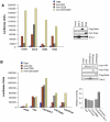
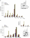
The co-receptor CD28 binds to several intracellular proteins including PI3 kinase, Grb-2, Gads and ITK. Grb-2 and PI3 kinase binding has been mapped to the pYMNM motif within the cytoplasmic tail of CD28 and has been shown to play a role in co-stimulation. In this study, we demonstrate that amongst the Grb-2 family adapter proteins, CD28 precipitated Grb-2 and specifically co-operated in the up-regulation of NFAT/AP-1 transcription. By contrast, Gads and Grap either failed or only weakly collaborated with CD28 ligation. Further, the loss of Grb-2 binding interferes with the ability of Vav1 to co-operate with CD28. Anti-CD28 ligation alone was capable for co-operating with Grb-2 or Grb-2-Vav1. Our findings define a pathway involving CD28 binding to Grb-2 and its co-operativity with Vav1 in the regulation of T-cell co-stimulation.
Figures


Similar articles
-
Grb2 and Gads exhibit different interactions with CD28 and play distinct roles in CD28-mediated costimulation.J Immunol. 2006 Jul 15;177(2):1085-91. doi: 10.4049/jimmunol.177.2.1085. J Immunol. 2006. PMID: 16818765
-
Itk Promotes the Integration of TCR and CD28 Costimulation through Its Direct Substrates SLP-76 and Gads.J Immunol. 2021 May 15;206(10):2322-2337. doi: 10.4049/jimmunol.2001053. Epub 2021 Apr 30. J Immunol. 2021. PMID: 33931484 Free PMC article.
-
T cell antigen CD28 binds to the GRB-2/SOS complex, regulators of p21ras.Eur J Immunol. 1995 Apr;25(4):1044-50. doi: 10.1002/eji.1830250428. Eur J Immunol. 1995. PMID: 7737275
-
Bridging the Gap: Modulatory Roles of the Grb2-Family Adaptor, Gads, in Cellular and Allergic Immune Responses.Front Immunol. 2019 Jul 25;10:1704. doi: 10.3389/fimmu.2019.01704. eCollection 2019. Front Immunol. 2019. PMID: 31402911 Free PMC article. Review.
-
Independent CD28 signaling via VAV and SLP-76: a model for in trans costimulation.Immunol Rev. 2003 Apr;192:32-41. doi: 10.1034/j.1600-065x.2003.00005.x. Immunol Rev. 2003. PMID: 12670393 Review.
Cited by
-
Crystal Structures and Thermodynamic Analysis Reveal Distinct Mechanisms of CD28 Phosphopeptide Binding to the Src Homology 2 (SH2) Domains of Three Adaptor Proteins.J Biol Chem. 2017 Jan 20;292(3):1052-1060. doi: 10.1074/jbc.M116.755173. Epub 2016 Dec 6. J Biol Chem. 2017. PMID: 27927989 Free PMC article.
-
TCR and CD28 activate the transcription factor NF-κB in T-cells via distinct adaptor signaling complexes.Immunol Lett. 2015 Jan;163(1):113-9. doi: 10.1016/j.imlet.2014.10.020. Epub 2014 Oct 23. Immunol Lett. 2015. PMID: 25455592 Free PMC article.
-
CD28 co-signaling in the adaptive immune response.Self Nonself. 2010 Jul;1(3):231-240. doi: 10.4161/self.1.3.12968. Epub 2010 Jul 12. Self Nonself. 2010. PMID: 21487479 Free PMC article.
-
CD28 Costimulation: From Mechanism to Therapy.Immunity. 2016 May 17;44(5):973-88. doi: 10.1016/j.immuni.2016.04.020. Immunity. 2016. PMID: 27192564 Free PMC article. Review.
-
Cell Type-Specific Regulation of Immunological Synapse Dynamics by B7 Ligand Recognition.Front Immunol. 2016 Feb 4;7:24. doi: 10.3389/fimmu.2016.00024. eCollection 2016. Front Immunol. 2016. PMID: 26870040 Free PMC article. Review.
References
-
- June CH, Bluestone JA, Nadler LM, Thompson CB. The B7 and CD28 receptor families. Immunol. Today. 1994;15:321–331. - PubMed
-
- Riley JL, June CH. The CD28 family: a T-cell rheostat for therapeutic control of T-cell activation. Blood. 2005;105:13–21. - PubMed
-
- Greenwald RJ, Freeman GJ, Sharpe AH. The B7 family revisited. Annu. Rev. Immunol. 2005;23:515–548. - PubMed
-
- Rudd CE, Schneider H. Unifying concepts in CD28, ICOS and CTLA4 co-receptor signalling. Nat. Rev. Immunol. 2003;3:544–556. - PubMed
Publication types
MeSH terms
Substances
Grants and funding
LinkOut - more resources
Full Text Sources
Molecular Biology Databases
Research Materials
Miscellaneous

