. 2008 Apr 1;316(1):62-73.
doi: 10.1016/j.ydbio.2008.01.012. Epub 2008 Jan 26.

A distinct cohort of progenitor cells participates in synovial joint and articular cartilage formation during mouse limb skeletogenesis

Affiliations

A distinct cohort of progenitor cells participates in synovial joint and articular cartilage formation during mouse limb skeletogenesis

Eiki Koyama et al. Dev Biol. .

Abstract

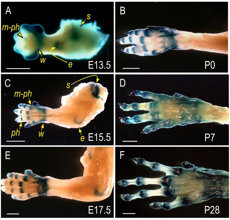
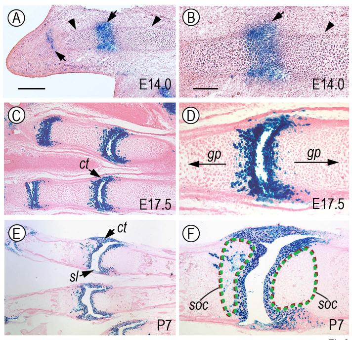
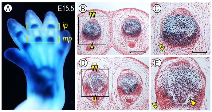
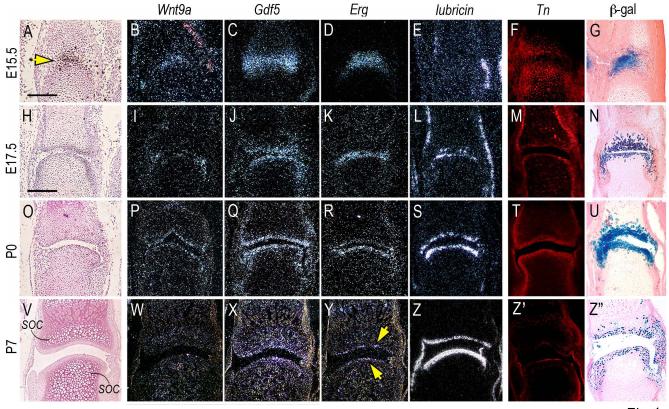
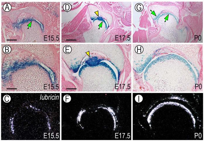
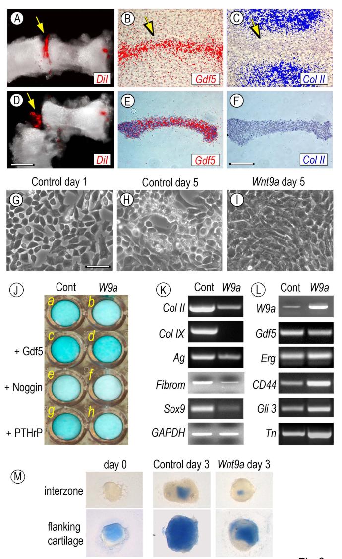
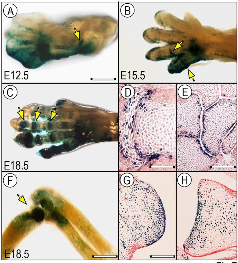
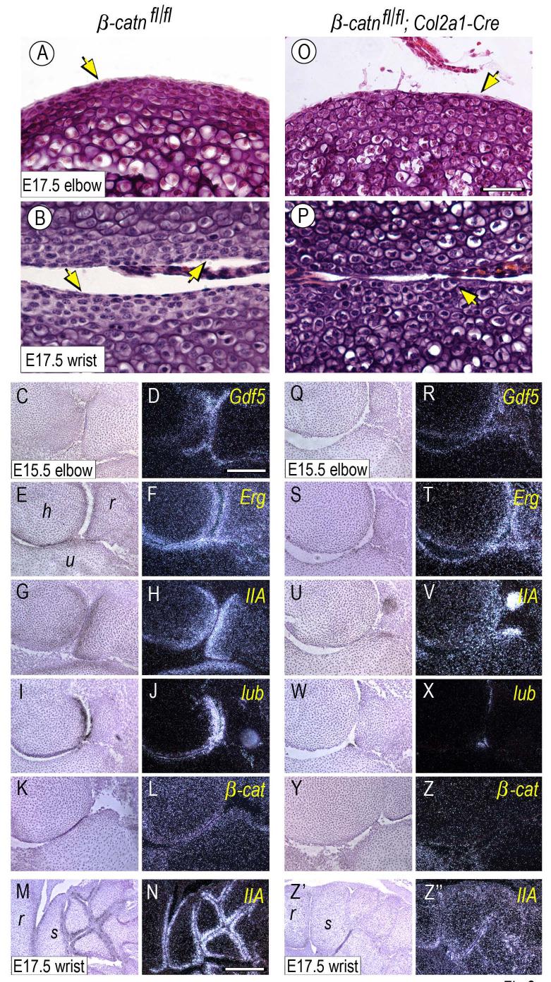
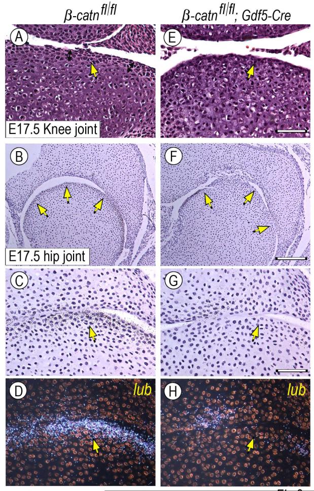
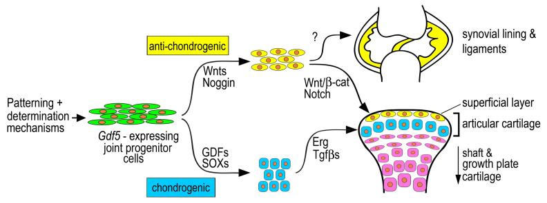
The origin, roles and fate of progenitor cells forming synovial joints during limb skeletogenesis remain largely unclear. Here we produced prenatal and postnatal genetic cell fate-maps by mating ROSA-LacZ-reporter mice with mice expressing Cre-recombinase at prospective joint sites under the control of Gdf5 regulatory sequences (Gdf5-Cre). Reporter-expressing cells initially constituted the interzone, a compact mesenchymal structure representing the first overt sign of joint formation, and displayed a gradient-like distribution along the ventral-to-dorsal axis. The cells expressed genes such as Wnt9a, Erg and collagen IIA, remained predominant in the joint-forming sites over time, gave rise to articular cartilage, synovial lining and other joint tissues, but contributed little if any to underlying growth plate cartilage and shaft. To study their developmental properties more directly, we isolated the joint-forming cells from prospective autopod joint sites using a novel microsurgical procedure and tested them in vitro. The cells displayed a propensity to undergo chondrogenesis that was enhanced by treatment with exogenous rGdf5 but blocked by Wnt9a over-expression. To test roles for such Wnt-mediated anti-chondrogenic capacity in vivo, we created conditional mutants deficient in Wnt/beta-catenin signaling using Col2-Cre or Gdf5-Cre. Synovial joints did form in both mutants; however, the joints displayed a defective flat cell layer normally abutting the synovial cavity and expressed markedly reduced levels of lubricin. In sum, our data indicate that cells present at prospective joint sites and expressing Gdf5 constitute a distinct cohort of progenitor cells responsible for limb joint formation. The cells appear to be patterned along specific limb symmetry axes and rely on local signaling tools to make distinct contributions to joint formation.


Figures

Fig. 1
Fig. 1
Reporter-expressing cells are predominant in developing joints pre-natally and post-natally. Limbs isolated from the progeny of ROSA R26R mice mated with Gdf5-Cre mice were processed for whole mount detection of β-galactosidase activity. (A) At E13.5, reporter activity is distinctly visible in incipient joints at the prospective metacarpal-phalangeal (m-ph), wrist (w), elbow (e) and shoulder (s) sites. (C and E) By E15.5 and E17.5, reporter activity is clearly restricted at sites of joint formation that now include also the inter-phalangeal (ph) site. (B, D and F). Preferential location of reporter activity persists at every postnatal time point examined including P0, P7 and P28. Bars, 1 mm.
Fig. 2
Fig. 2
Reporter-expressing cells are predominant in articulating layers and accessory joint tissues. Longitudinal sections from representative forelimb autopod specimens similar to those in Fig. 1 were processed for histochemical detection of β-galactosidase activity. In each joint, proximal side is on the right and distal side is on the left. (A) At E14.0, reporter-positive cells are predominant in nascent metacarpal-phalangeal and inter-phalangeal joint sites (arrows), but are undetectable in the flanking cartilaginous shafts (arrowheads). (C and E) By E17.5 and P7, the reporter-positive cells clearly constitute the 3 to 4 articular cell layers facing the incipient joint space and cavity and are present in accessory joint tissues such as synovial lining (sl) and capsule tissue (ct). (B, D and F) These are higher magnification views of images shown in panels A, C and E, respectively. Note that the reporter-positive cells are limited to nascent joint tissue at E14.0 (B) and to joint-facing articular layers and associated joint tissues at E17.5 and P7 (D and F). Note also that while the positive cells are excluded from growth plates and incipient secondary ossification center (soc) on the distal side, a few positive cells are also present in the sub-articular epiphyseal/soc area on the proximal side. Apparent size of synovial space may have become enlarged during tissue processing, particularly in specimen shown in E and F. Bar for A, C and E, 200 μm; bar for B, D and F, 75 μm.
Fig. 3
Fig. 3
Reporter-expressing cells are more numerous ventrally than dorsally. (A) Whole mount staining of a E15.5 Gdf5-Cre/ROSA forelimb in which developing metacarpal-phalangeal (mp) and inter-phalangeal (ip) joints are strongly positive. Histological cross sections of ip joints (B-C) and mp joints (D-E) show that there are more numerous reporter-positive cells in the ventral half (arrow) than dorsal half (double arrow). Note also: the presence of tightly-packed ventro-lateral cell masses (double arrowhead) possibly representing prospective capsule precursors; and initiation of cavitation process in the ventral side (arrowhead). Bar for B and D, 250 μm; bar for C and E, 110 μm.
Fig. 4
Fig. 4
Reporter-expressing cells acquire articular chondrocyte characteristics over developmental time. Longitudinal serial sections from fetal and postnatal autopods were processed for histology and in situ hybridization. Sections from companion specimens were processed for immunohistochemical detection of tenascin-C (Tn) or histochemical detection of β-galactosidase-positive (β-gal) cells. (A-G) At E15.5, interzone cells (yellow arrow in A) express Wnt9a, Gdf5 and Erg, are part of tenascin-C-containing epiphyseal tissue (F), display strong and distinct β-galactosidase activity (G), but lack detectable lubricin expression (E). (H-N and O-U) At E17.5 and P0, expression of Wnt9a, Gdf5 and Erg is clearly restricted to articular cells that display also lubricin transcripts (L and S), distinct tenascin-C content (M and T) and strong β-galactosidase activity (N and U). (V-Z”) By P7, Wnt9a and Gdf5 expression is markedly down-regulated (W-X), Erg expression is maintained in cells abutting the synovial space (Y, arrows), while lubricin expression remains strong throughout the articulating layer (Z) that also contains tenascin-C (Z’) and β-galactosidase-expressing cells (Z”). Note in (V) the prominent secondary ossification centers (soc) visible immediately below the articular layers. Bar for A-G, 150 μm; bar for H-Z”, 100 μm.
Fig. 5
Fig. 5
Cell fate-map analysis during hip joint development. Cross sections of hip joint region from E15.5, E17.5 and P0 animals were processed for histochemical detection of β-galactosidase-positive cells. Similar sections from companion animals were processed for in situ hybridization analysis of lubricin gene expression. (A, D and G) Positive cells are restricted to the articular layers facing the synovial cavity (arrows) and constitute also the teres which is the main femoral head’s ligament (arrowhead). (B, E and H) Higher magnification view of the above sections clearly shows that the reporter-positive cells are confined to 3 to 4 layers facing the joint cavity. (C, F and I) Lubricin gene expression is confined to joint associated cells. Distribution of lubricin-expressing cells is clearly reminiscent and virtually over-lapping that of reporter-positive cells. Bar for A, 175 μm; bar for B-C, 75 μm; bar for D and G, 350 μm; bar for E-F and H-I, 150 μm.
Fig. 6
Fig. 6
Isolation, culturing and testing of interzone-associated cells. Metatarsal and first inter-phalangeal interzones present in Day 6.5 chick embryo leg buds were microinjected with DiI (A, arrow) and were dissected out by means of custom-made tungsten needles (D, arrow). In situ hybridization on companion specimens shows that prior to dissection, the interzone tissue strongly expressed Gdf5 (B, arrow) but not collagen II (C, arrow); note that hybridization signal is pseudo-colored. These patterns were maintained in isolated tissue (E-F). On day 1 of culture, interzone tissue-derive cells displayed a fibroblastic-mesenchymal morphology (G), acquired a chondrocytic phenotype by day 5 (H), but retained a mesenchymal appearance after infection with Wnt9a-encoding RCAS virus (I). In J, companion control and Wnt9a-overexpressing cultures in multi-well plates were treated with 100 ng/ml rGdf5 (c-d), 200 ng/ml Noggin (e-f) or 0.1 μM PTHrP (g-h) or were left untreated (a-b). Fresh cytokines were given on day 1 and all cultures were stained with alcian blue on day 5. Note that exogenous rGdf5 increases proteoglycan content (c) and counteracts the Wnt9a-induced loss of proteoglycan staining (d), while Noggin treatment leads to a marked decrease in staining in both cultures (e-f). (K-L) RT-PCR analysis of genes expressed in day 5 control or Wnt9a-over-expressing cultures. Note that Wnt9a-overexpression leads to a marked down-regulation of chondrocyte-characteristic genes including collagen II (Col II) collagen IX (Col IX), aggrecan (Ag), fibromodulin (Fibrom) and Sox9, but causes an up-regulation of early joint/interzone-characteristic genes including CD44, Gli3 and tenascin-C (Tn) and a modest increase in Erg expression. (M) Explant cultures of interzones and flanking cartilage tissue stained with alcian blue after isolation (day 0) or after 3 days in culture with infection with insert-less or Wnt9a RCAS virus. Bar for A and D, 300 μm; bar for B-C and E-F, 150 μm; bar for G-I, 30 μm.
Fig. 7
Fig. 7
Wnt/β-catenin signaling is active at multiple stages of joint formation. Limbs from BATlacZ (A-E) and TOPGAL (F-H) Wnt-β-catenin reporter mouse embryos were processed for whole mount β-galactosidase (β-gal) staining (A-C, F) or section staining (D-E, G-H). (A) At E12.5, β-gal activity is already detectable in developing large joints such as the elbow (arrow). (B-E) By E15.5 and E18.5, activity is clearly visible in digit joint also (B-C, arrows) and is particularly strong in joint cells abutting the nascent interdigital and wrist joint cavities (D-E). (F-H) At E18.5, activity remains strong in large joints such as elbow (F) and characterizes articular cells all along the joint surface and neighboring cells (G-H). Bar for A-C, 1 mm; bar for D, 150 μm; bar for E, 300 μm, bar for F, 0.8 mm; bar for G-H, 250 μm.
Fig. 8
Fig. 8
Superficial flat cell layer organization and gene expression patterns are defective in conditional Col2a1-Cre β-catenin-deficient joints. Elbow and wrist joints from E15.5 and E17.5 control (β-catn fl/fl) and β-catenin-deficient (β-catn fl/fl/Col2a1-Cre) mouse embryo littermates were processed for standard H&E histology and in situ hybridization. Mutant joints display an appreciable reduction in the high-density flat cell layers all along the joint surface (O-P, arrows) that are quite evident in control joints (A-B, arrows). (C-N) Control joints display characteristic, prominent and expected patterns of expression of joint markers including Gdf5, Erg, collagen IIA (IIA) and lubricin (lub) (D, F, H, J, N). There is also appreciable expression of endogenous β-catenin (β-cat) (L). (Q-Z”) In contrast, mutant joints exhibit obvious decreases in gene expression, particularly collagen IIA and lubricin (V, X, Z”) and to a lesser extent Gdf5 and Erg (R, T). Endogenous β-catenin expression was barely detectable (Z). Bar for A-B and O-P, 35 μm; bar for C-L and Q-Z, 150 μm; bar for M-N and Z’-Z”, 250 μm.
Fig. 9
Fig. 9
Defects in superficial flat cell layer and gene expression patterns in conditional Gdf5-Cre β-catenin-deficient joints. E17.5 knee and hip joints from control (β-catn fl/fl) and β-catenin-deficient (β-catn fl/fl/Gdf5-Cre) mouse embryo littermates were analyzed by standard histology and for expression of lubricin. (A-D) Control joints display the high cell density flat cell layers along the joint perimeter (A-C, arrows) and lubricin (lub) transcripts (D). (E-H) Instead, the superficial high-density layers are much less obvious in mutant joints (E-G, arrows) and lubricin expression is much reduced (H). Bar for A and E, 50 μm; bar for B and F, 175 μm; bar for C-D and G-H, 60 μm.
Fig 10
Fig 10
Model of possible roles of Gdf5-expressing progenitor cells in joint formation and maintenance. Upstream patterning and determination mechanisms would initially induce emergence of the Gdf5-expressing cells at prospective limb joint sites. Spatio-temporally restricted action by members of Gdf, Bmp and Sox gene families would commit some of the cells to the chondrogenic lineage, and subsequent action by factors such as Erg (Iwamoto et al., 2007) and TGFβ (Serra et al., 1997; Spagnoli et al., 2007) would promote formation of the bulk of articular cartilage. Anti-chondrogenic factors including Wnt/β-catenin and Notch signaling and BMP inhibitors such as Noggin would instead direct some of the cells toward fibrogenic and mesodermal lineages, resulting in formation of synovial lining, intra-joint ligaments, inner capsule and articular cartilage’s superficial layer. The superficial layer would be maintained through life to serve as a source of lubricating molecules and as a possible source/recruiter of progenitor cells. Genes delineating the model above are not meant to be comprehensive.

References

    1. Barna M, Pandolfi PP, Niswander L. Gli3 and Plzf cooperate in proximal limb patterning at early stages of limb development. Nature. 2005;436:277–281. - PubMed
    1. Bird HA. Controversies in the treatment of osteoarthritis. Clin. Rheum. 2003;22:165–167. - PubMed
    1. Bland YA, Ashhurst d. E. Development and ageing of the articular cartilage of the rabbit knee joint: distribution of fibrillar collagens. Anat. Embryol. 1996;194:607–619. - PubMed
    1. Brandt KD. Non-surgical treatment of osteoarthritis: a half century of “advances”. Annu. Rheum. Dis. 2004;63:117–122. - PMC - PubMed
    1. Brunet LJ, McMahon JA, McMahon AP, Harland RM. Noggin, cartilage morphogenesis, and joint formation in the mammalian skeleton. Science. 1998;280:1455–1457. - PubMed

Publication types

MeSH terms