

The enhancement of chondrogenic differentiation of human mesenchymal stem cells by enzymatically regulated RGD functionalities
- PMID: 18295878
- PMCID: PMC2346439
- DOI: 10.1016/j.biomaterials.2008.01.035
The enhancement of chondrogenic differentiation of human mesenchymal stem cells by enzymatically regulated RGD functionalities
Abstract
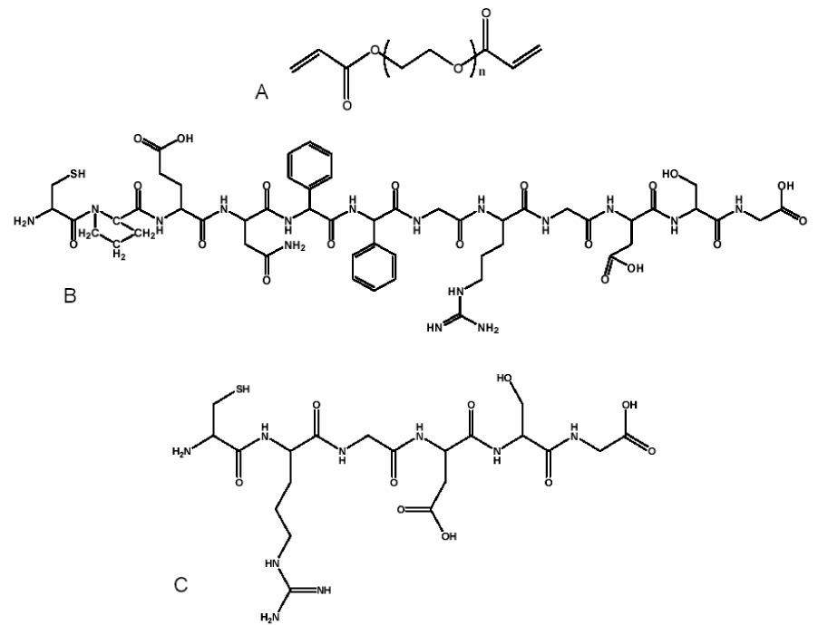
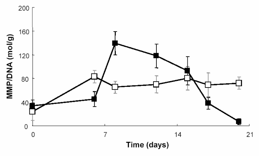
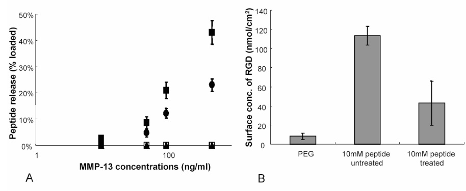
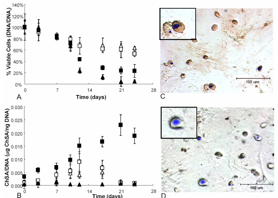
A thiol-acrylate photopolymerization was used to incorporate enzymatically cleavable peptide sequences into PEG hydrogels to induce chondrogenic differentiation of encapsulated human mesenchymal stem cells (hMSCs). An adhesive sequence, RGD, was designed with an MMP-13 specific cleavable linker. RGD promotes survival of hMSCs encapsulated in PEG gels and has shown to induce early stages of chondrogenesis, while its persistence can limit complete differentiation. Therefore, an MMP-13 cleavage site was incorporated into the peptide sequence to release RGD mimicking the native differentiation timeline. Active MMP-13 production of encapsulated hMSCs was seen to increase from day 9 to 14 and only in chondrogenic differentiating cultures. Seeded hMSCs attached to the material prior to enzymatic cleavage, but a significant population of the cells detach after cleavage and release of RGD. Finally, hMSCs encapsulated in RGD-releasing gels produce 10 times as much glycosaminoglycan as cells with uncleavable RGD functionalities, by day 21 of culture. Furthermore, 75% of the cells stain positive for collagen type II deposition where RGD is cleavable, as compared to 19% for cultures where RGD persists. Collectively, these data provide evidence that temporal regulation of integrin-binding peptides is important in the design of niches in differentiating hMSCs to chondrocytes.
Figures
References
-
- Rice MA, Anseth KS. Encapsulating chondrocytes in copolymer gels: bimodal degradation kinetics influence cell phenotype and extracellular matrix development. J Biomed Mater Res A. 2004;70(4):560–568. - PubMed
-
- Nuttelman CR, Henry SM, Anseth KS. Synthesis and characterization of photocrosslinkable, degradable poly(vinyl alcohol)-based tissue engineering scaffolds. Biomaterials. 2002;23(17):3617–3626. - PubMed
-
- Bryant SJ, Anseth KS. Controlling the spatial distribution of ECM components in degradable PEG hydrogels for tissue engineering cartilage. J Biomed Mater Res A. 2003;64(1):70–79. - PubMed
-
- Levesque SG, Shoichet MS. Synthesis of enzyme-degradable, peptide-cross-linked dextran hydrogels. Bioconjug Chem. 2007;18(3):874–885. - PubMed
-
- Rizzi SC, Hubbell JA. Recombinant protein-co-PEG networks as cell-adhesive and proteolytically degradable hydrogel matrixes. Part I: Development and physicochemical characteristics. Biomacromolecules. 2005;6(3):1226–1238. - PubMed
Publication types
MeSH terms
Substances
Grants and funding
LinkOut - more resources
Full Text Sources
Other Literature Sources
