. 2008 Jun;45(6):861-77.
doi: 10.1016/j.fgb.2008.01.001. Epub 2008 Jan 26.

Candida albicans VPS1 contributes to protease secretion, filamentation, and biofilm formation

Affiliations

Candida albicans VPS1 contributes to protease secretion, filamentation, and biofilm formation

Stella M Bernardo et al. Fungal Genet Biol. 2008 Jun.

Abstract

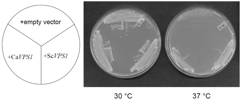
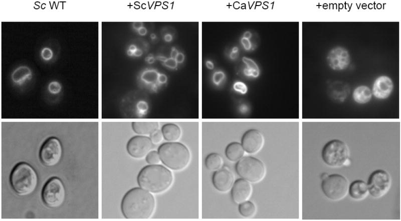
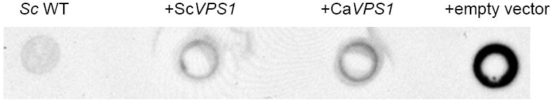
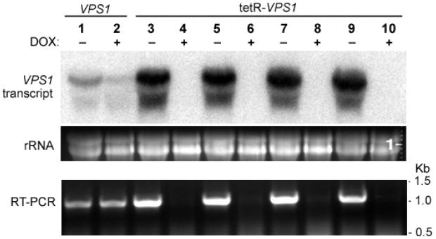
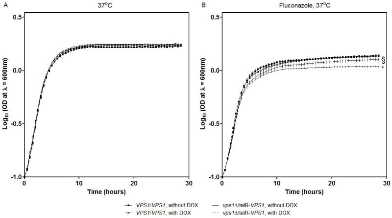
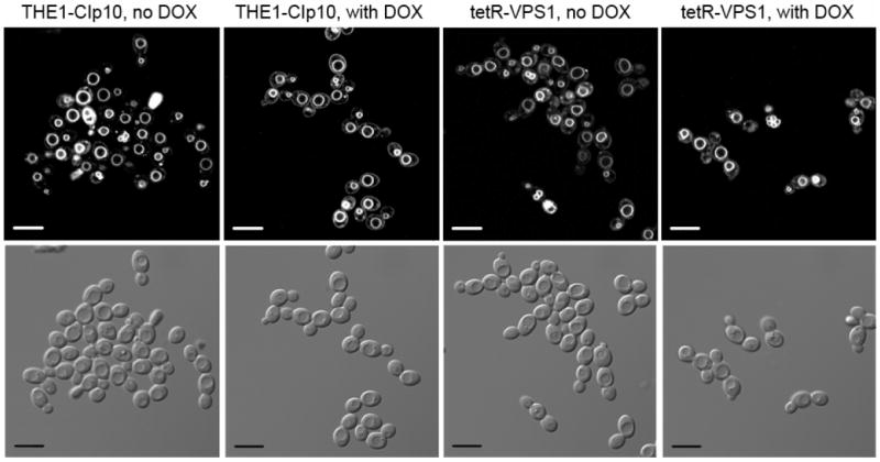
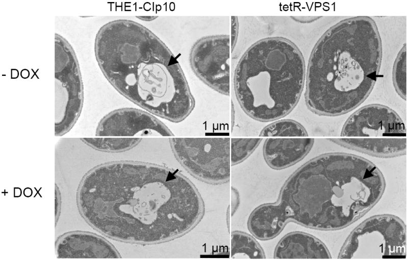
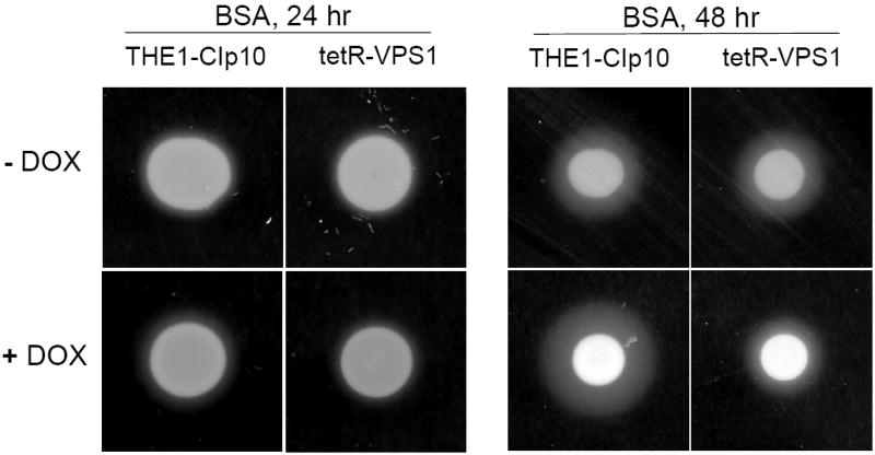
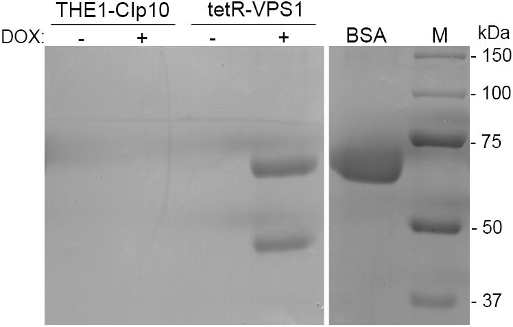
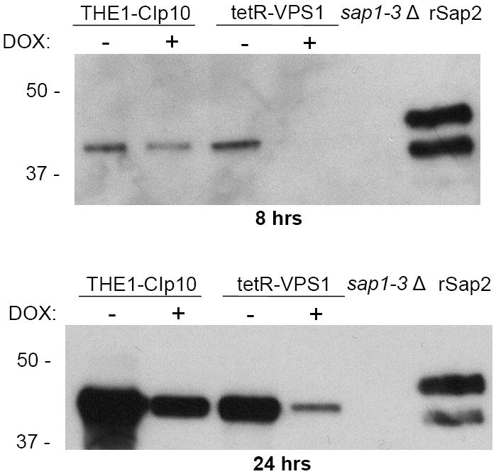
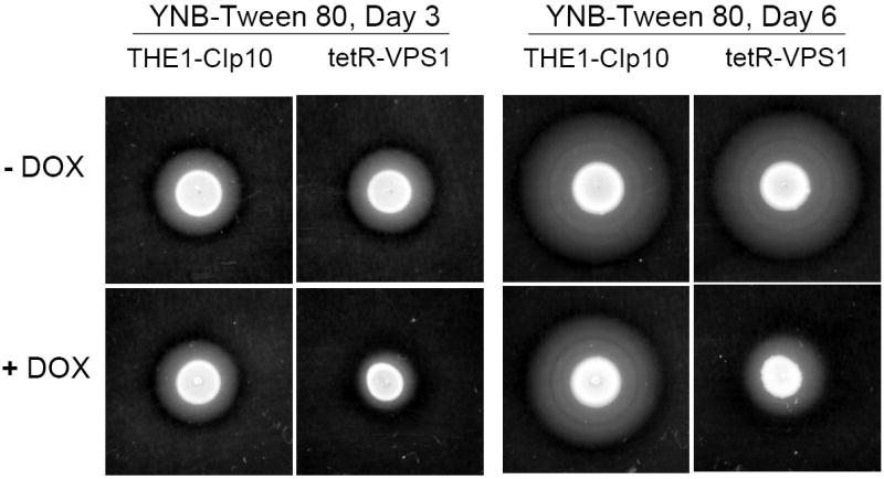
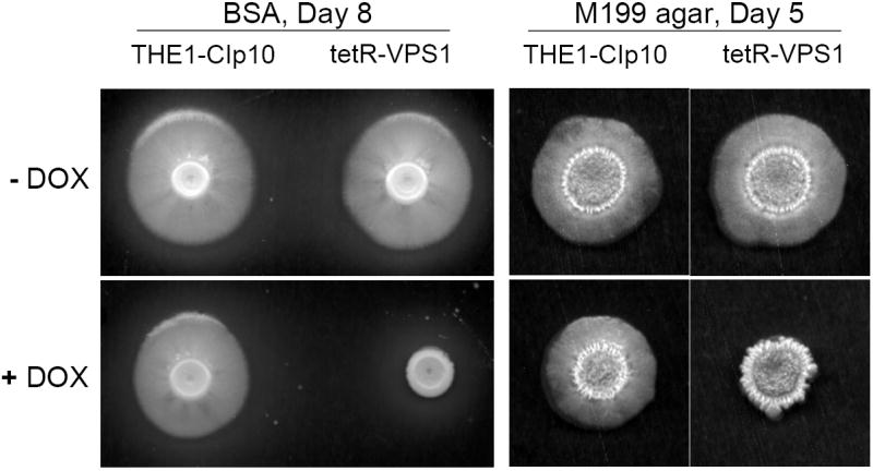
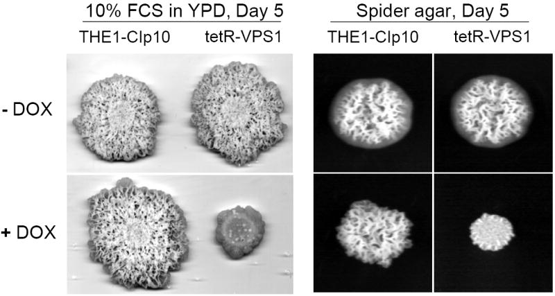
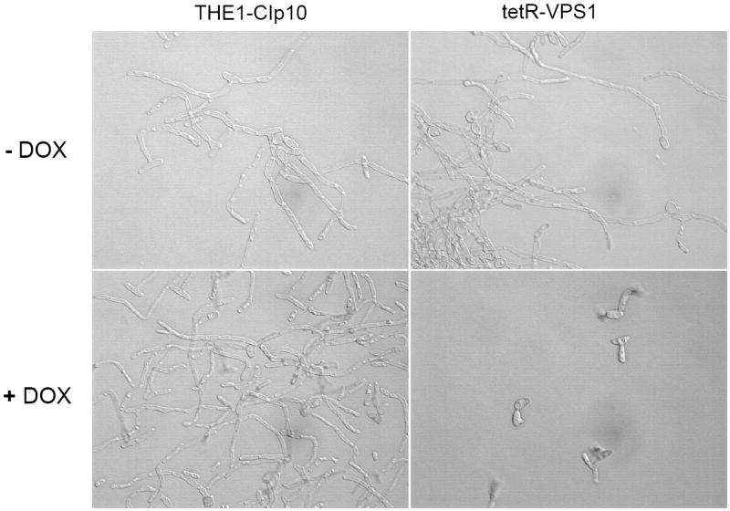
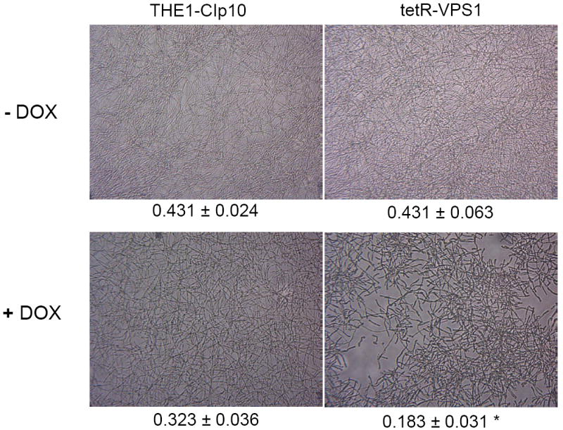
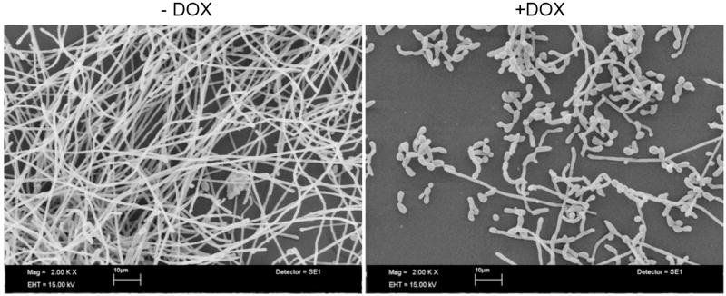
To investigate the pre-vacuolar secretory pathway in Candida albicans, we cloned and analyzed the C. albicans homolog of the Saccharomyces cerevisiae vacuolar protein sorting gene VPS1. C. albicans VPS1 encodes a predicted 694-aa dynamin-like GTPase that is 73.3% similar to S. cerevisiae Vps1p. Plasmids bearing C. albicans VPS1 complemented the temperature-sensitive growth, abnormal class F vacuolar morphology, and carboxypeptidase missorting of a S. cerevisiae vps1 null mutant. To study VPS1 function in C. albicans, a conditional mutant strain (tetR-VPS1) was generated by deleting the first allele of VPS1 and placing the second allele under control of a tetracycline-regulatable promoter. With doxycycline, the tetR-VPS1 mutant was hyper-susceptible to sub-inhibitory concentrations of fluconazole, but not amphotericin B, 5-fluorocytosine, or non-specific osmotic stresses. The repressed tetR-VPS1 mutant was defective in filamentation and secreted less extracellular protease activity. Biofilm production and filamentation within the biofilm were markedly reduced. These results suggest that C. albicans VPS1 has a key role in several important virulence-related phenotypes.


Figures

Fig. 1
Fig. 1. Complementation of a S. cerevisiae vps1 null mutant
Over-expression of C. albicans VPS1 complements the temperature-sensitivity of a S. cerevisiae vps1 null mutant (A). Low copy number yeast shuttle plasmids (pRS316) containing C. albicans VPS1 (Ca VPS1) and S. cerevisiae VPS1 (Sc VPS1) conferred on a temperature-sensitive S. cerevisiae vps1 mutant the ability to grow at 37 °C, but empty vector alone did not. Over-expression of C. albicans VPS1 complements the S. cerevisiae vps1 null mutant vacuolar phenotype (B). Low copy number plasmids (pRS316) containing Sc VPS1, Ca VPS1, or no insert were introduced into a S. cerevisiae vps1 null mutant strain. Overnight cultures of strains grown in rich media were shifted to fresh media and examined during late exponential phase and stained with the vacuolar dye FM4-64. Live cells were examined by epifluorescence and DIC microscopy. The strain bearing vector alone displays a characteristic class “F” vacuolar morphology. Over-expression of C. albicans VPS1 complements carboxypeptidase (CPY) missorting of a S. cerevisiae vps1 null mutant (C). S. cerevisiae vps1 mutants missort CPY to the extracellular space, in contrast to wild-type strains, which do not. Therefore, overnight cultures of a S. cerevisiae wild-type strain and S. cerevisiae vps1 null mutant strains transformed with a vector bearing either Ca VPS1, Sc VPS1, or vector alone were spotted on solid media. Next, a colony immunoblot was performed by overlaying the spotted colonies with a nylon filter, followed by Western blotting using monoclonal antibody to S. cerevisiae CPY. A positive signal indicates missorting of CPY to the extracellular space.
Fig. 2
Fig. 2. Transcriptional analyses of VPS1 in the tetR-VPS1 strains
A Northern blot was probed with a 32P-labeled PCR product amplified from strain SC5314 using primers VPS1-5NB and VPS1-3NB (Table 2). There is complete repression of the VPS1 transcript in the tetR-VPS1 strains when doxycycline is present. RT-PCR on mRNA isolated from the same total RNA used in the Northern blot also showed complete repression of VPS1 transcription in the tetR-VPS1 strains in the presence of doxycycline. An ethidium bromide stained gel shows normalized loading of the RNA samples used in the Northern blot.
Fig. 3
Fig. 3. Characterization of growth of the tetR-VPS1 strain
Growth of the tetR-VPS1 mutant strain was tested in the presence of sub-inhibitory concentrations of fluconazole. In complete synthetic medium supplemented with uridine at 37 °C, growth of the tetR-VPS1 strain was similar to the parental strain THE1 (A). However, growth of the tetR-VPS1 strain was inhibited in the presence of 10 μg ml-1 fluconazole (B). This sensitivity to fluconazole was increased when transcription of VPS1 was completely repressed with the addition of doxycycline. Growth curves were performed in triplicate, and corresponding error bars are indicated. The differences in growth between strain tetR-VPS1 without doxycycline and with doxycycline is statistically significant (* p < 0.0001). Similarly, the differences in growth between strain tetR-VPS1 without doxycycline and THE1-CIp10 is statistically significant (§ p < 0.0001). Statistical significance was tested by fitting growth curves to lognormal models which yielded R-squares of 99%.
Fig. 4
Fig. 4. Vacuolar morphology of the tetR-VPS1 strain
Vacuolar morphology was visualized by FM4-64 staining (A). A 10 μm bar is shown in either white or black. Thin-section electron microscopy images are shown in (B). A solid arrowhead is used to designate the vacuole. In either set of images, no major differences in vacuolar morphology were seen between the URA3-complemented wild-type strain THE1-CIp10 and the tetR-VPS1 strain, with or without doxycycline.
Fig. 5
Fig. 5. Secretion of proteolytic enzymes
Proteolysis assay on BSA plates (A). Overnight cultures were spotted onto BSA plates and incubated at 30 °C for 24 and 48 hours. The relative amount of extracellular protease activity is indicated by the halo surrounding the fungal colony. In the absence of doxycycline, strains THE1-CIp10 and tetR-VPS1 produced similar amounts of extracellular BSA degradation. In the presence of doxycycline, the tetR-VPS1 strain produced much less BSA degradation than strain THE1-CIp10. Proteolysis assay in liquid BSA (B). Overnight cultures were shifted to medium containing BSA as a sole nitrogen source and incubated at 30 °C for 24 hours, with and without doxycycline. The degree of proteolysis of BSA was analyzed by reducing SDS-PAGE and Coomassie blue staining. The tetR-VPS1 strain produced much less proteolysis in the presence doxycycline compared to tetR-VPS1 without doxycycline or the control strain. Lanes from the same gel containing standard protein markers (M) and intact BSA are shown for reference. Western analysis of Sap2p secretion (C). Cell-free supernatants prepared as described in (B) were analyzed by Western blotting using anti-Sap2p antibodies (from M. Monod). Compared to the parental strain THE1-CIp10 (with and without doxycycline) and tetR-VPS1 (without doxycycline), much less Sap2 protein was secreted by the tetR-VPS1 strain when doxycycline was present. The triple deletion mutant strain sap1-3Δ (from B. Hube) was used as a negative control. rSap2 indicates purified recombinant Sap2p (from M. Monod) used as a positive control. Analysis of lipase secretion (D). Overnight cultures were spotted onto YNB-Tween 80 agar plates and incubated at 37 °C. The relative amount of lipolytic degradation after 3 and 6 days is indicated by the halo surrounding the fungal colony. The tetR-VPS1 strain produced much less extracellular lipolytic activity in the presence of doxycycline than corresponding controls. In contrast, no major differences in phospholipase activity were seen when assayed on egg-yolk agar plates (data not shown).
Fig. 6
Fig. 6. Filamentation of strains grown in hyphal-inducing media
Filamentation on BSA and M199 agar plates (A). Overnight cultures of strains THE1-CIp10 and tetR-VPS1 were spotted onto BSA plates and incubated at 30 °C for 8 days. Robust filamentous structures emerging from the edge of each colony were observed in all strains, except in the tetR-VPS1 strain in the presence of doxycycline. Similar results were observed when these strains were spotted onto Medium 199 agar containing Earle’s salts, buffered with 150 mM HEPES to pH 7.5, and incubated at 37 °C for 5 days. Filamentation on Spider and 10% FCS agar plates (B). Overnight cultures were spotted onto either YPD containing 10% fetal calf serum (FCS) or Spider agar medium and incubated at 37 °C for 5 days. On Spider agar, robust filamentation extending above the surface of the plate can be seen in all strains except for the repressed tetR-VPS1 strain, which did not produce a raised colony. Similarly, on YPD agar with 10% FCS, filamentation was robust in all strains except in the tetR-VPS1 strain grown in the presence of doxycycline. Filamentation in liquid RPMI-1640 (C). Filamentation was assessed in RPMI-1640 medium buffered to pH 7.3 with 165 mM MOPS, with or without doxycycline. Medium was inoculated with 5 × 10⁶ cells/ml and incubated at 37 °C for 24 hr with constant shaking at 200 rpm. Strain THE1-CIp10 produced hyphae when grown in the presence or absence of doxycycline. In contrast, strain tetR-VPS1 produced masses of hyphae when grown under derepressing conditions, but not under repressing conditions. Filamentation in YPD containing 10% FCS (D). Pre-warmed medium was inoculated with 5 × 10⁶ cells/ml and incubated at 37 °C with constant shaking at 200 rpm. Induction of filamentation was followed by light microscopy at fixed time points from the time of inoculum, with a four hour time point shown. The tetR-VPS1 strain remained predominantly in the yeast form when grown in the presence of doxycycline, whereas, in contrast the other strains produced longer and greater numbers of filamentous structures.
Fig. 7
Fig. 7
Light microscopy of biofilms formed by strains THE1-CIp10 and tetR-VPS1 (A). The gross morphology of biofilms formed by strains THE1-CIp10 and tetR-VPS1, grown with and without doxycycline, were visualized with light microscopy. Relative values from the XTT-reduction assay are indicated. A statistically significant reduction (* p < 0.001) in biofilm mass was observed in the tetR-VPS1 strain grown under repressed conditions (with doxycycline). Scanning electron microscopy of biofilms formed by the tetR-VPS1 strain (B). Biofilms formed by the tetR-VPS1 strain were visualized using scanning electron microscopy. Under derepressing conditions (without doxycycline), the tetR-VPS1 strain produced a thick biofilm that consisted predominantly of mature hyphae. Under repressing conditions (with doxycycline) the tetR-VPS1 strain produced a biofilm that was markedly less dense, and composed predominantly of yeast and pseudohyphal cells. The figures shown depict representative structures at 2,000x magnification with a 10 μm bar indicated.
Fig. 7
Fig. 7
Light microscopy of biofilms formed by strains THE1-CIp10 and tetR-VPS1 (A). The gross morphology of biofilms formed by strains THE1-CIp10 and tetR-VPS1, grown with and without doxycycline, were visualized with light microscopy. Relative values from the XTT-reduction assay are indicated. A statistically significant reduction (* p < 0.001) in biofilm mass was observed in the tetR-VPS1 strain grown under repressed conditions (with doxycycline). Scanning electron microscopy of biofilms formed by the tetR-VPS1 strain (B). Biofilms formed by the tetR-VPS1 strain were visualized using scanning electron microscopy. Under derepressing conditions (without doxycycline), the tetR-VPS1 strain produced a thick biofilm that consisted predominantly of mature hyphae. Under repressing conditions (with doxycycline) the tetR-VPS1 strain produced a biofilm that was markedly less dense, and composed predominantly of yeast and pseudohyphal cells. The figures shown depict representative structures at 2,000x magnification with a 10 μm bar indicated.

References

    1. Ausubel FM, Brent R, Kingston RE, Moore DD, Seidman JG, Smith JA, Struhl K. Current protocols in molecular biology. Wiley; New York: 1993.
    1. Bates S, Hughes HB, Munro CA, Thomas WP, MacCallum DM, Bertram G, Atrih A, Ferguson MA, Brown AJ, Odds FC, Gow NA. Outer chain N-glycans are required for cell wall integrity and virulence of Candida albicans. J Biol Chem. 2006;281:90–98. - PubMed
    1. Braun BR, Johnson AD. Control of filament formation in Candida albicans by the transcriptional repressor TUP1. Science. 1997;277:105–109. - PubMed
    1. Braun BR, Johnson AD. TUP1, CPH1 and EFG1 make independent contributions to filamentation in Candida albicans. Genetics. 2000;155:57–67. - PMC - PubMed
    1. Brown AJ, Gow NA. Regulatory networks controlling Candida albicans morphogenesis. Trends Microbiol. 1999;7:333–338. - PubMed

Publication types

MeSH terms