. 2008 Apr 25;283(17):11772-84.
doi: 10.1074/jbc.M707572200. Epub 2008 Feb 22.

HIV-1 Nef binds PACS-2 to assemble a multikinase cascade that triggers major histocompatibility complex class I (MHC-I) down-regulation: analysis using short interfering RNA and knock-out mice

Affiliations

HIV-1 Nef binds PACS-2 to assemble a multikinase cascade that triggers major histocompatibility complex class I (MHC-I) down-regulation: analysis using short interfering RNA and knock-out mice

Katelyn M Atkins et al. J Biol Chem. .

Abstract

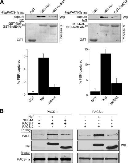
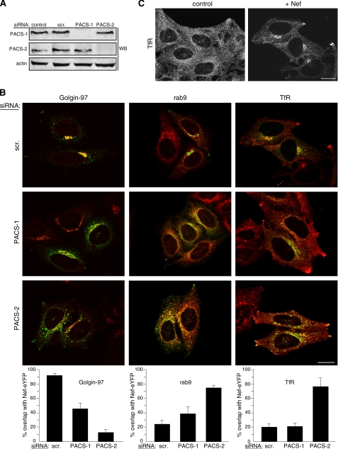
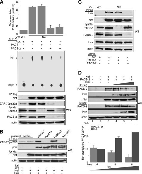
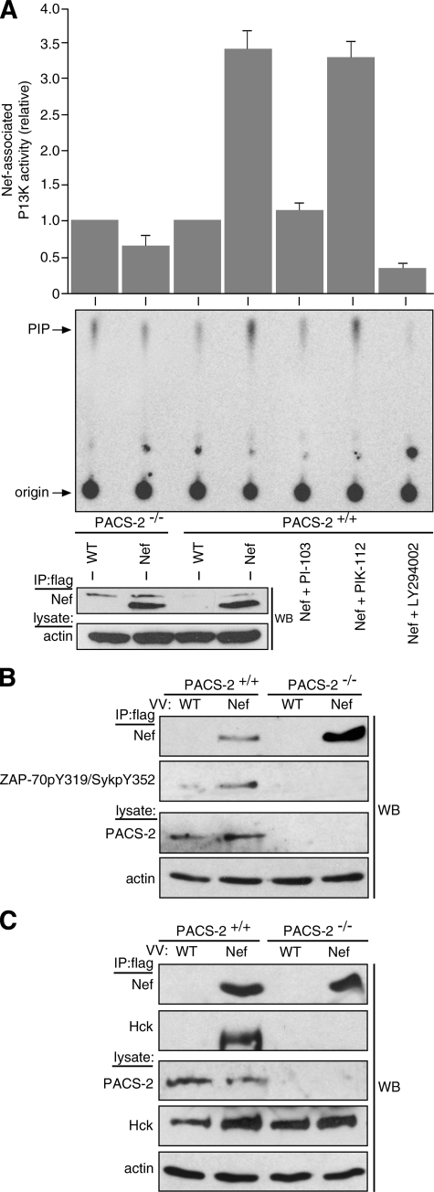
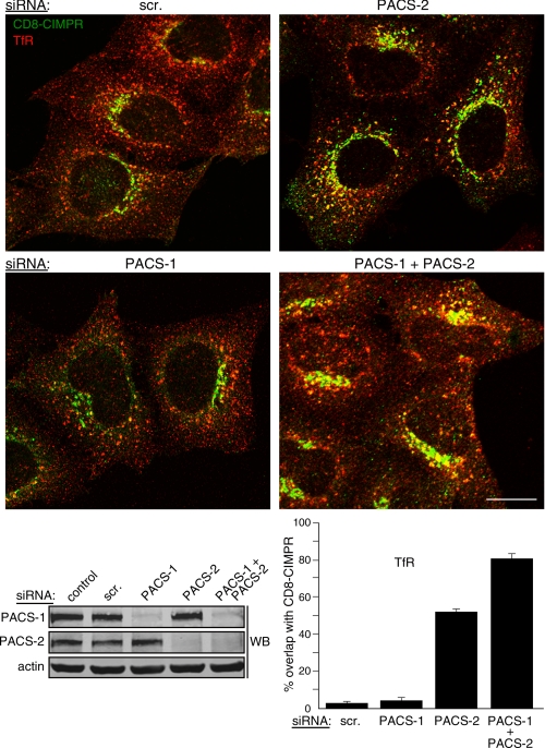
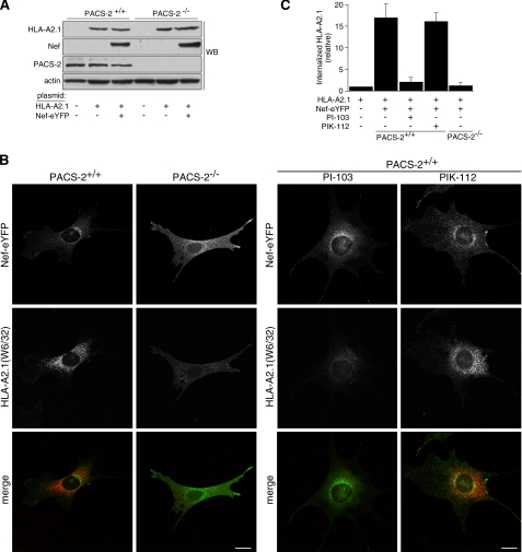
Human immunodeficiency virus, type 1, negative factor (Nef) initiates down-regulation of cell-surface major histocompatibility complex-I (MHC-I) by assembling an Src family kinase (SFK)-ZAP70/Syk-phosphoinositide 3-kinase (PI3K) cascade through the sequential actions of two sites, Nef EEEE(65) and PXXP(75). The internalized MHC-I molecules are then sequestered in endosomal compartments by a process requiring Nef Met(20). How Nef assembles the multikinase cascade to trigger the MHC-I down-regulation pathway is unknown. Here we report that EEEE(65)-dependent binding to the sorting protein PACS-2 targets Nef to the paranuclear region, enabling PXXP(75) to bind and activate a trans-Golgi network (TGN)-localized SFK. This SFK then phosphorylates ZAP-70 to recruit class I PI3K by interaction with the p85 C-terminal Src homology 2 domain. Using splenocytes and embryonic fibroblasts from PACS-2(-/-) mice, we confirm genetically that Nef requires PACS-2 to localize to the paranuclear region and assemble the multikinase cascade. Moreover, genetic loss of PACS-2 or inhibition of class I PI3K prevents Nef-mediated MHC-I down-regulation, demonstrating that short interfering RNA knockdown of PACS-2 phenocopies the gene knock-out. This PACS-2-dependent targeting pathway is not restricted to Nef, because PACS-2 is also required for trafficking of an endocytosed cation-independent mannose 6-phosphate receptor reporter from early endosomes to the TGN. Together, these results demonstrate PACS-2 is required for Nef action and sorting of itinerant membrane cargo in the TGN/endosomal system.


Figures

FIGURE 1.
FIGURE 1.
Nef EEEE65-dependent binding to PACS-1 and PACS-2. A, purified His6PACS-1FBR (residues 117-294) or His6PACS-2FBR (residues 38-217) was incubated with GST, GST-Nef, or GST-NefE4A pre-bound to glutathione-Sepharose. Top, bound proteins were analyzed by Western blot (WB) with anti-His6 antibody showing a 10% input. Bottom, Ponceau S stain of input proteins. Quantification showed that GST-Nef captured 5.8-fold more His6PACS-1FBR and 4.7-fold more His6PACS-2FBR than did GST-NefE4A. Data are representative of three independent experiments. B, H9 CD4+ T-cells were co-infected with VV:PACS-1 (m.o.i. = 3, 8 h) or VV:PACS-2 (m.o.i. = 5,8h) and either VV:WT, VV:Nef/f, or VV:NefE4A/f (m.o.i. = 5, 8 h). FLAG-tagged Nef proteins were immunoprecipitated (IP), and co-precipitating PACS proteins were analyzed by Western blot with antibody 17703 (PACS-1) or antibody 18193 (PACS-2). Quantification showed that Nef/f co-precipitated 3.7-fold more PACS-1 and 6.3-fold more PACS-2 than did NefE4A/f.
FIGURE 2.
FIGURE 2.
Nef-mediated MHC-I down-regulation in primary CD4+ T-cells requires PACS-1 and PACS-2. A, primary human CD4+ T-cells isolated from healthy donors were nucleofected (Amaxa) with a control siRNA (scr) or siRNAs specific for PACS-1 or PACS-2. At 72 h post-transfection, the cells were either harvested for Western blot (WB) of PACS proteins (bottom) or infected with VV:WT or VV:Nef (m.o.i. = 10, overnight), and cell-surface HLA-A2.1 was analyzed by flow cytometry using mAb BB7.1 (top). The expression of Nef is shown in the insets for each graph. B, top, H9 cells were nucleofected (Amaxa) with pmaxGFP together with a control siRNA or siRNAs specific for PACS-1 or PACS-2. At 72 h post-transfection, the GFP+ cells were enriched by FACS and then infected with VV:WT or VV:Nef (m.o.i. = 10, 5 h) and then fixed and stained with W6/32 to visualize MHC-I down-regulation. Scale bar = 20 μm. Bottom, Western blot of lysates from parallel samples of nucleofected cells.
FIGURE 3.
FIGURE 3.
Paranuclear localization of Nef-eYFP requires PACS-2. A, Western blot (WB) of extracts from HeLa cells nucleofected (Amaxa) with a control siRNA (scr) or siRNAs specific for PACS-1 or PACS-2. B, cells in A were nucleofected a second time with pNef-eYFP (green). At 16 h post-transfection the cells were fixed, stained with antibodies for Golgin 97, Rab9, and TfR followed by incubation with fluorescently labeled secondary antibodies (red), and analyzed by confocal microscopy. Morphometric analysis was performed as described under “Experimental Procedures.” Error bars represent mean and S.D. from 20 cells per marker in five independent experiments. Scale bar = 20 μm. C, untreated HeLa cells or HeLa cells expressing Nef-eYFP were fixed, stained with anti-TfR, and analyzed by confocal microscopy. Scale bar = 20 μm.
FIGURE 4.
FIGURE 4.
PACS-2 is required for Nef to assemble the SFK—ZAP-70/Syk—PI3K cascade. A, H9 CD4+ cells were nucleofected (Amaxa) with a control (scr) siRNA or siRNAs specific for PACS-1 or PACS-2 on day 1 and again on day 3. On day 5, cells were infected with either VV:WT or VV:Nef/f (m.o.i. = 10, 8 h). Nef was immunoprecipitated (IP), and co-immunoprecipitating Tyr(P)292Zap70 was detected by Western blot (WB). The amount of Nef-associated PI3K activity was quantified as described under “Experimental Procedures.” Bottom, Western blot showing expression of PACS-1, PACS-2, and actin. Error bars represent the mean ± S.D. of three independent experiments. B, H9 cells were transfected on day 1 with plasmids expressing p85α/f, p85αR358A/f, p85αR649A/f, or p85αRARA/f. On day 3, the cells were infected with VV:WT (m.o.i. = 10, 8 h) or co-infected with VV:Nef (untagged, m.o.i. = 10, 8 h) and VV:Hck (m.o.i. = 2, 8 h). Cells were harvested, and FLAG-tagged p85α was immunoprecipitated with mAb M2 and co-precipitating Tyr(P)292ZAP-70 detected by Western blot. Data are representative of two independent experiments. C, H9 cells were transfected with siRNAs as described in A. On day 5, the cells were co-infected with VV:WT or VV:Nef/f (m.o.i. = 10, 8 h) and VV:Hck (m.o.i. = 2, 8 h). Nef was immunoprecipitated, and co-immunoprecipitating Hck was detected by Western blot. Data are representative of three independent experiments. D, parallel plates of H9 CD4+ cells were infected or not with VV:Nef/f (m.o.i. = 6 each) and VV:PACS-2ha (m.o.i. = 5 each) and with decreasing amounts of VV:WT (m.o.i. in lane 2 = 14, lane 3 = 3, lane 4 = 2, and lane 5 = 1) or VV:Hck (m.o.i. in lane 4 = 1, lane 5 = 2, lane 6 = 3; total m.o.i. = 14, 18 h) and harvested as indicated under “Experimental Procedures.” Nef/f was immunoprecipitated with mAb M2, and co-precipitating Hck and PACS-2ha were detected by Western blot. Error bars represent mean and S.D. from three independent experiments.
FIGURE 5.
FIGURE 5.
Pacs-2 gene knock-out prevents Nef assembly of the SFK—ZAP-70/Syk—PI3K cascade in mouse splenocytes. A, splenocytes isolated from WT C57Bl/6 or PACS-2-/- C57Bl/6 mice were infected with VV:WT or VV:Nef/f (m.o.i. = 25, 8 h). Nef was immunoprecipitated (IP), and PI3K activity was measured as described under “Experimental Procedures.” Western blot (WB) depicts immunoprecipitated Nef/f. A replicate immunoprecipitate from Nef/f-expressing WT C57Bl/6 splenocytes was aliquoted and treated with 0.1 μm PI-103, PIK-112, or 5 μm LY294002, and Nef-associated PI3K activity was measured. Error bars represent the mean ± S.D. of five independent experiments (10 mice total). B, primary splenocytes isolated from WT or PACS-2-/- C57Bl/6 mice were infected as described in A. Nef/f was immunoprecipitated and co-immunoprecipitating Tyr(P)319Zap-70/Tyr(P)352Syk was detected by Western blot. C, primary splenocytes isolated from three paired sets of WT or PACS-2-/- C57Bl/6 mice were co-infected with VV:WT or VV:Nef/f (m.o.i. = 10, 18 h) and VV:Hck (m.o.i. = 2, 18 h). Nef was immunoprecipitated, and co-immunoprecipitating Hck was detected by Western blot. The lower m.o.i. in the VV:WT-infected samples does not affect the result (see Fig. 4D).
FIGURE 6.
FIGURE 6.
PACS-1 and PACS-2 mediate the sorting of endocytosed CD8-CIMPR. HeLa-CD8CIMPR cells were nucleofected (Amaxa) with a control siRNA (scr) or siRNAs specific for PACS-1 or PACS-2. After 48 h, anti-CD8 (mAb UCHT-4, green) was added to the medium for 1 h at 37 °C. The media were removed and replaced with fresh culture medium for 30 min, and the cells were fixed and stained with anti-TfR (red) followed by isoform-specific fluorescent secondary antibodies. The cells were then visualized by confocal microscopy. Morphometric analysis was performed as described under “Experimental Procedures.” Error bars represent mean ± S.D. from 20 cells per marker in eight independent experiments. Bottom, Western blot (WB) of lysates from parallel samples of nucleofected cells. Scale bar = 20 μm.
FIGURE 7.
FIGURE 7.
Nef-eYFP and endogenous CI-MPR are mislocalized in PACS-2-/- MEFs. A, PACS-2+/+ and PACS-2-/- MEFs were transfected (Amaxa) with pNef-eYFP. After 18 h the cells were fixed and stained with anti-mannosidase II or anti-EEA1 (red) and visualized by confocal microscopy. Western blot (WB) in upper right-hand corner of PACS-2 in lysates from PACS-2+/+ and PACS-2-/- MEFs is shown. Inset, Nef-eYFP in dispersed EEA1-postive compartments in PACS-2-/- MEFs. B, PACS-2+/+ and PACS-2-/- MEFs were transfected (Amaxa) or not with pGalT-CFP. After 18 h the cells were fixed, stained with anti-EEA1 (red) or anti-CI-MPR 8738 (red in GalT-expressing cells; green in EEA1 co-stained cells (bottom)), and visualized by confocal microscopy. Inset, CI-MPR in EEA1-positive compartments. Morphometric analysis was performed as described under “Experimental Procedures.” Error bars represent mean ± S.D. from 20 cells per marker in four independent experiments for Nef-eYFP and three independent experiments for CI-MPR. Scale bar = 20 μm.
FIGURE 8.
FIGURE 8.
PACS-2 and class I PI3K are required for Nef to down-regulate MHC-I in MEFs. A, Western blot (WB) of lysates from PACS-2+/+ and PACS-2-/- MEFs co-transfected (Amaxa) with pUHD10.1HLA-A2.1 and pNef-eYFP. B, after 48 h, anti-MHC-I (mAb W6/32, red) was added to the media for 1 h at 37 °C. Where indicated the cells were pretreated with 1 μm PI-103 or PIK-112 for 1 h prior to antibody uptake, and the inhibitors were maintained in the culture medium for the duration of the antibody uptake. Following uptake, the media were removed and replaced with fresh culture media for 30 min in the absence or presence of inhibitor. The cells were fixed, stained with a fluorescent secondary antibody, and then visualized by confocal microscopy. C, morphometric quantitation of internalized HLA-A2.1 (W6/32). The amount of internalized antibody was the same in WT or PACS-2-/- cells transfected with pUHD10.1HLA-A2.1 alone, and therefore the same value was used for normalization of uptake in lane 1. The error bars represent the mean ± S.D. from 20 cells per marker from three independent experiments. Scale bar = 20 μm.

References

    1. Das, S. R., and Jameel, S. (2005) Indian J. Med. Res. 121 315-332 - PubMed
    1. Peterlin, B. M., and Trono, D. (2003) Nat. Rev. Immunol. 3 97-107 - PubMed
    1. Stevenson, M. (2003) Nat. Med. 9 853-860 - PubMed
    1. Stove, V., Naessens, E., Stove, C., Swigut, T., Plum, J., and Verhasselt, B. (2003) Blood 102 2925-2932 - PubMed
    1. Thoulouze, M. I., Sol-Foulon, N., Blanchet, F., Dautry-Varsat, A., Schwartz, O., and Alcover, A. (2006) Immunity 24 547-561 - PubMed

Publication types

Substances