. 2008 Jun;94(11):4493-503.
doi: 10.1529/biophysj.107.121186. Epub 2008 Feb 22.

Measuring the adsorption of Fatty acids to phospholipid vesicles by multiple fluorescence probes

Affiliations

Measuring the adsorption of Fatty acids to phospholipid vesicles by multiple fluorescence probes

Jeffrey R Simard et al. Biophys J. 2008 Jun.

Abstract

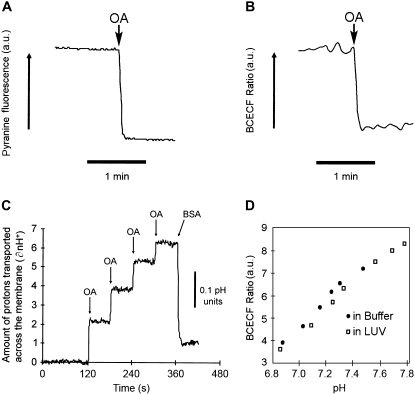
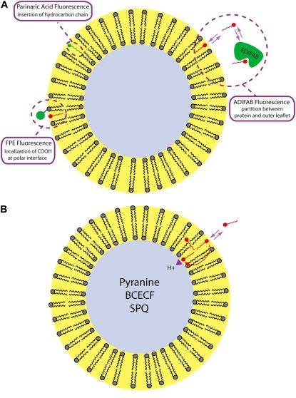
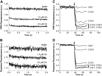
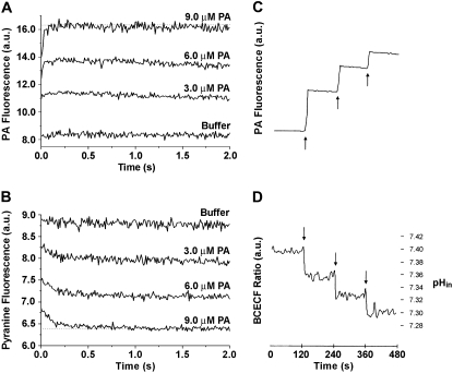
Fatty acids (FA) are important nutrients that the body uses to regulate the storage and use of energy resources. The predominant mechanism by which long-chain fatty acids enter cells is still debated widely as it is unclear whether long-chain fatty acids require protein transporters to catalyze their transmembrane movement. We use stopped-flow fluorescence (millisecond time resolution) with three fluorescent probes to monitor different aspects of FA binding to phospholipid vesicles. In addition to acrylodan-labeled fatty acid binding protein, a probe that detects unbound FA in equilibrium with the lipid bilayer, and cis-parinaric acid, which detects the insertion of the FA acyl chain into the membrane, we introduce fluorescein-labeled phosphatidylethanolamine as a new probe to measure the binding of FA anions to the outer membrane leaflet. We combined these three approaches with measurement of intravesicular pH to show very fast FA binding and translocation in the same experiment. We validated quantitative predictions of our flip-flop model by measuring the number of H(+) delivered across the membrane by a single dose of FA with the probe 6-methoxy-N-(3-sulfopropyl) quinolinium. These studies provide a framework and basis for evaluation of the potential roles of proteins in binding and transport of FA in biological membranes.


Figures

FIGURE 1
FIGURE 1
Measuring the flip-flop of fatty acids to the inner membrane leaflet using pH-sensitive fluorophores. LUV were prepared containing entrapped pyranine (A), BCECF acid (B), and SPQ (C). Vesicles were suspended in buffer (pH 7.4) to a final PC concentration of 500 μM. Single or multiple doses of oleic acid (10 nmol) caused the fluorescence of each probe to change rapidly with a t1/2 < 2 s, indicating rapid diffusion of OA to the inner leaflet. The final concentration of OA after each addition was below the solubility limit (3.3 μM per dose). In C, SPQ fluorescence showed the instantaneous influx of 2.5 nmol H+ per 10.0 nmol of added OA. The subsequent addition of BSA (2.8 mol BSA per 1 mol OA) resulted in extraction of OA from the LUV and an instantaneous efflux of H+ (t1/2 < 2 s). (D) Shows that the fluorescence titration of trapped and untrapped BCECF is not significantly different.
FIGURE 2
FIGURE 2
Schematic diagram of the fluorescence probes and assays used to discriminate FA binding and flip-flop in membranes. (A). ADIFAB, FPE, and cis-parinaric acid are used to monitor LCFA binding to the outer leaflet. ADIFAB measures the concentration of unbound LCFA in equilibrium with the membrane and cis-parinaric acid, the insertion of the acyl chain into the hydrophobic bilayer. FPE detects the arrival of the charged LCFA carboxyl at the membrane surface. These charged FA molecules shift the pKa of FPE in the membrane and induce a decrease in its fluorescence emission intensity. Most LCFA are ionized in solution but become 50% un-ionized on binding to membranes due a shift in their pKa to ∼7.5. (B). Un-ionized LCFA rapidly diffuse to the inner leaflet, reach ionization equilibrium, and release H+ to the internal volume that can be detected by entrapped pH-sensitive fluorophores such as pyranine, BCECF, and SPQ. It is important to note that entrapped probes measure the combined steps of binding and flip-flop. In addition to sensing pH changes, SPQ can also be used to quantitate H+ flux across the membrane in response to the addition of LCFA to the external buffer. Adapted from (43).
FIGURE 3
FIGURE 3
Partitioning of different chain length fatty acids into SUV using the surface potential probe FPE. For the stopped flow measurements (A and B), a suspension of SUV (containing 0.1 mM pyranine) in 20 mM Hepes buffer at pH 7.4 was mixed rapidly with increasing amounts of OA (1.5 and 3.0 μM final concentration) in the same buffer. To minimize potential complications from the precipitation of OA, the initial concentration of OA (before mixing) was below the its estimated solubility limit of 5–6 μM at pH 7.4 (19). Vesicles were mixed with only buffer as a mixing control. The final PC concentration after mixing was 700 μM. Separate measurements were carried out while monitoring the fluorescence of FPE (A) and pyranine (B). All fluorescence traces are the average of 4–6 measurements. On-line FPE experiments were carried out by delivering a single dose of fatty acid (20 μM; arrow) to SUV (500 μM) labeled with 0.2 mol % FPE (containing 0.1 mM pyranine) in 50 mM Hepes buffer at pH 7.4. The fatty acids used were octanoate (C8:0), decanoate (C10:0), laurate (C12:0), myristate (C14:0), palmitate (C16:0), oleate (C18:1), and linoleate (C18:2). FPE (C) and pyranine (D) were monitored simultaneously and the fluorescence of each probe changed rapidly with t1/2 < 2 s. The magnitude of the fluorescence change reflects increased partitioning of LCFA into the membrane.
FIGURE 4
FIGURE 4
Dual fluorescence measurement of fatty acid binding and flip-flop using a pH-sensitive fluorophore and cis-parinaric acid. For the stopped flow measurements (A and B), a suspension of SUV (containing 0.1 mM pyranine) in 20 mM Hepes buffer at pH 7.4 was rapidly mixed with increasing amounts of PA (3, 6, 9 μM final concentration) in the same buffer. Vesicles were mixed with only buffer as a mixing control. The final PC concentration after mixing was 700 μM. Separate measurements were carried out while monitoring the fluorescence of PA (A) and pyranine (B). All fluorescence traces are the average of 4–6 measurements. Parallel on-line experiments were carried out by delivering sequentially three equal doses of PA (20 μM PA; arrows) to a cuvette containing a suspension of SUV (500 μM) containing entrapped 0.2 mM BCECF in 100 mM Hepes buffer at pH 7.4. PA (C) and BCECF (D) were monitored simultaneously and the fluorescence of each probe changed rapidly with t1/2 < 3 s. The calibration of BCECF fluorescence with pHin is described in the Materials and Methods.
FIGURE 5
FIGURE 5
Dual fluorescence measurement of fatty acid binding and flip-flop using a pyranine and ADIFAB. For the stopped flow measurements (A and B), a suspension of ADIFAB and SUV (containing pyranine) in 20 mM Hepes buffer at pH 7.4 was rapidly mixed with increasing amounts of OA in the same buffer (1.5 and 3.0 μM final concentration). Vesicles were mixed with only buffer as a mixing control. The final PC concentration after mixing was 700 μM. Separate measurements were carried out while monitoring the fluorescence of ADIFAB (A) and pyranine (B). All fluorescence traces are the average of 4–6 measurements. Parallel on-line experiments were carried out by delivering five equal doses of OA (10 nmol OA; arrows) sequentially to a cuvette containing a suspension of ADIFAB (0.2 μM) and SUV containing BCECF (500 μM) in “Measuring Buffer” at pH 7.4 (20 mM Hepes/KOH, 150 mM NaCl, 5 mM KCl, 1 mM Na2HPO4). ADIFAB (C) and BCECF (D) were monitored simultaneously and the fluorescence of each probe changed rapidly with t1/2 < 3 s. The concentration of unbound OA per total amount of added OA was plotted (E). A linear fit of these data yield the partitioning coefficient (Kp) of OA into PC membranes. The calibration of ADIFAB and BCECF fluorescence with unbound OA and pHin, respectively, is described in the Materials and Methods.

References

    1. Hamilton, J. A., and F. Kamp. 1999. How are free fatty acids transported in membranes? Is it by proteins or by free diffusion through the lipids? Diabetes. 48:2255–2269. - PubMed
    1. Kleinfeld, A. M. 2000. Lipid phase fatty acid flip-flop, is it fast enough for cellular transport? J. Membr. Biol. 175:79–86. - PubMed
    1. Pownall, H. J., and J. A. Hamilton. 2003. Energy translocation across cell membranes and membrane models. Acta Physiol. Scand. 178:357–365. - PubMed
    1. Cupp, D., J. P. Kampf, and A. M. Kleinfeld. 2004. Fatty acid-albumin complexes and the determination of the transport of long chain free fatty acids across membranes. Biochemistry. 43:4473–4481. - PubMed
    1. Kampf, J. P., D. Cupp, and A. M. Kleinfeld. 2006. Different mechanisms of free fatty acid flip-flop and dissociation revealed by temperature and molecular species dependence of transport across lipid vesicles. J. Biol. Chem. 281:21566–21574. - PubMed

MeSH terms

LinkOut - more resources