. 2008 May;190(9):3118-28.
doi: 10.1128/JB.01784-07. Epub 2008 Feb 22.

Roles of effectors in XylS-dependent transcription activation: intramolecular domain derepression and DNA binding

Affiliations

Roles of effectors in XylS-dependent transcription activation: intramolecular domain derepression and DNA binding

Patricia Domínguez-Cuevas et al. J Bacteriol. 2008 May.

Abstract

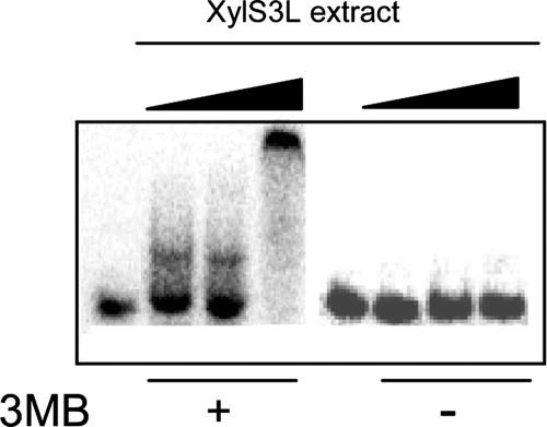
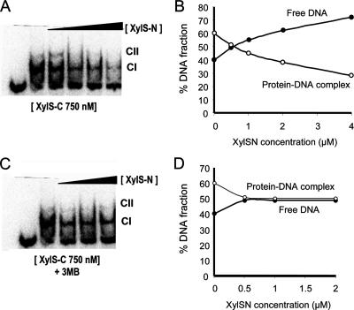
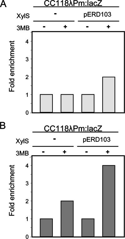
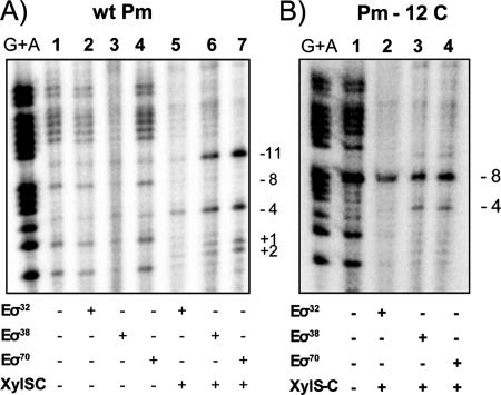
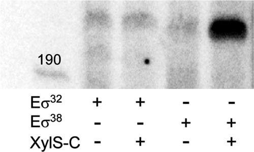
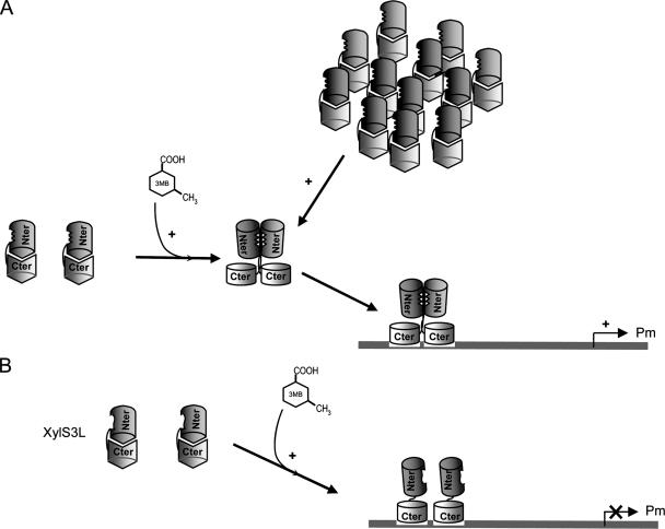
XylS, an AraC family protein, activates transcription from the benzoate degradation pathway Pm promoter in the presence of a substrate effector such as 3-methylbenzoate (3MB). We developed a procedure to obtain XylS-enriched preparations which proved suitable to analyze its activation mechanism. XylS showed specific 3MB-independent binding to its target operator, which became strictly 3MB dependent in a dimerization-defective mutant. We demonstrated that the N-terminal domain of the protein can make linker-independent interactions with the C-terminal domain and inhibit its capacity to bind DNA. Interactions are hampered in the presence of 3MB effector. We propose two independent roles for 3MB in XylS activation: in addition to its known influence favoring protein dimerization, the effector is able to modify XylS conformation to trigger N-terminal domain intramolecular derepression. We also show that activation by XylS involves RNA polymerase recruitment to the Pm promoter as demonstrated by chromatin immunoprecipitation assays. RNA polymerase switching in Pm transcription was reproduced in in vitro transcription assays. All sigma(32)-, sigma(38)-, and sigma(70)-dependent RNA polymerases were able to carry out Pm transcription in a rigorous XylS-dependent manner, as demonstrated by the formation of open complexes only in the presence of the regulator.


Figures

FIG. 1.
FIG. 1.
(A) Schematic representation of XylS-C structure. The predicted α-helixes in the sequence are depicted as gray boxes. The relative sizes of the different helixes are drawn to scale. DNA-contact helixes α3 and α6 are in white. (B) Pm promoter sequence organization. The bold arrows indicate the two XylS binding sites (proximal and distal), each composed of conserved A1/A2 and B1/B2 boxes. The −10 and −35 hexamers are in bold and double-underlined. A right-angled arrow indicates the transcription initiation site.
FIG. 2.
FIG. 2.
Heparin chromatography of XylS-containing extracts and estimation of XylS concentration in the extracts. (A) XylS was purified from inclusion bodies after solubilization with 6 M guanidium, renaturing, and His-affinity chromatography. It is worth noting that XylS protein obtained with this protocol was inactive. Samples of known XylS concentration (5, 3.5, 2.5, 1.5, 1, 0.5, 0.1, 0.01, and 0.001 μg loaded in lanes 1 to 9, respectively) were separated by denaturing sodium dodecyl sulfate-PAGE, transferred to a nitrocellulose membrane, and probed with antibodies at a dilution of 1/1,000 against XylS (76). (B) Cell extracts (170 μg of total protein) of E. coli CC118 (pLOW2::XylS) (lanes 1 and 2) or E. coli CC118 (pLOW2) (lanes 3 and 4) were loaded in a 1-ml heparin column, and samples were eluted as indicated in Materials and Methods. Lanes 1 and 3, whole extract; lanes 2 and 4, extract eluted from the column. Molecular weight markers were BSA (66 kDa), ovalbumin (45 kDa), pepsin (36 kDa), carbonic anhydrase (29 kDa), trypsinogen (24 kDa), and trypsin inhibitor (20 kDa).
FIG. 3.
FIG. 3.
DNA binding of XylS to Pm promoter. EMSA for binding of 32P-labeled wild-type Pm DNA fragment (A) or mutant Pm245 DNA fragment (B) by purified extracts containing wild-type XylS protein. EMSA was performed as described in Materials and Methods with either no protein added (lane 1) or increasing amounts (0.5, 1, 2, 5, 10, and 15 μg) of XylS-enriched extracts (lanes 2 to 7). An excess of specific (0.5 μg of wild-type Pm DNA fragment; lane 8) or nonspecific competitor [1 μg of poly(dI-dC) DNA; lane 9] was added to reaction mixtures that also contained 15 μg of crude extract. Wild-type and mutant Pm sequences used in panels A or B, respectively, are shown. A and B box sequences are shown in bold, and mutations in Pm245 nucleotides are underlined.
FIG. 4.
FIG. 4.
DNA binding of mutant XylS(3L) to wild-type Pm promoter. EMSA for binding of 32P-labeled wild-type Pm DNA fragment in the presence (+) or absence (−) of 3MB by purified extracts containing mutant XylS(3L) protein. Mobility shift assays were performed as described in Materials and Methods with either no protein added (first and fifth lanes from the left) or increasing amounts (0.5, 1, and 10 μg) of extracts that contained XylS(3L) (second to fourth lane and sixth to eighth lane). Controls with an excess of nonlabeled Pm DNA strongly reduced the amount of shifted DNA, while the addition of excess unspecific DNA did not modify the shift.
FIG. 5.
FIG. 5.
N-terminal domain repression of XylS-C binding to Pm DNA. EMSA for binding of 32P-labeled wild-type Pm DNA fragment by XylS-C in the presence and absence of increasing concentrations of purified XylS-N in the absence (A and B) or presence (C and D) of 1 mM 3MB. EMSAs were performed as described in Materials and Methods with either no protein added or a fixed amount of purified XylS-C (750 nM) in the presence of increasing concentrations of XylS-N (from 0.5 to 4 μM). (B and D) The fraction of total radiolabel in each band from each lane was quantified and plotted as a function of XylS-N concentration: black circles, free DNA; white circles, XylS-C-DNA complex.
FIG. 6.
FIG. 6.
ChIP analysis of RNA polymerase and XylS binding to the Pm promoter. ChIP was carried out as described in Materials and Methods. XylS binding to the Pm promoter in the presence and absence of 3MB was determined. E. coli CC118λPm::lacZ with or without the plasmid pERD103 was grown in the presence or absence of 3MB. Data show the ratio of real-time PCR quantitation of anti-XylS (A) or anti-β-subunit (B) antibody immunoprecipitate to nonspecific precipitate without antibody, generated by the Pm promoter sequence and corrected with reference to the mlt sequence, in each strain and under each growth condition. Data represent the enrichment of QT-PCR product relative to the control CC118λPm::lacZ strain in the absence of 3MB. Results are the average of three independent experiments.
FIG. 7.
FIG. 7.
Potassium permanganate footprinting of the Pm promoter. The DNA (bottom strand) in the presence of either no protein (lane 1) or RNA polymerase with different sigma factors in the presence (lanes 5 to 7 in A and lanes 2 to 4 in B) or absence (lanes 2 to 4 in A) of XylS-C was modified with potassium permanganate (10 mM) and cleaved with piperidine. The numbers (−11, −8, −4, +1, and +2) indicate the cleavage sites. Wt, wild type.
FIG. 8.
FIG. 8.
Effect of XylS on Pm transcription. Transcription was performed using reconstituted RNA polymerase with either σ32 or σ38 and XylS-C in the presence of [α-32P]UTP-labeled nucleotide. RNA products were resolved by urea-PAGE. The template was a linear DNA fragment containing Pm and was obtained as indicated in Materials and Methods.
FIG. 9.
FIG. 9.
Model for XylS activation of the Pm promoter. (A) Under basal conditions, XylS DNA-binding domains (light gray barrels) are unable to make contact with DNA. The addition of 3MB produces a conformational change with two important consequences: (i) dimerization interactions are favored between monomers, probably because monomer dimerization regions are exposed; and (ii) the regulator DNA-binding domains are opened, which favors contacts with DNA. Under physiological conditions (i.e., in vivo), XylS does not dimerize unless 3MB is present. When overexpressed or in purified preparations, high concentrations of XylS favor dimerization in the absence of effector. Dimerization might also lead to conformational changes which make DNA-binding domains more available for interactions with target DNA sites. In both cases, XylS binding to Pm in the presence of RNA polymerase activates the promoter. (B) In XylS(3L), 3MB generates the corresponding conformational change, but the result in this case is only the opening of DNA-binding sites, since this mutant is unable to dimerize. Accordingly, the bound protein is not able to promote transcription.

References

    1. Ausubel, F. M., R. Brent, R. E. Kingston, D. D. Moore, J. G. Seidman, J. A. Smith, and K. Struhl. 1991. Current protocols in molecular biology. Wiley, New York, NY.
    1. Bhende, P. M., and S. M. Egan. 1999. Amino acid-DNA contacts by RhaS: an AraC family transcription activator. J. Bacteriol. 1815185-5192. - PMC - PubMed
    1. Bhende, P. M., and S. M. Egan. 2000. Genetic evidence that transcription activation by RhaS involves specific amino acid contacts with sigma 70. J. Bacteriol. 1824959-4969. - PMC - PubMed
    1. Bradford, M. M. 1976. A rapid and sensitive method for the quantitation of microgram quantities of protein utilizing the principle of protein-dye binding. Anal. Biochem. 72248-254. - PubMed
    1. Busby, S., and R. H. Ebright. 1997. Transcription activation at class II CAP-dependent promoters. Mol. Microbiol. 23853-859. - PubMed

Publication types

MeSH terms

LinkOut - more resources