. 2008 Mar 4;105(9):3339-44.
doi: 10.1073/pnas.0800165105. Epub 2008 Feb 22.

Glucocorticoids differentially regulate degradation of MyoD and Id1 by N-terminal ubiquitination to promote muscle protein catabolism

Affiliations

Glucocorticoids differentially regulate degradation of MyoD and Id1 by N-terminal ubiquitination to promote muscle protein catabolism

Liping Sun et al. Proc Natl Acad Sci U S A. .

Abstract

Accelerated protein degradation via the ubiquitin-proteasome pathway is the principal cause of skeletal muscle wasting associated with common human disease states and pharmacological treatment with glucocorticoids. Although many protein regulatory factors essential for muscle development and regeneration are degraded via the ubiquitin system, little is known about the mechanisms and regulation of this pathway that promote wasting muscle. Here, we demonstrate that, in differentiated myotubes, glucocorticoid, via the glucocorticoid receptor, selectively induces a decrease in protein abundance of MyoD, a master switch for muscle development and regeneration, but not that of its negative regulator Id1. This decrease in MyoD protein results from accelerated degradation after glucocorticoid exposure. Using MyoD and Id1 mutants deficient in either N terminus-dependent or internal lysine-dependent ubiquitination, we further show that these ubiquitination pathways of MyoD degradation are regulated differently from those of Id1 degradation. Specifically, glucocorticoid activates the N-terminal ubiquitination pathway in MyoD degradation in myotubes, without concomitant effects on Id1 degradation. This effect of glucocorticoid on MyoD and Id1 protein degradation is associated with the distinct cellular compartments in which their degradation occurs. Taken together, these results support a key role for the N terminus-dependent ubiquitination pathway in the physiology of muscle protein degradation.


Conflict of interest statement

The authors declare no conflict of interest.

Figures
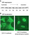
Fig. 1.
Fig. 1.
Protein expression of glucocorticoid receptor (GR) (A) during C2C12 myogenic differentiation and localization of GR (B) in C2C12 myoblasts with or without dexamethasone treatment (1 μM for 16 h).
Fig. 2.
Fig. 2.
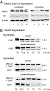
MyoD and Id1 protein expression (A) and MyoD protein degradation half-lives (t1/2 h) (B) in C2C12 myoblasts (A) and myotubes (B) with or without dexamethasone treatment. 1, control; 2, dexamethasone at 1 μM for 16 h; 3, mifepristone (RU486) at 1 μM for 16 h; and 4, dexamethasone and RU486 at 1 μM each for 16 h. For half-life in myotubes, C2C12 myoblasts were transiently transfected with MyoD encoding plasmid DNA. Upon confluence, transfected C2C12 cells were switched to differentiation medium and maintained for 4 days with medium changed every 24 h. Protein half-life analysis was then performed by using cycloheximide (CHX) and proteasome inhibitor MG132 as described in Materials and Methods. The lower MyoD band represents the hypophosphorylated form (18).
Fig. 3.
Fig. 3.
Half-lives of wild-type MyoD (anti-MyoD), MyoD-LL-HA (anti-HA), and 6×Myc-MyoD (anti-Myc) in myoblasts (A) and in myotubes (B) with or without dexamethasone treatment. Protein half-lives were determined in a similar fashion as described in the Fig. 2 legend.
Fig. 4.
Fig. 4.
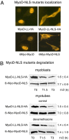
Subcelluar localization (A) of MyoD-LL-NLS-HA and 6×Myc-MyoD-NLS in C2C12 myoblasts and protein degradation half-lives (B) of MyoD-LL-NLS-HA and 6×Myc-MyoD-NLS in C2C12 myoblasts and myotubes with or without dexamethasone treatment. (A) Localization of various MyoD protein forms was determined with immunofluorescent staining and fluorescence microscopy. (B) MyoD half-lives were determined in a similar fashion as described in the Fig. 2 legend. MG132 stabilized the degradation of these MyoD mutants (data not shown).
Fig. 5.
Fig. 5.
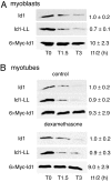
Half-lives of wild-type Id1(anti-Id1), Id1-LL (anti-Id1) and 6×Myc-Id1 (anti-Myc) in C2C12 myoblasts (A) and myotubes (B) with or without dexamethasone treatment. Id1 half-lives were determined in a similar fashion to that described in the Fig. 2 legend.
Fig. 6.
Fig. 6.
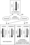
A proposed model for the ubiquitin–proteasome-mediated degradation of MyoD and Id1 in muscle cells. For Id1 degradation, the N-terminal ubiquitination pathway is dominantly active in both myoblasts and myotubes with or without glucocorticoid administration. For MyoD degradation in myoblasts and myotubes without exposure to excess glucocorticoids, the internal lysine-dependent ubiquitination pathway is much more active in the nucleus, whereas, in the cytoplasm, the N-terminal ubiquitination pathway is more active. In myotubes upon glucocorticoid administration, the N-terminal ubiquitination pathway is activated and becomes dominant in MyoD degradation in the nucleus, whereas no significant change is induced regarding the activities of the two ubiquitination pathways within the cytoplasm. The thickness of the arrows indicates relative pathway activity.

References

    1. Hong DH, Forsberg NE. Effects of dexamethasone on protein degradation and protease gene expression in rat L8 myotube cultures. Mol Cell Endocrinol. 1995;108:199–209. - PubMed
    1. Thompson MG, et al. Stimulation of myofibrillar protein degradation and expression of mRNA encoding the ubiquitin–proteasome system in C (2)C(12) myotubes by dexamethasone: Effect of the proteasome inhibitor MG-132. J Cell Physiol. 1999;181:455–461. - PubMed
    1. Wang L, Luo GJ, Wang JJ, Hasselgren PO. Dexamethasone stimulates proteasome- and calcium-dependent proteolysis in cultured L6 myotubes. Shock. 1998;10:298–306. - PubMed
    1. Bodine SC, et al. Identification of ubiquitin ligases required for skeletal muscle atrophy. Science. 2001;294:1704–1708. - PubMed
    1. Combaret L, et al. Glucocorticoids regulate mRNA levels for subunits of the 19 S regulatory complex of the 26 S proteasome in fast-twitch skeletal muscles. Biochem J. 2004;378:239–246. - PMC - PubMed

Publication types